Section: Skin and soft tissue Sub-section: Skin cancer Curriculum: Curriculum, page 52
Definition
- Aggressive cutaneous malignancy
- Arise from multipotent stem cells which undergo neuroendocrine transformation when they become malignant
- The cell of origin is debated - it was traditionally believed that they arose from Merkel cells which are located in the basal layer of the epidermis - these are a/w with dermal papillae and skin mechanoreceptor
- Also known as:
- Cutaneous Neuroendocrine Carcinoma
- Small Cell Tumour of the Skin
- Primary undifferentiated carcinoma of the skin
- Anaplastic carcinoma of the skin
- Murky cell carcinoma
Incidence
- Rare: 0.3 / 100,000
- < 1% of skin malignancies
- Older adult 60-80 years of age, whites, M≈F
-
50% occur in the head and neck
Risk factors
- Lighter skin colour
- Increasing age
- Male sex
- Immunosuppression - especially organ transplant patients, HIV, and hematological malignancies.
- Other malignancies increase risk - CLL, multiple myeloma, melanoma.
- UV radiation exposure - more common in sun-exposed areas.
- Merkel cell polyomarvirus infection
- Germline nutations in BRCA1, BRCA2, TP53, ATM, are a/w Merkels
Merkel Cell polyomavirus
- 60-80% of patients have detectable MCPyV in their specimens.
- Merk Cell polyomavirus integrates two oncogenes into the host genome - which encode proteins large tumour antigen and small tumour antigen, which help with immune evasion.
Clinical Presentation
- AEIOU
- Asymptomatic
- Expanding rapidly
- Immunosuppressed
- Older than 50yrs
- UV exposed/fair skin
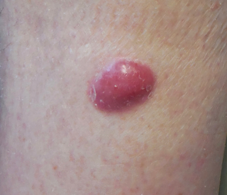
- Firm, painless, shiny lump (raised intradermal nodule)
- Can be red, pink, or blue (“violaceous”)
- Overlying Epidermis intact
- Ulceration rare; suggestive of rapid growth
- Grows rapidly & often metastasizes → Lymphatics → Satellite Lesions → LNs
- 70-80% present with localized disease
- 1-4% present with metastatic disease
Differential diagnosis
- For rapidly growing lesions:
- Small cell carcinoma of the lung with cutaneous metastasis.
- Small cell melanoma - this would stain positive for HMB-45, Melan-A, and S-100
- Basal cell carcinoma
- Lymphoma
- Dermatofibroma
- Squamous cell carcinoma
Regional disease
- Must examine nodal basins
- High propensity for regional and distant mets
- 15% <0.5, 25% <1cm, 40% <2cm
- Spread via lymphatics
- Mets to LN and liver > bone > brain > lung > skin
- May completely regress (replaced by a lymphocytic infiltrate)
Pathology
- Immunohistochemical
- Stains are usually required to differentiate it from other poorly differentiated tumours.
- Express AE1/AE3, CAM 5.2, and CK20
- Also express various neuroendocrine markers on histochemistry including chromogranin, synaptophysin, calcitonin, VIP, and somatostatin
- Microscopically
- Dermal mass which extends into the subcutaneous tissue.
- It rarely involves the epidermis.
- Small round blue cells with neuroendocrine features
- Nests of monotonous, round, blue cells, containing large basophilic nuclei and minimal cytoplasm.
Investigations
- Excision biopsy
- Narrow margins or incisional/punch biopsy
- Immunohistochemistry (Cytokeratin 20 +ve)
- Stain with K20, neuron specific enolase (neuroendocrine marker)
- FNA any suspicious LN
- Staging
- All patients should undergo CT-PET
- In NZ, it is approved for patients prior to radical therapy for Merkel Cell Carcinoma
- All patients should undergo CT-PET
- Others tests
- Merkel Cell polyomarvirus serology - should be measured.
- Patients who are seronegative have an increased risk of recurrence
- Merkel Cell polyomarvirus serology - should be measured.
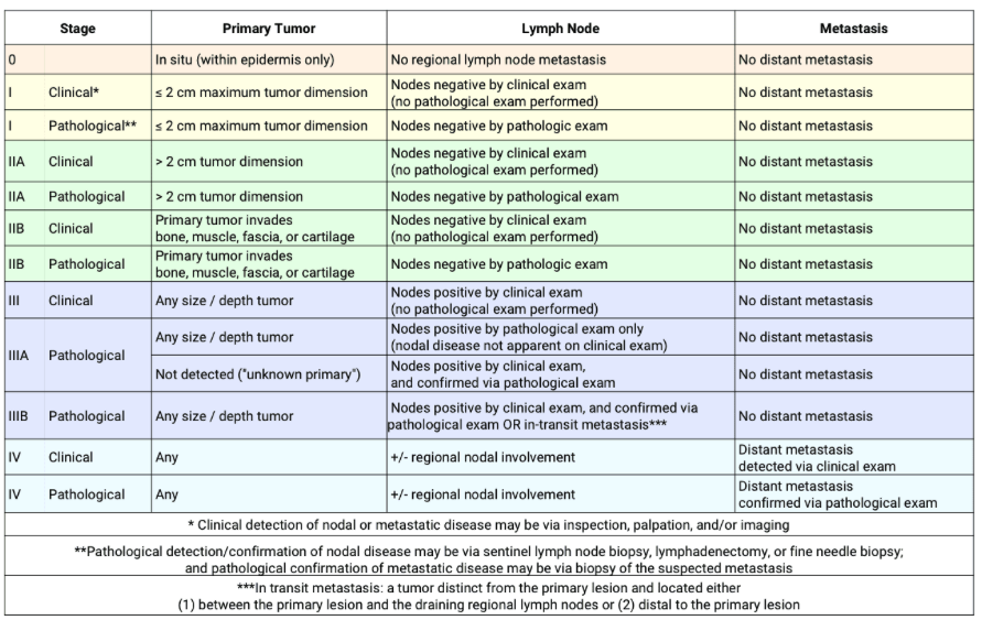
Staging
T Staging
Clinical and pathological are the same
| T category | T criteria |
|---|---|
| TX | Primary tumor cannot be assessed (eg, curetted) |
| T0 | No evidence of primary tumor |
| Tis | In situ primary tumor |
| T1 | Maximum clinical tumor diameter ≤2 cm |
| T2 | Maximum clinical tumor diameter >2 but ≤5 cm |
| T3 | Maximum clinical tumor diameter >5 cm |
| T4 | Primary tumor invades fascia, muscle, cartilage, or bone |
N Staging
Clinical
| N category | N criteria |
|---|---|
| NX | Regional lymph nodes cannot be clinically assessed (eg, previously removed for another reason, or because of body habitus) |
| N0 | No regional lymph node metastasis detected on clinical and/or radiologic examination |
| N1 | Metastasis in regional lymph node(s) |
| N2 | In-transit metastasis (discontinuous from primary tumor; located between primary tumor and draining regional nodal basin, or distal to the primary tumor) without lymph node metastasis |
| N3 | In-transit metastasis (discontinuous from primary tumor; located between primary tumor and draining regional nodal basin, or distal to the primary tumor) with lymph node metastasis |
| Pathological |
| N category | pN criteria |
| pNX | Regional lymph nodes cannot be assessed (eg, previously removed for another reason or not removed for pathological evaluation) |
| pN0 | No regional lymph node metastasis detected on pathological evaluation |
| pN1 | Metastasis in regional lymph node(s) |
| pN1a(sn) | Clinically occult regional lymph node metastasis identified only by sentinel lymph node biopsy |
| pN1a | Clinically occult regional lymph node metastasis following lymph node dissection |
| pN1b | Clinically and/or radiologically detected regional lymph node metastasis, microscopically confirmed |
| pN2 | In-transit metastasis (discontinuous from primary tumor; located between primary tumor and draining regional nodal basin, or distal to the primary tumor) without lymph node metastasis |
| pN3 | In-transit metastasis (discontinuous from primary tumor; located between primary tumor and draining regional nodal basin, or distal to the primary tumor) with lymph node metastasis |
M stage
Clinical and pathological the same
Clinical
| M category | M criteria |
| M0 | No distant metastasis detected on clinical and/or radiologic examination |
| M1 | Distant metastasis detected on clinical and/or radiologic examination |
| M1a | Metastasis to distant skin, distant subcutaneous tissue, or distant lymph node(s) |
| M1b | Metastasis to lung |
| M1c | Metastasis to all other visceral sites |
Pathological
| M category | M criteria |
| M0 | No distant metastasis detected on clinical and/or radiologic examination |
| pM1 | Distant metastasis microscopically confirmed |
| pM1a | Metastasis to distant skin, distant subcutaneous tissue, or distant lymph node(s), microscopically confirmed |
| pM1b | Metastasis to lung, microscopically confirmed |
| pM1c | Metastasis to all other distant sites, microscopically confirmed |
TNM stage
- Clinical
- Stage 1
- T1
- Stage 2
- A - T2-T3
- B - T4
- Stage 3
- N1-N3
- Stage 4
- M1
- Stage 1
- Pathological
- Stage 1
- T1
- Stage 2
- A - T2-3
- B - T4
- Stage 3
- A - N1a, N1a(sn)
- B - N1b-3
- Stage 4
- M1
- Stage 1
Break that down
- Stage 1 - T1 <2cm
- Stage 2 - T2-T4, >2cm +/- invasion
- Stage 3 - positive lymph nodes
- Stage 4 - distant mets
Management
Clinically negative regional lymph nodes
- WLE of the primary tumour with SLNB
- SLNB
- Negative
- Observe
- Positive
- Either completion lymph node dissection or RT to regional nodes.
- If the completion lymph node dissection reveals multiple involved nodes or extracapsular extension - should consider adjuvant RT.
- Either completion lymph node dissection or RT to regional nodes.
- Note
- Some centres = No SLNB if > 2cm - because the risk of LN positivity is so high the patient should be given RT regardless.
- If a patient has a negative SLNB, they may still receive RT if they are perceived high risk
- Negative
- SLNB
Clinically positive regional nodes
- Should confirm diagnosis with FNA or excision biopsy
- Patients should undergo therapeutic lymph node dissection or RT.
- If there are multiple positive nodes OR extracapsular extension - should receive adjuvant RT
Management of the primary tumour
- WLE with a margin of 1-3cm
- Or
- Primary RT
- Peter Mac Lecture: Surgical excision rarely performed now and main treatment is primary radiotherapy + adjuvant PD-1 Inhibitor
- Tumours with high risk features should also receive adjuvant RT
High risk features
- Tumour >2cm
- Microscopically positive or limited surgical margins.
- LVI
- Head and neck primary
- Immunosuppression.
Unresectable disease
- Definitive RT
Treatment of systemic disease
- Pembrolizumab and Nivolumab are used
Outcomes
- 5 year survival of 30-50%.