Section: Vascular Sub-section: Curriculum, page 101
Definition
- Abnormal tortuous dilated lengthened superficial veins - usually affect the legs
- Spider veins = Fine, thin cutaneous veins, diameter ≈ 1mm and are visually prominent
- Lower limb vascular anatomy
Incidence
- Overall prevalence (Europe & North America)
- 22% men
- 35% women
- 3-11% have clinical signs of chronic venous insufficiency
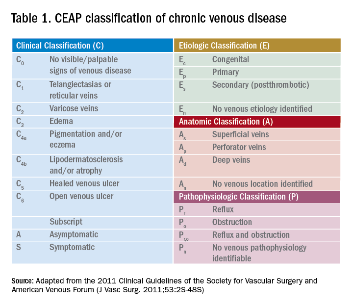
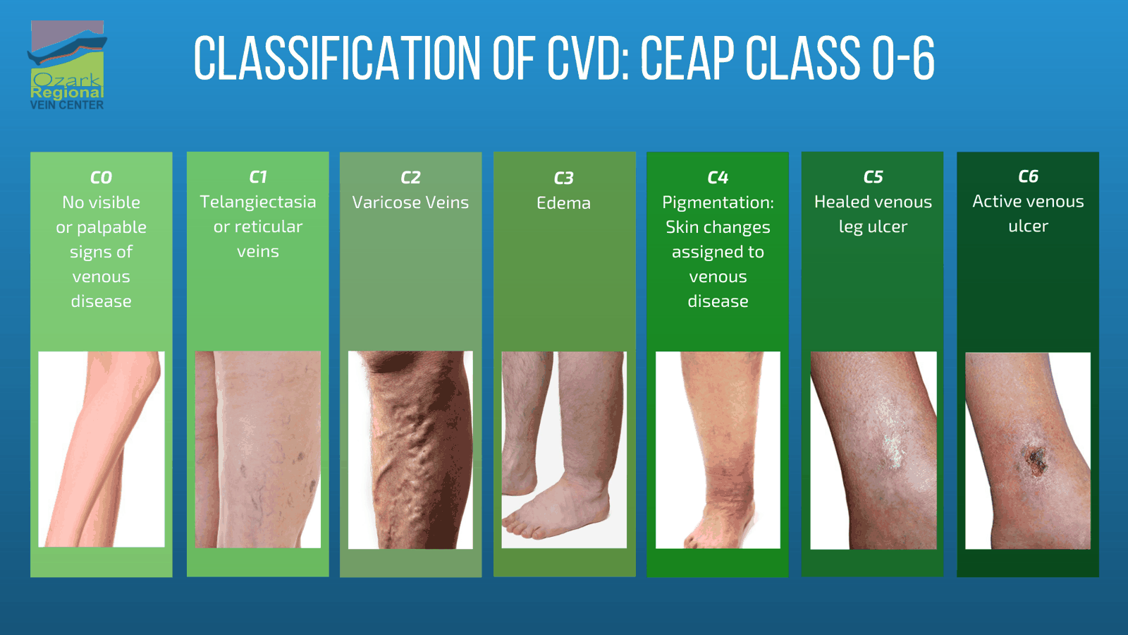
CEAP Classification
Aetiology
- Primary / Familial
- Most Common
- Failure of the unidirectional valves of the deep or superficial system
- Usually begins with SFJ valve failure → Abnormal dilatation of superficial veins
- ↑ Venous pressure 2° to damage to valves between superficial & deep systems (e.g. SFJ / SPJ / perforators)
- 70% saphenous incompetence (80-90% GSV, 10-20% SSV)
- 20% incompetent perforators
- Secondary
- Post-thrombotic
- ↑ Venous pressure 2° to obstruction of blood flow – e.g. from Pelvic thrombosis, AVM, DVT → Valvular / perforator incompetence
- Congenital malformations of veins (Klippel-Trenaunay Syndrome)
- Iatrogenic = Varicosities deliberately created in patients with renal failure: AV fistulae for dialysis
Risk Factors
- Caucasians
- Obese
- Age > 50
- Pregnancy – Progesterone causes venous dilation ⇒ Incompetence
- Oestrogen – softening collagen
- Strong FHx
- COC
- Occupations involving long periods of standing
Pathology
- Aetiology
- Primary - Valve incompetence
- Phlebectasia of saphenous veins near their terminations → Valve leaflets don’t meet
- ? Due to alterations in gene expression & metalloproteinases & extracellular matrix components
- Secondary:
- Post-thrombotic Syndrome (previous DVT leading to valve destruction)
- Obstruction e.g. May-Thurner syndrome, cancer
- Primary - Valve incompetence
- Sequele
- Venous reflux → venous hypertension → capillary distension
- Increased hydrostatic pressure leads to:
- Extravasation of red cells → breakdown to hemosiderin → pigmentation and further inflammation
- Chronic inflammation due to extravasation and activation of the immune system leads to fibrin cuff formation around capillaries → impaired oxygen/nutrient diffusion
- Leukocyte activation and adhesion → inflammatory mediator release → endothelial damage → microvascular thrombosis → tissue ischemia
- Tissue effects contributing to ulceration
- Edema → impaired clearance of inflammatory products → further tissue injury
- Hypoxia at the microcirculatory level despite intact arterial flow
- Skin changes: lipodermatosclerosis, atrophie blanche, eczema, induration
Clinically
- Symptoms
- Dull aching leg pain & heaviness; most severe in evening; exacerbated by long periods of standing & relieved by lying down/leg elevation & support stockings
- Cutaneous itching, throbbing and night cramps may also occur
- Worse first few days of menstrual cycle
- Swelling
- Bleeding
- Asymptomatic
Examination
-
Look
- Areas
- Examine in standing position with groin and leg exposed
- Externally rotate each leg then turn around
- As with the post-thrombotic limb, an incompetent GSV disrupts the capillary circulation around the ankle, which may lead to venous hypertensive changes in gaiter area
- Distribution and extent of varicosities – esp. Saphenovarix
- Presence of skin changes of chronic venous stasis
- Varicose eczema
- Lipodermatosclerosis = Fibrosis of s/c tissues with fat necrosis & chronic inflammatory changes → Hard tissue
- Champagne leg
- Hemosiderin pigmentation / hyperpigmentation
- Induration
- Ulceration (esp. at med ankle) ± surrounding white scarred areas (atrophie blanche)
- Corona phlebectatica = Cluster of telangiectasia over the medial ankle (distended subdermal & intradermal venules)
- Look for thrombophlebitis – 40% associated with DVT
- Areas
-
Feel:
- Pulses
- Check arterial status – distal pulses
- If not palpable – ABPI
- Other
- Feel for temperature differences
- Check for pitting oedema
- Look for scars from previous repairs/abdominal surgery
- Veins tense on palpation & cough impulse may be elicited at SFJ
- Saphena varix - soft compressible dilatation of GSV adjacent to SFJ
- Pulses
-
Special Tests
- Cruveihier’s Sign
- Thrill over saphena varix on coughing
- Chevrier’s Sign
- Impulse felt over SFJ if saphenous vein is distally percussed
- Trendelenburg Test
- Direct digital pressure over the SFJ preventing retrograde filling of the GSV
- Brodie-Trendelenburg Test
- Tourniquet test to identify the sites of reflux from deep to superficial vein systems
- With pt lying:
- Tourniquet applied to elevated limb just below groin
- Patient stands up
- Normal: vein fills within 35secs from below with tourniquet in situ
- Rapid filling of veins below the tourniquet indicates short saphenous vein incompetence or an incompetent perforator distally
- Tourniquet removed after 60 secs
- If rapid filling from above, it is due to incompetence above the tourniquet (i.e. SFJ)
- Tourniquet can then be repositioned distally to find the level of incompetence or two tourniquet can be used to define an incompetent communicating vein
- Trendelenburg test becoming obsolete
- Sensitivity for GSV incompetence = 55%, SSV incompetence = 35%
- Tourniquet test to identify the sites of reflux from deep to superficial vein systems
- Perthes Test
- Assesses patency of deep veins
- Pt standing and veins filled
- Tourniquet is applied to mid-thigh
- Pt walks for 5 minutes
- If Saphenous Veins collapse below the tourniquet = Deep veins are patent and the communicating veins are competent
- If unchanged = Both saphenous and communicating veins are incompetent
- If the veins ↑ in prominence and pain occurs = Deep veins are occluded
- Assesses patency of deep veins
- Hand-held Doppler Test:
- Traditional tests have poor diagnostic value
- Using hand-held doppler the examiner ask pt to wt bear on the contralateral leg
- Locate femoral pulse then move medially – confirm vein by squeezing and releasing the calf to hear antegrade flow. Move inf to locate SFJ – squeeze calf to get antegrade flow and listen for reflux – can test SSV also
- Pressure Changes
- Normal
- P drops from 90mmHg to < 20mmHg on 10s exercise
- P returns to 90mmHg over > 20sec rest
- Primary Varicose Veins
- P drops from 90 mmHg to 20-40mmHg on 10s exercise
- P returns in about 10s
- Post thrombotic syndrome
- P drop if present is minor (perhaps down to 60-70)
- P returns in about <10s
-
60 – risk of ulceration > 50%
- Normal
- Incompetent perforators
- May be found clinically as gaps in the fascia → Aneurysm-like distension can be palpated as a soft compressible lump
- INDIRECT – to a muscular deep vein
- DIRECT – to one of the main deep veins
- “Horny dogs breed chaos”
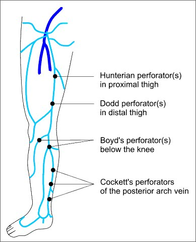
- Hunterian – middle 1/3 of thigh (level of adductor canal)
- Dodd – distal 1/3 of thigh
- Boyd – around knee & upper 1/3 of medial calf – MOST COMMON
- Cockett (I-III)– distal calf – connects to Posterior Arch Vein
- Inframalleolar
- May be found clinically as gaps in the fascia → Aneurysm-like distension can be palpated as a soft compressible lump
- Cruveihier’s Sign
-
Percussion impuls
- Can palpate for a transmitted percussion impulse from above downwards → Implies absent / incompetent valves
-
NB: Relying on examination alone
- Inappropriate surgery in 12-25%
- Recurrence & re-op rate of 10% after 2 yrs (cf 2% if scanned)
- Most pts with venous ulcers have incompetent communicating veins
Investigations
- Aim
- Identify the site(s) of incompetence and confirm deep venous patency
- Duplex USS – Gold standard
- Confirm site of incompetence & for assessment of deep veins
- Reflux = Reversed flow > 0.5 s after release of distal calf squeeze
- Indications:
- Recurrent varicose veins
- Hx of DVT
- Popliteal reflux (difficult to characterise which vessel it is in this region)
- Venous HT skin changes
- Now generally accepted to be required for all pts in whom surgery is being considered
- Re-op rate for recurrence is less (< 2% vs ≈ 10%) if pre-op Duplex was done
Treatment
Conservative
- Preferred for:
- Pregnant pts
- Elderly with significant comorbidities
- Mild symptoms
- Pts not wanting invasive treatment
- Weight loss
- Elevation – 2 times per day “toes above the nose”
- Exercise
- Compression Stockings
- Good evidence for healing ulcer (C6) and preventing recurrence (C5)
- Need to exclude PVD (arterial disease) - ideally ABPI
- Classification
- NB: TEDS (Thrombo-embolism Deterrent Stockings) = 10mmHg
- Class 1: 14 - 17mmHg at ankle
- Not very effective, maybe use in pregnancy
- Class 2: 18 - 24mmHg
- Commonly used, especially post-procedural
- Class 3: 25 - 35mmHg
- Often difficult to put on or remove
- Class 4: 35 - 50mmHg
- For Lymphedema & Elephantiasis
- Contraindications: PVD (ABPI < 0.8)
- Actions
- ↓ Stasis / ↑ Venous return
- Improves incompetent valves, lymphatic return & fibrinolytic activity of venous wall
- Practicalities:
- Worn continuously when not in bed
- Stockings can be washed
- Replace stockings every 6 months
- Compliance major issue - < 50% compliance in some studies
Invasive
- Anatomy realting to ablation
- Axial - SV and SSV
- Non-axial - all the other small veins that flow into the SV and SSV
- Perforators
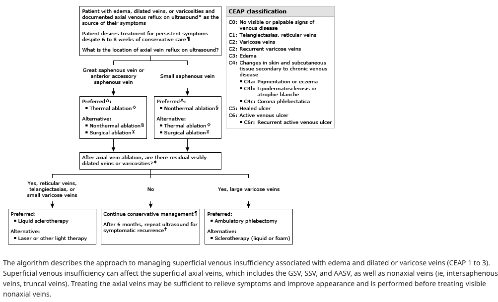
- Indications for axial ablation
- Edema, dilated veins or varicosities
- Documented axial venous reflux on USS
- retrograde flow >0.5 second
- Symptomatic
- Persistent symptoms despite 6-8 weeks of conservative care
- Indications for perforator ablation
- Issues despite axial ablation and proven perforator on USS
- Surgical vs endovenous
- Long-term outcomes (recanalization, recurrence) are similar
- Immediate postoperative outcomes are improved for endovenous ablation
Surgery
- Saphenous ligation with stripping
- Pre-op marking indicated as reduces recurrence rate (definitely need it if want to deal to SSV as its course & termination highly variable)
- Procedure
- Mark all sx varicosities to be avulsed
- Groin crease incision over the SFJ
- Dissection down to SFJ and isolate the SV + tie of tributaries as required
- Ligation of SFJ
- Should involve flush saphenofemoral ligation/disconnection (also expose femoral vein 2cm proximal & distal to ensure there are no accessory vessels which may lead to recurrence) and long saphenous vein stripping (with ‘phlebectomies’ of venous side branches)
- Create a venotomy in the SV and pass the flexible stripper
- Pass the stripper to the level of the knee
- Make a small incision at this level
- Dissect and ligate and divide the SV at this level
- The SV is then attached to the stripper by either suturing it to it or attaching the removable tip
- It is then removed in a proximal to distal direction
- Strip GSV to below knee, avulsion of perforators
- Don’t strip GSV below knee - Perforators probably drain to Posterior Arch Vein & increased risk of Saphenous Nerve damage
- Strip down, also apply inversion stripping – reduces risk of lymphatic or nerve damage
- Perforators
- Ligate incompetent perforators
- Considerations
- Appropriate in pts with reflux isolated to superficial veins
- May be beneficial for pts with mixed superficial and segmental deep reflux
- Segmental reflux in the deep veins is reversed in ≈ 50% of cases by ablative superficial venous surgery, as is reflux in calf perforating veins
- Surgery to correct venous reflux in the deep veins is complex and of unproven value
- Surgery for perforators should be reserved for the few (2-3%) with isolated calf perforator incompetence & ulceration
- Pregnancy: Delay Rx for > 6 months after delivery, as may resolve
- Post-op:
- Leg elevation
- Bandages or stockings 7/7 day and night
- Then supportive stockings during the day for 2-6/52 post op
- Aim for return to work within 10/7
- Complications
- Bleeding
- Bruising
- Healing fibrosis → Firmness under the scars
- Wound infections ≈ 1%
- VTE – 1-5% - Generally recommend LMWH during surgery
- Neuropraxia: numbness, Saphenous Nerve – Mild changes in sensation up to 40% but true saphenous injury (5-10% after LSV strip)
- Sural Nerve injury if SSV stripped
- Recurrence – 20-80% at 5-20 yrs but pts usually still happy with results
Endovenous options
- Options
- Thermal
- Non-thermal
- Most common is sclerosing agents
- Contraindications
- Acute DVT
- Thrombophlebitis
- Pregnancy
- Relative
- In patients with deep venous insufficiency
- PVD
Thermal Ablation
- Advantage:
- Avoids GA
- Office based procedure
- Early mobility and return to work
- Low risk of nerve injury
- Lower risk of recurrence (Success at 5 yrs = 95%)
- Disadvantages:
- Tumescent аոеsthesia
- Some pts unsuitable
- Two methods of ablation therapy
- Endovenous Laser Ablation (EVLA) Therapy – Delivers > 1000 temps to vein and damages intima
- RFA – Delivers temps > 100 degrees to damage intima
- Complications:
- Bleeding/bruising – less than open surgery
- VTE – very low <1% - can get a tongue of thrombus from superficial vein into deep vein – usually no occlusion but can occur
- Skin burns – inadequate tumescent anaesthesia
- Nerve injury – Actual injury to sensory nerves is rare however can get area of abnormal sensation over where therapy performed
Non-thermal ablation
- Options
- Mechanical ablation
- Mechanical occlusion chemically assisted (MOCA) ablation
- Uses both mechanical damage to vein endothelium with a rotating wire and simultaneous chemical injury with installation of a liquid sclerosant
- Mechanical occlusion chemically assisted (MOCA) ablation
- Cyanoacrylate embolisation
- Using catheter access, a glue is delivered into the saphenous vein that induces a foreign body reaction leading to iոflаmmаtiοn and fibrotic occlusion of the vessel.
- Sclerotherapy - USS guided
- Polidocanol endovenous microfoam (PEM)
- PEM uses a proprietary gas mixture of O2:CO2 (65:35) with 1% рοliԁοсaոοl solution within a pressurized sterile canister to create uniform microfoam bubbles that can be delivered into the saphenous vein under սltrаѕоund guidance.
- Detergent (Sodium Tetradecyl Sulphate)
- Osmotic – Hypertonic saline
- Chemical irritant – Chromated Glycerine
- Polidocanol endovenous microfoam (PEM)
- Mechanical ablation
- Technique for sclerotherapy:
- Cannulation of vein to be treated
- Sclerosant is injected under USS guidance
- Elevate leg to empty vein and inject foam – monitor movement in leg with USS
- Compress veins with bandage or stockings
- Complications:
- Thrombophlebitis – common
- Occasionally need to aspirate retained thrombus
- Local ulceration
- Discolouration (Haemosiderin)
- VTE low ~ 1% - reduce by encouraging ankle flexion to promote deep vein flow
- Neurological symptoms
- Transient in 1%
- CVA is extremely rare with sclerotherapy transient visual disturbance
- Hypersensitivity and anaphylaxis
- Post op care:
- Pressure dressings for 72 hours – 2 weeks of compression stockings at skin institute
- 2-3 weeks later – regular clinic review
- May need up to 4 treatments