Venous Anatomy
- Deep + Superficial system + Perforators
- Deep veins run with arteries (but often double)
Deep System
- Beneath the deep fascia
- Mostly follow the arteries
- Dorsal venous arch
- Posterior tibial and fibular veins
- Popliteal vein
- Profunda femoris vein
- Femoral Vein
Superficial system
Great Saphenous Vein
- Superficial to deep fascia but deep to superficial fascia
- Starts at medial side of foot from Medial Marginal Vein
- Courses in front of Medial Malleolus
- Runs up behind medial border of Tibia
- Passes knee joint just behind medial condyles, hands breadth behind patella
- Enters Femoral Vein at Saphenofemoral Junction (SFJ) via Fossa Ovalis (Cribriform fascia) 3-4cm lateral & inferior to Pubic Tubercle
- Termination highly constant
- Valves: 5 to 25 (every 7cm)
- Most numerous distally
- Deep External Pudendal Artery runs medially behind the Saphenous vein near its termination
- Variations: Joins mid-thigh, doesn’t join, bifid (10%)
Important Tributaries (superficial to superficial fascia)
- Join at knee
- Anterior Tributary Vein
- Posterior Tributary Vein
- Posterior Arch Vein (of Leonardo)
- At knee common to have communication between GSV & SSV (usually flow SSV → GSV)
- Join in thigh
- Anterolateral Vein
- Posteromedial Vein (very large – can be mistaken for the GSV)
- Can be called Anterior & Posterior Accessory GSVs
- At SFJ (smaller tributaries)
- Identical to the Femoral Artery branches
- Superficial Epigastric (may join Thoracoepigastric = Communication between SVC/IVC)
- Superficial Circumflex Iliac
- Superficial External Pudendal (may have to divide to adequately expose SFJ)
- Deep External Pudendal (Variation: Drains directly into Femoral Vein)
- Identical to the Femoral Artery branches
Source: NETTER ATLAS 8th
Short Saphenous Vein
- Starts at the lateral aspect of Dorsal Venous Arch
- Ascends behind Lateral Malleolus
- Course to the midline behind calf
- Usually enters Popliteal Vein (70%) in Popliteal Fossa but will often penetrate fascia anywhere from middle of calf to upper 1/3
- May run within a tunnel of deep fascia
- Termination highly variable
- Saphenopopliteal junction may be 7cm above popliteal skin crease
- May enter on strange angle, lateral, anterior
- If doesn’t enter Popliteal vein may enter GSV, Femoral Vein, Profunda, Gluteal Veins
- Can send an Accessory Vein (Giacomini) to GSV making SPJ H-shaped
- Valves: 4-12
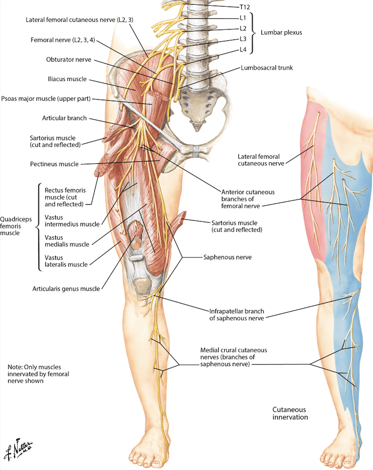
Saphenous Nerve
- Off Femoral nerve
- Saphenous Nerve usually anterior to GSV in lower leg but gets right up to vein 12cm below knee crease
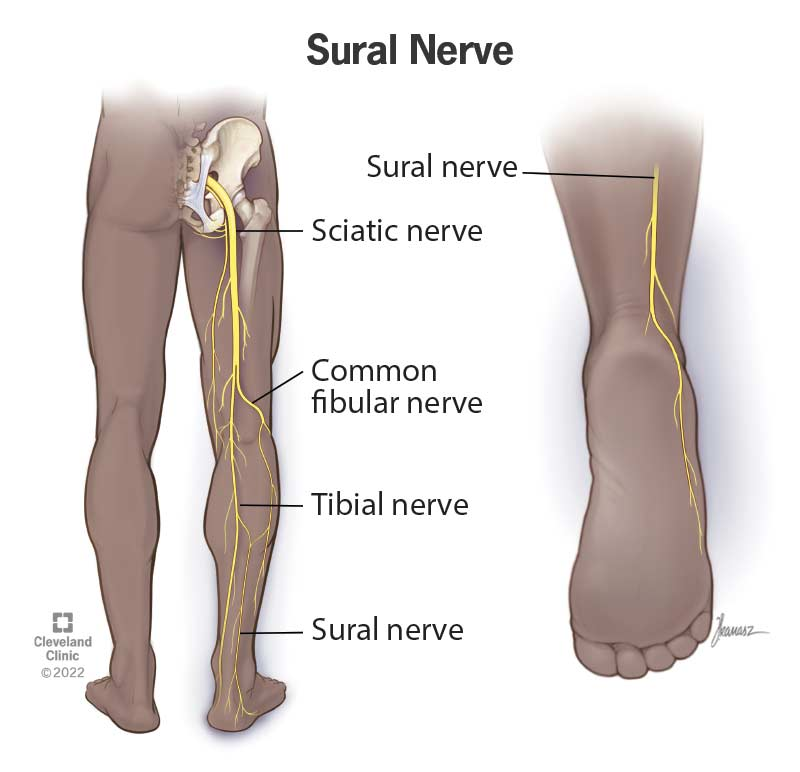
Sural Nerve
- Cutaneous branch off Tibial Nerve (S1/ S2)
- Supplies posterolateral sensation
- Lies in proximity to distal half of SSV
- Deep the SSV runs between heads of Gastrocnemius & can be in close relationship to Tibial Nerve
Perforators
- Clinically important in Varicose veins
- -Incompetent perforators may be found clinically as gaps in the fascia → Aneurysm-like distension can be palpated as a soft compressible lump
- INDIRECT – to a muscular deep vein
- DIRECT – to one of the main deep veins
- -Incompetent perforators may be found clinically as gaps in the fascia → Aneurysm-like distension can be palpated as a soft compressible lump
- “Horny dogs breed chaos”
- Hunterian – middle 1/3 of thigh (level of adductor canal)
- Dodd – distal 1/3 of thigh
- Boyd – around knee & upper 1/3 of medial calf – MOST COMMON
- Cockett (I-III)– distal calf – connects to Posterior Arch Vein
- Inframalleolar