Definition
- Compression of the left common iliac vein by the right common iliac artery against bony structures, leading to venous obstruction or thrombosis.
Epidemiology
- Prevalence is underestimated due to asymptomatic cases.
- Underlying cause of 2-5% of symptomatic DVTs.
Risk Factors
- Gender: Female, particularly postpartum, multiparous, or on oral contraceptives.
- Other Factors: Spinal abnormalities, prior aortoiliac stents, dehydration, hypercoagulable states.
Clinical Features
- Acute presentation: Extremity pain, swelling, and venous claudication.
- Chronic symptoms: Venous insufficiency.
- Typical case: Young females (2nd or 3rd decade) with acute left lower extremity swelling.
Diagnosis
- Initial Tests: Duplex ultrasound.
- Advanced Imaging:
- For non-thrombotic MTS: CT or MR venography.
- For suspected DVT-associated MTS: Presumptive clinical diagnosis; confirm with invasive venous imaging after thrombus removal.
- Definitive Diagnosis:
- Intravascular ultrasound (IVUS): High sensitivity and specificity, shows morphology and stenosis.
Treatment
- No Symptoms: No treatment needed.
- Mild Symptoms: Conservative management with compression stockings.
- Moderate-to-Severe Symptoms:
- Without DVT: Angioplasty with stenting preferred over angioplasty alone.
- With DVT:
- Catheter-directed thrombolysis or pharmacomechanical thrombolysis plus anticoagulation to uncover and confirm stenosis.
- Follow-up with angioplasty and stenting.
- Post-Treatment Management:
- Compression stockings (30-40 mmHg).
- Antiplatelet therapy if bleeding risk is low.
- Continued anticoagulation for DVT per guidelines.
- Surgical Treatment:
- Rarely needed; reserved for contraindications to thrombolysis.
- Open femoral venotomy can be performed to evacuate thrombus before angioplasty/stenting.
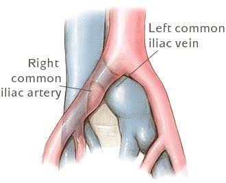
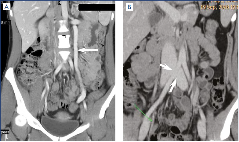
- Left pelvic congestion syndrome and right MTS