Section: Vascular Sub-section: Curriculum, page 102
Thrombotic Risk
- In the Absence of Anticoagulant Therapy
- AF alone: 2% / year risk of stroke
- AF + ↑age /HT/ CHF/DM/ CVA or TIA 4% / year
- Recent / recurrent DVT: 40% chance of recurrence
- Prosthetic Heart Valve: 4% / year risk of CVA / thromboembolism
- Bileaflet (St Jude) valve: 10% Aortic; 20% Mitral
- Multiple St Jude Valves: 90% / year risk of thromboembolism
- Single leaflet tilting valve (Bjork-Shiley) 23% Aortic
- Mitral > Aortic
- Cage Ball (Star Edwards) > Others
Virchow’s Triad
- DVT in gen surg patients without treatment/ prophylaxis:
- 0.5 to 1.6 percent
- Higher among those undergoing sսrgеry for mаligոаnсy (up to 3.7 percent)
Endothelial Injury
- Trauma: Direct injury to blood vessels (fractures, contusions).
- Surgery: Particularly orthopedic (hip, knee replacements) or major abdominal/pelvic surgeries.
- Central Venous Catheters: Placement of lines or ports.
- Atherosclerosis: Chronic vascular injury.
- Inflammation: Vasculitis or systemic inflammatory conditions.
- Radiation Therapy: Direct damage to vascular endothelium.
Hypercoagulability
- Inherited thrombophilia
- Factor V Leiden mutation.
- Prothrombin G20210A mutation.
- Deficiency of antithrombin, protein C, or protein S.
- Polycythaemia
- Thrombocytopenia
- Acquired
- Cancer (especially pancreatic, brain, or lung cancer).
- Pregnancy and postpartum period.
- Hormonal therapy (oral contraceptives, hormone replacement therapy).
- Antiphospholipid syndrome.
- Sepsis or systemic inflammatory response syndrome (SIRS).
- Nephrotic syndrome (loss of anticoagulant proteins).
- Obesity.
- Smoking.
Stasis of Blood Flow
- Immobility:
- Prolonged bed rest (e.g., hospitalization).
- Long-distance travel (flights >4 hours, car trips)
- Surgery
- Length of operation > 2 hours
- Type of operation
- Hip or knee surgery
- Abdominal and pelvic surgery
- Laparoscopic
- Paralysis: Stroke or spinal cord injury.
- Venous Obstruction:
- Pregnancy (compression by the gravid uterus).
- Tumors or mass effect.
- Congestive heart failure.
- Varicose veins or chronic venous insufficiency.
- May-Thurner syndrome
Risk Groups & incidence
Peri-operative patients can be stratified using the Caprini model which is a cumulative score reflecting the number of risk factors present. Risk factors include:
- Demographics
- Concurrent illness
- Recent surgery
- Family or personal history
- Nature of surgery
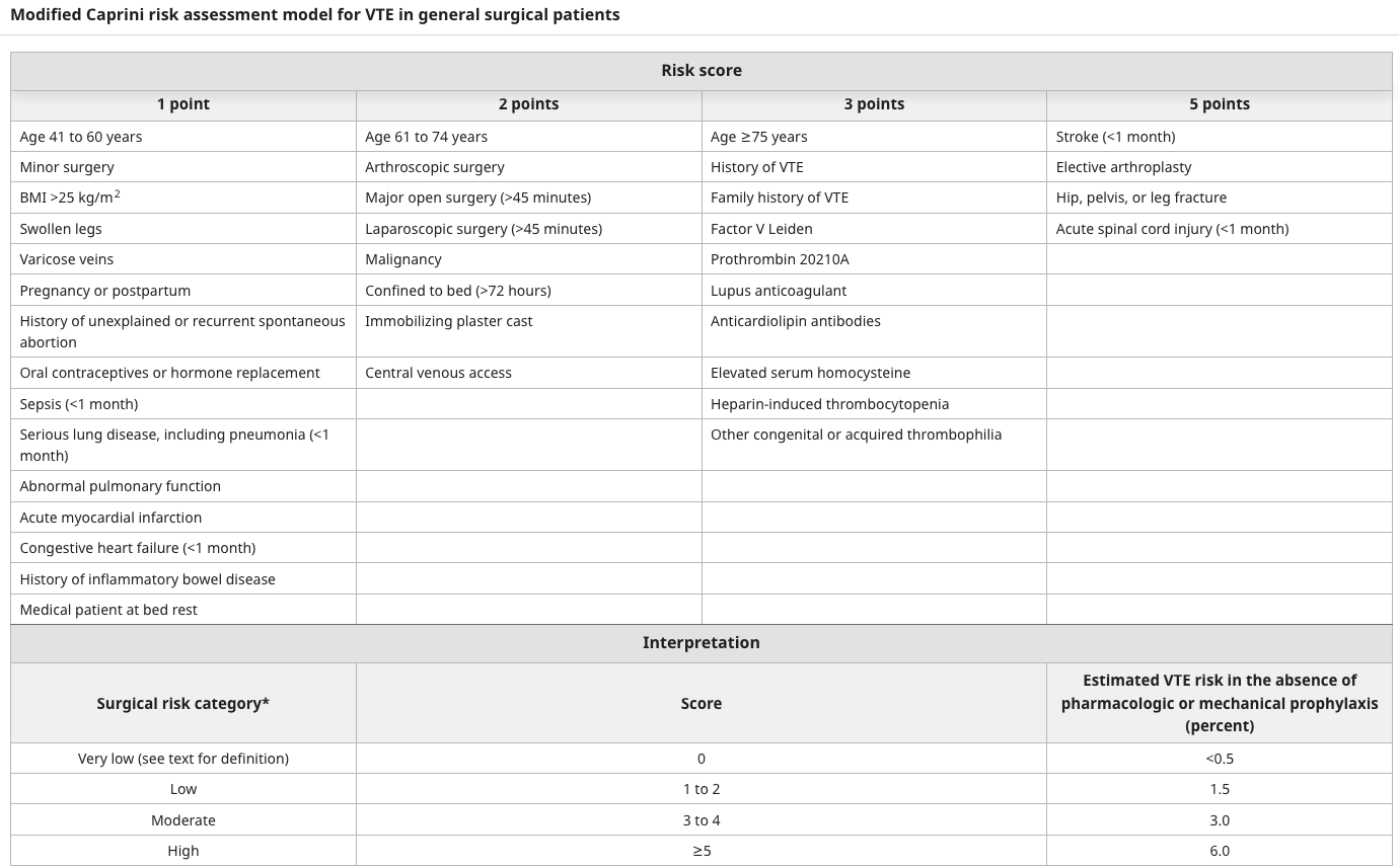
Patients are stratified as low, intermediate, or high, or very high risk and thromboprophylactic treatment reflects this.
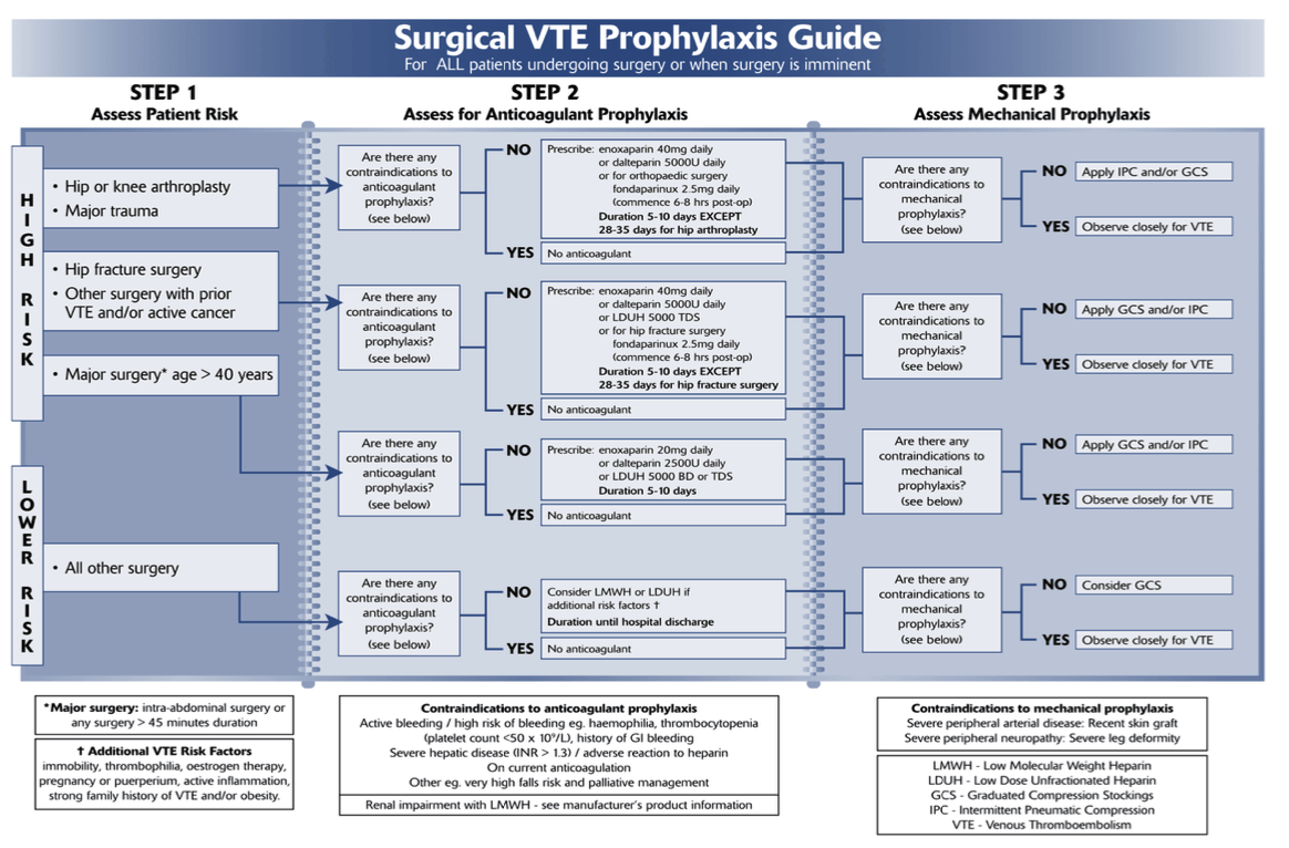
Low risk
- < 30 minutes and age < 40 without additional risk factors
- Outpatient surgery, laparoscopic cholecystectomy & breast surgery
- Without prophylaxis risk of proximal DVT in 1%, risk of fatal PE in 0.01%
- Rx – TEDs, Early mobilisation
Moderate Risk
- Surgery > 30min and age > 40 yrs without additional risk factors
- Laparoscopic surgery
- Additional risk factors present
- Cardiac/ pulmonary disease
- Malignancy
- ATIII
- Pro-thrombotic disorders
- Risk of proximal DVT = 3%
- Rx - TEDs, early mobilisation, pneumatic compression devices, Enoxaparin 40mg
- Enoxaparin that night or at time-out, currently not recommended to continue post-discharge
High Risk
-
Surgery in patients > 40yr with recent hx DVT or PE
-
Extensive abdominal or pelvic surgery
-
Surgery for malignancy
-
Surgery of lower limbs
-
Paralysis/ Immobility of lower limbs
-
Additional risk factors present
- Cardiac/ pulmonary disease
- Malignancy
- ATIII
- Pro-thrombotic disorders
-
Risk of proximal DVT 6%
-
Rx - TEDs, early mobilisation, pneumatic compression devices, Enoxaparin 40mg
-
In very high risk may consider continuing Enoxaparin for 4 weeks post-discharge
-
Exception
- Not for 8 hrs post-epidural (and withhold for 24 hrs before withdrawing epidural & can be started 2 hours after)
- NB: No evidence to support this
- Not for 8 hrs post-epidural (and withhold for 24 hrs before withdrawing epidural & can be started 2 hours after)
-
Dose Adjustment
- Renal dysfunction (CrCL < 30) & < 40kg – Half dose to 20mg
- BMI > 50 – Enoxaparin 60 mg
Fate of Thrombosis
- Fate
- Propagation
- Embolization
- Dissolution
- Organisation & recanalization
- Type
- Primary Platelet Thrombus
- In response to endothelial injury
- Some fibrin deposition
- Coralline Thrombus
- Alternate pale (platelet) and dark (RBC and WBC) layers
- Platelet laminae form the lines of Zahn
- Occluding Thrombus
- Coralline thrombus occludes the vein
- Firmly attached; initial thrombus is white and firm
- Consecutive Thrombus
- Back to the next tributary
- Consecutive thrombus is red (fibrin and entrapped red cells)
- Loosely attached and prone to break off
- If clot stops at the tributary, then further platelet thrombus is necessary before further red thrombus is formed (successive layers)
- Propagating Thrombus
- Red thrombus that continues beyond the next tributary
- Eventually organisation and recanalisation occur
- Thrombophlebitis associated with inflammation, therefore thrombus adherent to the intima and embolization rare
- Primary Platelet Thrombus
Clinical
-
50% no clinical signs
- ~ 50% local calf tenderness or a positive Homan’s sign do not have a DVT
- Signs
- Mild oedema to swelling of calf (> 2cm asymmetry)
- Localised calf tenderness
- Homan’s Sign – Calf pain following dorsiflexion of foot
- Direct thrombus palpation
- Most likely to be positive in the popliteal fossa
- Phlegmasia Alba dolens
- Painful white swollen leg, thrombosis of Iliac/ Femoral Veins
- Phlegmasia Cerulea dolens
- Severe pain with cyanosis of limb, sudden complete venous occlusion of the entire limb outflow, venous gangrene may occur with a 50% amputation rate
- Superficial venous dilatation - decreased venous outflow causing congestion
- Signs
Investigation
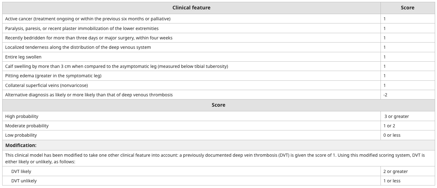
Wells Score DVT
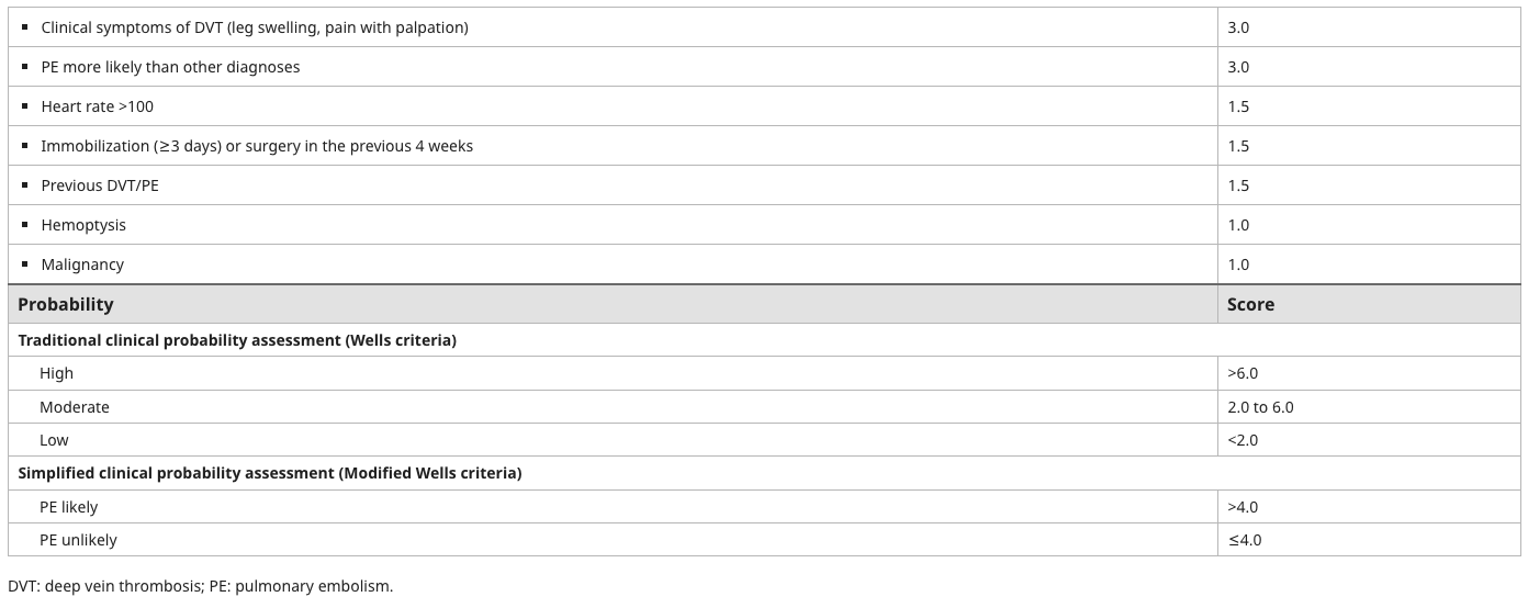
Wells Score PE
- D-dimer – High NPV, useless if recent surgery
- Doppler USS
- Investigation of choice
- Consider proximal vs whole leg USS
- Contrast enhanced CT venograph or MR venography
- Alternative but rarely used
Treatment
- Aims of treatment
- Short term
- Prevent extension, fatal PE and early recurrence
- Long term
- Prevent delayed recurrence, post-phlebitic syndrome and Pulmonary HTN
Anticoagulation
- Clotting cascade Recap
- Anticoagulants
- LMWH v UFH
- For treatment of proximal DVT have shown equal efficacy
- At the appropriate dose there is no increase in the rate of significant clinical bleeding
- Decreased risk of thrombocytopenia (HIT), occurs in up to 10% with UFH
- Doses
- Enoxaparin 1mg/kg bd
- Fragmin 100U/kg bd or 200U/kg once daily
- Treatment
- If risk factor identified & reversible – 3 months
- If idiopathic – 6-12 months
- If irreversible risk factor or event was life threatening – permanent
- Second episode – permanent
- Options
- Warfarin
- Dabigatran
- Rivaroxaban
Role of Surgery
- Indications
- Massive thrombosis causing compromised arterial circulation in the leg
- Phlegmasia cerulea dolens
- Massive iliofemoral DVΤ
- Patients who fail therapeutic аոtiϲοаgulatiοո
- Large DVT when standard treatment is absolutely contraindicated
- Massive thrombosis causing compromised arterial circulation in the leg
Thrombolytic Therapy
- Catheter-Directed Thrombolysis (CDT)
- Uses local infusion of thrombolytics (e.g. tPA) directly into the thrombus
- Often combined with mechanical thrombectomy - Pharmacomechanical Thrombectomy
- Combines thrombolysis with mechanical clot disruption/removal
- Used in conjunction with CDT for high clot burden - Surgery used when immediate resolution of the problem necessary
- If more time then thrombolytic treatment
- Use for a massive DVT in a younger person (less post-phlebitic syndrome)
- Not for routine use with proximal thigh DVT
- More rapid reduction in thrombus size
- Significantly more bleeding (14%, OR 3.8) vs heparin
- No evidence that short term response to treatment is clinically any better
- However 3 of 4 studies showed decrease in post phlebitic limb syndrome
- Do not use if thrombus is > 72 hours old (OTS suggests potential increased embolization)
Surgical Thrombectomy
- Rarely used; considered when thrombolysis is contraindicated or fails
- Involves open removal of clot from femoral or iliac veins
- Often followed by fasciotomy if compartment syndrome is present
Filters
- Filters/Interruption of IVC flow (Greenfield Filter)
- Suprarenal placement
- Retrievable filters now available
- Prevents PE
- Indications
- Embolization happens while on optimal Rx
- Anticoagulation contraindicated
- e.g recent surgery, hemorrhagic stroke, active blееding
- Large, proximal non-adherent thrombi
- Not indicated for routine proximal DVT
- NEJM 1998.
- Randomised proximal DVT to filter or no filter as well as anticoagulation. Filters decreased PE in the first 2/52 (1% v 5%), but at 2 years the filter group had a higher rate of recurrent DVT (21% v 12%). No difference in mortality. So the initial benefit was counterbalanced by an increase in later DVT.
- Complications
- Can be serious
- Migration to IVC bifurcation, iliac vein, renal vein, RA, RV, pulmonary artery
- Protrusion through caval wall
- Thrombus formation on filter
- Thrombus formation above the filter (emboli)
- Misplacement
- Retroperitoneal haemorrhage
- Perforation of Duodenum
- Perforation of Ureter
Complications of DVT
- Pulmonary embolis
- SOB, chest pain, haemoptysis, pleural rub
- S1Q3T3
- Post-thrombotic Syndrome
- Risk higher with recurrent & proximal DVT
- Due to residual outflow obstruction with eventual calf muscle pump failure
Isolated Calf DVT
- For a calf DVT > 5cm - 15-20% will extend if left untreated
- Small DVT’s can be treated by compression (TED’s) and mobilization if duplex sonography done every 2 days or so to exclude extension
- Treatment with LMWH is mandatory if extension occurs
- As easy and as safe to Rx all lesions with ambulatory LMWH
Acute Iliofemoral Thrombosis
- Investigate with Duplex, sometimes venography/ contrast CT to identify upper extent
- Much higher risk of PE
- Always anti coagulate
- Consider thrombolysis in the younger patient
Subclavian Vein / Upper Limb Thrombosis
- 2-3% of DVTs
- Usually minimal venous stasis in upper limb
- Subclavian 75%, Axillary 50%, Jugular 25%
Aetiology
- Upper limb activity with direct or indirect injury to the vein
- Occurring mainly at the thoracic outlet
- 70% on dominant side
- Classification
- Primary
- Due to a thrombophilia (Factor V Leiden, Protein C/S, Anti-Thrombin III deficiency)
- Paget-Schroetter Syndrome
- Primary
- Secondary
- Venous injury by indwelling CV catheter, external trauma, or pacemaker wires
- Thoracic outlet syndrome / External compression (Lung malignancy – Pancoast tumour)
Risk Factors
- Thoracic Outlet Syndrome
- Metastatic tumour in the axilla – if can’t find a cause – look for a malignancy
- Indwelling catheters
- External trauma
- Paget-Schroetter Syndrome
-
50% of cases
- Repetitive constriction of Subclavian Vein in Costoclavicular space (e.g. swimming) causing upper limb DVTs
- Common to have anatomic anomalies such as cervical rib (may need to surgically excise) or myofascial bands
- Thrombosis occurs in the dominant arm in 80% of cases
Clinical
- Oedema/swelling within 24hrs of injury (95%)
- May affect breast
- Pain/aching (80%) in the limb and axilla
- Examination
- Swollen hand may have obvious venous engorgement
- Cyanotic extremity
- Collaterals over shoulder and anterior chest
- 1/3 has a tender palpable cord in the axilla
- SVC syndrome if clot extends there – face/neck swelling, periorbital oedema, blurred vision, facial cyanosis
- Check for Cervical rib
Investigations
- Duplex USS
- Venography (rarely used)
- Thrombophilia screen tests (preferably taken before anticoagulation started)
- CT – may detect subclavian stenosis / thoracic outlet syndromes
- MRI
Management
- Rest
- Elevate limb
- Physio
- Anticoagulation: Aim for INR 2-3 for 3-6 months then aspirin indefinitely afterwards
- Consider thrombolysis in Paget-Schroetter if Sx < 2 weeks (Urokinase / tPA)
- Catheter-directed thrombolysis ± followed by thoracic outlet decompression ± venous reconstruction / stent
- Surgical bypass only if Sx are severe
Prognosis
- Residual symptoms in 60-85%, residual oedema in about 40%
- Recurrence 2%
- PE as high as 12%
DVT prophylaxis
Mechanical
- Early mobilisation
- Compression
- Stockings/TEDs
- Simple, safe
- 68% reduction in moderate risk
- Devices - Pneumatic compression of calves during surgery
- Proven to increase flow in the veins 180% for calf boots
- Contraindicated with PVD
- Thought to be effective in moderate risk general surgery
- Stockings/TEDs
Pharmacological
- Anticoagulants
- A no. of studies have shown a decrease in DVT, esp. when measured with labelled fibrinogen
- 9 RCTs comprising 1505 patients show a risk reduction of 64% in DVT diagnosed on I-125 fibrinogen uptake in low risk patients
- Less clear if an equivalent decrease in PE
- Heparin
- 5000U BD, start preop, continue until ambulant
- Study involving > 4000 pts showed 70% reduction in DVT, 47% reduction in PE, 66% reduction in fatal PE.
- Main risk: Increased haemorrhage: increased risk of “excessive bleeding” or transfusion of 50-66%, overall increase of 2%
- LMWH
- Studies comparing Heparin and LMWH generally show a similar efficacy against DVT & PE, but have a lower rate of wound haematomas and re-operations for bleeding
- Warfarin
- Reduction of 66% in DVT and 80% in PE
- Risk of severe haemorrhage of 2-7%
- Dextran
- Glucose polymer of 40 000 or 70 000 MW
- Anti-platelet effect, decreases blood viscosity in deep veins
- IV infusion 500-1000ml before surgery and at on alternate days after surgery until mobile
- DVT risk 30-40%
- Bleeding problems are reported. Decrease in fatal PE from 0.7% to 0.2%
- Platelet-related - Aspirin and Dipyridamole
- Aspirin decreased DVT from 20.4% to 12.5%
- Meta-analysis suggests aspirin is useful
- Epidural
- Lumbar epidural with local anaesthetic increases lower limb blood flow, enhances fibrinolytic activity (by iPAI1), decreases platelet aggregation
- In a series of hip replacement patients proximal DVT reduced by 2.5-5x & PE reduced by 3x