Definition
- Variety of disorders caused by abnormal compression of arterial / venous / neural structures in the base of the neck
Incidence
- uncertain (reports range 3-80 per 1000)
- F>M 3:1 – especially neurologic syndrome (but venous more common in males)
- 20-50yo
Aetiology
- Due to alteration of normal structural relationships (especially in the interscalene triangle) which may become more pronounced with advancing age, due to:
- Bony abnormality:
- Cervical rib (usually → arterial effects) 0.4% population
- Passes beneath BP & subclavian artery (no relationship to vein as vein is anterior to anterior scalene muscle
- Attaches to scalene tubercle
- Lower trunk of BP or artery stretched over this (or squeezed between cervical rib & scalenus medius
- Long transverse process of C7
- Abnormal insertion of first rib
- Cervical rib (usually → arterial effects) 0.4% population
- Muscular & Soft tissue abnormality
- Anomalous subclavius or anterior scalene muscle
- Scalenus anterior abnormal insertion with an extension running back along 1st rib under subclavian artery
- Fibrous anterior extension of scalenus anterior can obstruct axillary vein
- Hypertrophy of the scalenus muscle
- Fibromuscular bands (congenital) → usually cause neurological effects
- Anomalous ligaments
- Anomalous subclavius or anterior scalene muscle
- Other
- Cervical trauma – neck hematoma, bone dislocation
- Malignancy
- Bony abnormality:
Clinical
- DDx; Cervical spondylosis, Carpal tunnel syndrome
- Sx rarely develop until adulthood – 90% neurological (C8/T1)
- Cervical rib may be palpable
- Neurological features
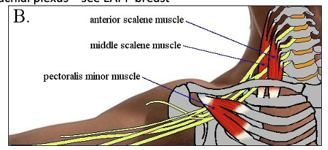
- Main Sx usually due to compression of one or more trunks of the brachial plexus – C8, T1 most commonly involved (most commonly ulnar distribution)
- Numbness of the hands wakes from sleep - - medial aspect of arm & hand, fifth finger & medial aspect of forth finger
- Light percussion over the brachial plexus in the supraclavicular fossa produces peripheral sensations (Tinel’s test) and reproduces Sx in pts with chronic neurologic impingement
- Muscular atrophy – hypothenar, interosseous
- Arterial features
- Fatigue, weakness, coldness, ischemic pain, paraesthesia
- Exercise or cold weather may exacerbate
- Unilateral Raynaud’s
- Cervical bruit
- May feel aneurysm
- Signs of embolization – splinter haemorrhage, gangrenous fingertips
- Compression of the subclavian artery can → stenosis & poststenotic dilatation → arterial occlusion / emboli
- Compression of the vein can → thrombosis → upper extremity pain & swelling
- Compression of vascular structures may be exaggerated with exercise → occlusion = “effort thrombosis” of axillary / subclavian vein = Paget-Schroetter syndrome (see Thromboembolic disease)
- Four provocative tests
- Adson (scalene) test
- Narrowing between scalene
- Full inspiration & hold
- Neck fully extended & turn to affected side
- Examining radial pulse
- Halsted (costoclavicular) test
- Narrow costoclavicular space
- Shoulders in military position (drawn backwards & downwards)
- Check radial pulse
- Wright (hyperabduction) test
- Subcoracoid region compression with pectoralis tendon, head of humerus or corocoid process
- Arm abducted to 180 degrees
- Check radial pulse
- Roos test
- Abduct arm to 90 degrees with external rotation of shoulder
- Open & close hand rapidly for 3 minutes
- ?Reproduce symptoms
- Other
- Tinel’s –provocative tapping of nerve
- Phalen’s - Provocative flexion of the elbow or wrist
- Adson (scalene) test
Pathology
- Pathogenesis probably involves an underlying anomaly + trauma / precipitant that leads to change in proteins / structure of the elements involved
- Structures affected may be:
- Brachial plexus (95%) → neurologic Sx
- Subclavian vein (4%) – thrombosis
- Subclavian artery (1%) – obstruction
- Interscalene triangle = space btwn ant & middle scalene + medial surface of 1st rib
- Costoclavicular triangle = space btwn middle third of clavicle, 1st rib & upper border of the scapula
- Arterial pathology – intimal damage, post-stenotic dilatation, aneurysmal degeneration, mural thrombus, embolization causing Raynaud’s syndrome
Anatomy
- At apex of thorax the subclavian vessels & nerves of the brachial plexus traverse the cervicoaxillary canal – divided into two portions by first rib
- 1st – scalene triangle – scalenus anterior anteriorly, scalenus medius posteriorly, first rib inferiorly
- 2nd –costoclavicular space - clavicle & first rib
- Subclavian artery exits chest behind the sternoclavicular joints & passes between the two scalene muscles as does the BP as the individual nerve roots exit the intervertebral foramina. The axillary vein passes posteriorly to the costocoracoid ligament and pectoralis muscle, it becomes the subclavian vein as it passes anteriorly over first rib, joins jugular vein after passing between scalenus anterior & clavicle (passing anterior to scalenus anterior)
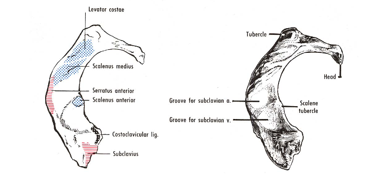
- Scalenus anterior
- Origin - anterior tubercles of the four ‘typical’ cervical vertebra (3-6)
- Insertion – narrow tendon into scalene tubercle on first rib
- Scalenus medius
- Origin – lateral ends of transverse processes of atlas & axis & posterior tubercles of all other cervical vertebrae
- Insertion – quadrangular area between neck & subclavian groove of first rib
-
Brachial plexus
-
Subclavian artery
-
Vessels can be compressed
- Interscalene space- artery/ nerve
- Costoclavicular space – vein
- Subcoracoid space – all three
Investigations
- Scalene block with local
- CXR/c-spine
- Cervical myelograms
- Doppler
- CT / MRI
- Arteriograms may demonstrate subclavian/axillary artery stenosis when arm is abducted
- Post-stenotic dilation of the artery is abnormal and indicates a definite lesion
- Nerve conduction studies / EMG
- Decreased or absent motor potentials in abductor pollicis brevis
- Sensory potential low to 5th finger (C8)
- Sensory potential normal across wrist
- Normal conduction in ulnar nerve from wrist to mid-arm
- EMG fibrillation potentials in 1st interosseous (chronic partial denervation)
Management
- For neurologic
- Postural correction
- Physio therapy – to strengthen the shoulder girdle
- Indications for surgery
- Failure of conservative measures
- Progression of sensory or motor symptoms
- Excessively prolonged ulnar or median nerve conduction velocities
- For vascular
- Anticoagulation
- Consideration of surgery if narrowed/occluded subclavian artery, thrombosis of axillary or subclavian vein
Surgery
- Cervical rib resected via supraclavicular approach (if there is one)
- Complete 1st rib excision - Transaxillary / supraclavicular/ infraclavicular/ transthoracic/ posterior approaches all described
- Complete 1st rib excision (includes excision of anterior scalene & any associated fibrous bands) ± arterial reconstruction
Complications
- BP injury
- Vascular injury
- Pleural effusion
- Winged scapula
- Infection
Prognosis / Natural Hx
- Neurologic syndromes: unlikely to progress, but also unlikely to resolve spontaneously
- Vascular syndromes: good outcome with adequate Rx
- 70% 5year success rate with surgery
- 1% recurrence post-surgery