- 5-15% of Breast Ca
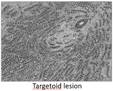
- Single file pattern – concentric circles around ducts (targetoid pattern)
- Invasive with predominant in situ component
- 10% of symptomatic breast ca
- Pathology:
- Rubbery
- Associated with E-cadherin inactivation
- Difficult to detect on mammogram
- 10% have co-existing ductal component
- More often multifocal; 10-20% bilateral
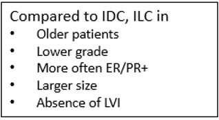
- Better prognosis cf ductal
- 70% ER+, 40% PR+
- 5 main subtypes:
- i. Classical
- small monotonous cells arranged in single file (Indian filing)
- Characteristic can make them clinically occult
- Targetoid lesions – files arranged around duct
- ii. Solid – sheet like, small & monotonous cells
- iii. Alveolar – globular aggregates, small & monotonous cells
- iv. Pleomorphic – diffuse infiltrate; nuclear pleomorphism
- v. Mixed