Section: Breast Curriculum: Curriculum, page 13
Workup and Investigation
- Overlying principle – TRIPLE ASSESSMENT
- If concordance, 99.5% sensitive for cancer
- If all 3 negative, chance of malignancy 1:10,000
- If concordance, 99.5% sensitive for cancer
Clinical History
- Symptomatic
- Lump
- How long it has been there,
- Constant in size, fluctuates, increasing
- Multiple
- Skin changes or nipple changes
- Discharge
- Colour and type, site, timing and provoking factors, breastfeeding, bilateral or multiduct
- Associated medications
- Pain – character, cyclical
- Sx of mets: bone pain, respiratory changes, weight loss, etc
- Lump
- Oestrogen exposure
- Age of Menarche/ Menopause
- Parity/ breastfeeding
- Including age at first full term pregnancy
- Oophorectomy, COC, HRT
- Previous breast Sx
- Previous Biopsies and results
- Screening – Mammograms and results
- FHx – breast and ovarian, pre-or post-menopausal
- Smoker
Physical Exam
- Begin Upright
- Inspect
- Masses, asymmetry, skin changes
- Nipple retraction, discharge, excoriation/Paget’s
- Peau D’Orange
- Obstruction of dermal lymphatic channels by emboli of carcinoma cells
- Palpate
- Axilla with arm supported, supra and infraclavicular regions
- NB: 30% of palpable LN are benign
- Inspect
- Lie down
- Palpate breast tissue with arm above head
- Size,
- Shape
- Consistency
- Location
- Fixed or mobile?
- Palpate breast tissue with arm above head
Bloods
- Tumour markers - Ca15-3 and Ca27-29 - markers of breast cancer mets (CEA and Ca125 - mets and worse prognosis)
- Ca15-3 - indicator of metastatic disease, low spec and sens
- Ca 27-29 - same
- CEA - indicator of metastatic disease
- Ca-125 - poor prognostic indicator, tumour burden, aggressive sub-types
Imaging
- < 30 years:
- USS (good for dense tissue) or
- MRI (for high-risk patients)
- NB: HRT increases breast density – so even though it increases risk of Breast Ca it also increase chance of recall therefore potential for earlier pick-up
-
30 years:
- Mammogram (± USS)
- Sensitivity age dependent
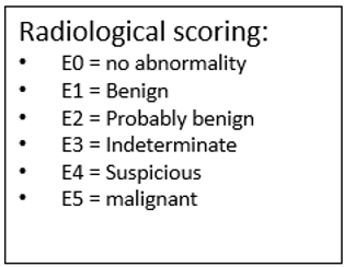
Mammography
- Radiation exposure of mammogram
- ≈ Background radiation on a flight LA → Auckland
- Less radiation than CXR
- Views
- MLO - med-lateral oblique (can see some of pectoralis major)
- CC - cranio-caudal (NB: name/markers usually placed laterally)
- If abnormalities, can add
- Magnification views (esp. for calcifications)
- Compression views (to further assess mass characteristics)
- Describe
- Density
- Calcifications
- Architectural distortion
- Masses/spicules, circumscribed or not?
- Mammogram can’t often differentiate between cystic & solid
- Cysts typically seen as very round dense (white) lesions
- Skin changes
- Asymmetry
- NB: Should be a ‘fat plane’ against the chest wall = No densities here
- Calcifications on Mammogram:
- Benign
- Rounded
- Homogeneous in size & density
- Regional or scattered distribution
- Malignant
- Increased number
- Heterogeneous density / shape / size
- Clustering, linear / vermicular forms, casting / branching → PPV of Ca is 88%
- Mostly dystrophic calcifications secondary to necrotic tumour cells
- If none of the above are present → PPV of Ca is 7%
- Require stereotactic biopsy (as often not palpable)
- If Bx is non-diagnostic / suspicious / malignant → Needs excision with hook-wire
- NB: Only 25% of Bx for calcifications yield carcinoma
- Benign
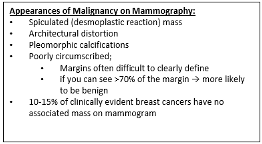
Additional Malignant Mammogram Features:
- Asymmetrical skin thickening
- Nipple inversion
USS
- High frequency (>10MHz)
- Safe, no ionising radiation
- Can use independently of complimentary to mammography
- Assists in tissue sampling
- Good for assessment of dense breast tissue (Young)
- Useful in determining if solid or cystic, contour, internal properties
- Poor screening tool
- Highly operator dependent
- Less sensitive than mammography for signs of Early Breast Ca
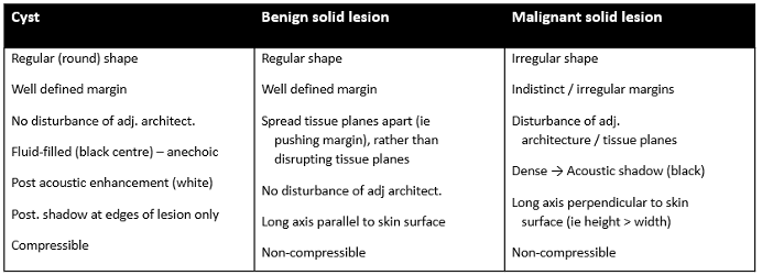
USS Features of Malignant Lymph Nodes
- Cortical bulging or thickening
- Rounded shape
- Loss of fatty hilum
- Indistinct margins
- Extra-capsular extension
- Matted nodes
- Peri-nodal Oedema
MRI
- Scan prone, IV contrast required
- Indications:
- Obtain accurate size of lesion
- HG DCIS, imaging inconclusive size
- Lobular Cancer (poor sensitivity with mammogram and ultrasound)
- Paget’s disease and no mass on mammography
- Young women < 40yo with cancer or that need screening
- Multifocal / bilateral disease / high risk (BRCA)
- Axillary LN metastasis + Occult primary (not seen on mammogram)
- Recurrence
- Differentiation from scar, esp. if breast reconstruction
- To assess silicone implant integrity and to survey patients with implants
- Assessing response of locally advanced cancer to Neoadjuvant therapy
- Assess spine for metastatic disease
- Obtain accurate size of lesion
- Disadvantage:
- Overly sensitive
- May pick up insignificant a/n
- May lead to over investigation / unnecessary biopsies
Tissue Diagnosis
FNA
- 22G needle, multiple passes, negative pressure
- Need ≥ 5 clumps of cells to be adequate
- Uses
- Evaluation of suspicious LNs
- Evaluation of second ipsilateral mass to
- ? Multifocal breast cancer
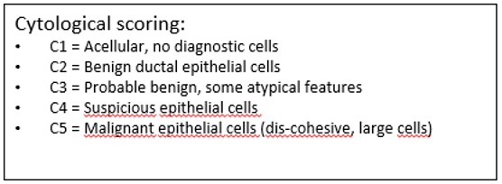
- Normal/benign cytological features
- Cohesive cells, monomorphic, in one plane, myoepithelial cell nuclei seen as ‘naked’/bare nuclei
- Malignant features
- Loss of cohesion, pleomorphism, cells in various planes, myoepithelial cell nuclei absent, nuclear atypia
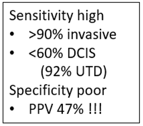
- Cytology false -ve 15%, false +ve 1% (Accuracy >90%)
- False +ves occur with cellular fibroadenoma or proliferative lesions, esp. during pregnancy / hormonal therapy
- False -ves: sampling error or well differentiated tumours with few epithelial cells
- Cytology does not distinguish invasive cancer from in situ cancer; a core biopsy is still required
Punch Biopsy
- For skin changes (Paget’s etc), commonly use 3mm
Core Biopsy
- Breast Core biopsy
- 11-14G needle, 4-5 cores taken from 4 quadrants of the lesion
- 10% inconclusive
- Accuracy 97-99%
- 10% that have DCIS will have carcinoma at definitive surgery
- 20% with “ADH” will have DCIS or carcinoma
- Stereotactic (Mammographic) Biopsy
- 2 views @ 15o tube shift to vertical
- Will often leave titanium clip afterwards
- 95-98% accurate
- Can also have USS and MRI guided
- Gold standard - Large core (≥ 14G) vacuum assisted (Mammotome)
- Can do IHC on core but not FNA
- Can’t do ER/PR status on FNA
Excisional Biopsy
- Rarely used, can delay definitive management
- Indications
- Excision of fibroadenoma ? Phyllodes
- Discordant imaging and biopsy results
- ADH in a field of microcalcifications, suspicious for DCIS
Summary of Investigations for Confirmed Cancer
- Triple assessment
- History and clinical exam
- Imaging
- Biopsy
- Staging:
- LFTs, UEC, FBC, Calcium
- ? CXR
- Stage III (T3/T4, N1-2) – bone scan, CT
- ± MRI if
- < 40yo or Lobular Ca – to assess multifocality / extent
- PET
- Not indicated in staging of Stage I – III disease (NCCN 2009)
- “PET or PET/CT should only be used in situations where other staging studies are equivocal or suspicious; even in these, Bx of the sites is more likely to provide useful info”
- Poor for axillary LN: sens 84.5% (ie too many false –ves), spec 98.5%
- Tumour markers: CA 15-3, CA 27-29, CEA
- Role unclear
Risk factors
Overview
- Risk Factors That Cannot be Modified
- Increasing age
- Female sex
- Estrogen
- Early age at menarche (before age 12 yr)
- Older age at menopause (> age 55 yr)
- Nulliparity
- Family hx of Breast Ca
- Genetic predisposition (BRCA1/2)
- Personal hx of Breast Ca
- Race, ethnicity (white women have increased risk compared with women of other races)
- History of radiation exposure
- Risk Factors That Can Be Modified
- Estrogen
- Age at first live birth (full-term pregnancy after age 30 yr)
- Parity
- Lack of breastfeeding
- Obesity
- Use of HRT
- Alcohol consumption
- Tobacco smoking
- Decreased physical activity
- Shift work (night shifts)
- Estrogen
- Histologic Risk Factors
- Proliferative breast disease
- Atypical ductal hyperplasia
- Atypical lobular hyperplasia
- Lobular carcinoma in situ
Age
- Most important risk factor
- Age related 5yr risk
- 20-40yrs - 0.5%
- 50yrs- 2%
- 60yrs- 4%
Sex
- Incidence men <1% of incidence in women
Personal history of Breast Cancer
- Increased risk of ca in contralateral side (RR = 5)
- Absolute risk 0.5-1% per year
- Dependent of age of first ca, ER status or original and ? Adjuvant therapy
Histologic Risk Factors
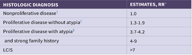
- Lobular carcinoma in situ
- Considered to be an index of risk for invasive Ca, rather than a premalignant lesion
- Risk equal in both breasts (1% per year)
- RR 5-12x – risk is highest if diagnosed before age 40yrs,
- Lobular up to 18x, ductal up to 4x
- Lobular up to 18x, ductal up to 4x
Family History of Breast Cancer
- First degree relative
- Relatives with pre-menopausal malignancy (RR = 2)
- Multiple relatives on the same side
- 20% of females diagnosed with breast CA have ≥ 1 affected relative
- 4-5% of Breast Ca due to Autosomal Dominant genetic predisposition
- 90% of these are BRCA 1 & 2
- Often bilateral, earlier onset/ premenopausal, multifocal
Reproductive Risk Factors
Related to Oestrogen Exposure
- Menarche < 12 yrs
- 10% risk reduction for each 2yr delay in menarche
- First live child > 30yrs
- First child <18 has ½ risk c.f. First child >30
- Nulliparity
- Menopause > 55yrs
- Doubles risk
- NB: RR of all of these is low compared to other factors, but does affect entire female population therefore does have large effect on prevalence in population
- Breastfeeding can reduce risk
- ? Related to decreased number of lifetime menstrual cycles
Exogenous Hormones
- OCP for premenopausal women
- Oestrogen based
- Small incr RR (1.7) if used for 8 yrs
- Risk diminishes as time since OCP stopped increases
- No increased risk at 10yrs post cessation
- No increased risk if started > 35yrs
- COC higher
- NB: Decreases Endometrial and Ovarian Ca
- Therefore overall reduced rates of Ca for woman
- Oestrogen based
- HRT for post-menopausal women
- Combination oestrogen + progesterone
- Increased Breast Ca risk if used for 5yrs
- Oestrogen alone (if previous hysterectomy)
- No increased risk
- Combination oestrogen + progesterone
Protective factors
- Lower lifetime oestrogen exposure
- Late menarche
-
15 yrs vs < 12 yrs RR = 1.3
-
- Early menopause
- < 45 yrs vs > 55 yrs RR = 1.2-1.5
- Many children
- Includes protective influence of early age of 1st birth (< 20 vs. nil >30yrs, RR=1.7)
- Low fat, high fibre diet
- Exercise
Risk Prediction
Overview
- First step is to determine risk
- The average lifetime risk of breast cancer is 15%. Moderate risk of 15-20, and high is > 20%.
- When determining what surveillance strategy you should use - you should determine what category a patient falls into (i.e. low, moderate or high).
- Woman with a BRCA1/2, PTEN, TP53, STK11 mutation, or history of previous radiotherapy are high risk.
- For other patients - a risk prediction model is often used to determine there risk.
Risk prediction tools available
- Models which look at family history alone to determine risk
- Claus model
- BOADICEA (now called CanRisk)
- Models which look family history + hormonal and reproductive factors
- Breast Cancer Surveillance Consortium risk calculator
- Tyrere-Cuzick model
- Age, age at menarche, age at first birth, weight/height, age at menopause, presence of proliferative breast conditions, and family hx and use of HRT
- Gail Model
- Tool calculates a woman’s risk of developing breast cancer over the next 5 years and within her lifetime
- Age, age at menarche, age at first birth, previous breast pathology, family history, ethnicity
Management of Risk Groups
Average risk screening
- In NZ the funded screening program is 2 yearly mammography from 45-69
- Screening woman in there 40’s results in more false positive biopsy results, and mammography is less sensitive at picking up breast cancers.
- The major benefit from screening is the reduction in breast cancer mortality, the major harm, is overdiagnosis - overdiagnosis refers to the detection of cancers which never would have become clinically apparent.
- Screening is beneficial for woman as long as there life expectancy is > 10 years.
- Mammography is the only imaging technique which has been shown to decrease breast cancer mortality (although it is the most studied).
Moderate risk screening
- Average risk is considered 15-20% lifetime risk.
- Up to date recommends that woman with moderate risk of breast cancer be offered the same screening as average risk.
- NICE guidelines recommend that yearly mammography be performed from age 40-60
High risk screening
- Those with a calculated lifetime risk of > 20% should be considered high risk.
- There are no guidelines I could find in New Zealand but below are the NICE guidelines.
- Patients with risk 20-30%
- Yearly mammography from age 40-60 then general screening.
- Patients with >30% risk OR BRCA, tP53 or other known high risk gene, or chest wall radiation
- Yearly MRI and yearly mammogram from aged 30
- Patients with risk 20-30%
Category 1: Average/Slightly Above Risk Group
-
95% of women
- No confirmed FHx
- 1 x 1◦ > 50yrs
- 1 x 2◦ any age
- 2 x 2° > 50yrs on same side of family
- 1x 1◦ + 1x 2◦ > 50yrs on different sides
-
90% of women in this group will not get Breast Ca
- Lifetime risk between 1/8 – 1/11
- RR 1.5
- Management:
- Breast Screen Aotearoa
Category 2: Moderate Risk Group
- 4% of females
- 1 x 1◦ < 50yr
- 2 x 1◦ on same side of family (Breast or Ovary)
- 2 x 2◦ on same side of family, 1 < 50yr
- Dense breasts (4-5 x risk)
- 75-90% of women in this group will not get Breast Ca
- Lifetime risk between 1/4 – 1/8
- RR 1.5-3
- Lifetime risk between 1/4 – 1/8
- Management:
- Annual mammograms from age 40 or 5 yrs before the diagnosis of Breast Ca in 1° relative
- GP to give annual breast examination
Category 3: Potentially High-Risk Group
- < 1% of females
- 2 x 1◦ or 2◦ same side of family (Breast or Ovary) AND
- Additional relatives (Breast or Ovary)
- Any < 40
- Bilateral
- Breast + Ovary same woman
- Ashkenazi Jew
- Any male
- A relative who has tested positive for high-risk gene mutation, e.g. BRCA1/2
- 50-75% of women in this group will not get Breast Ca
- Lifetime risk: 1:2 to 1:4
- RR > 3
- Management
- Annual Mammograms +/- MRI starting at age 30 or least 5 years before Dx of Ca in relative (lower limit 30)
- Refer for genetics r/v
Breast cancer syndromes
BRCA1
- Autosomal dominant
- Mutation in the Breast Cancer Type 1 gene on 17q21
- Penetrance 80-100%
- Cancer susceptibility gene
- Regulates gene expression, probably tumour suppressing
- Mutations generally inactivates the tumour suppressor gene
- Not all mutations are pathogenic
- 1:800 of population are carriers of mutation
- Up to 1% in Ashkenazi Jew population
- Risk
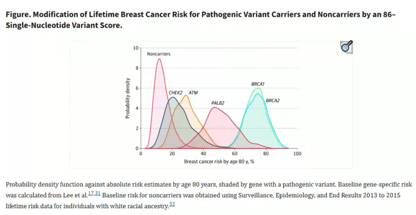
- Cumulative risk of breast cancer
- 20% at 40
- 50% at 50
- 70% at 70
- 60% risk of Contralateral Ca (higher than non-BRCA Ca)
- Cumulative risk of ovarian cancer
- 40-60% at 70
- Cumulative risk of other cancers
- Colorectal Ca 6% by age 70
- Prostate Ca 15-20% by age 70
- Pancreatic 1-3%
- Cumulative risk of breast cancer
- Accounts for 45% hereditary early onset breast cancer
- 4-5% of all breast cancers
- Median age of onset 45, frequently bilateral, worse histoprognostic factors
- More likely to be ER/PR –ve
- 70% chance of triple negative high grade cancers
- BUT - No difference in recurrence or death rate c.f. non-BRCA tumours
BRCA2
- Autosomal Dominant
- Mutation in the Breast Cancer Type 1 gene on 13q12/13
- Penetrance 80%
- Cancer susceptibility gene
- Tumour suppressor, mutations inactivate this function
- Accounts for 70% of remaining (non-BRCA1) hereditary breast cancer
- Risk
- Cumulative risk of breast cancer
- 45-70% at 70
- 60% risk of Contralateral Ca (higher than non-BRCA Ca)
- Cumulative risk of ovarian cancer
- 10-20% at 70
- Cumulative risk of other cancers
- Pancreatic Ca 2-5% by age 70
- Prostate Ca 30-40% by age 70
- Male Breast Ca - 80% of inherited (as opposed to BRCA 1)
- Lifetime Risk: 7%
- Laryngeal
- Bladder
- Cumulative risk of breast cancer
- Cancer histological subtypes not as poor as BRCA 1
- More likely to be ER/PR+
BRCA1 and BRCA2
- Life-time risk of developing Breast Ca 70% (Ovarian Ca 10-40%)
- Combined CA risk 4% per year
- Incidence of Breast Ca in BRCA carriers is high in pre-menopausal women
- NB: This approaches the population incidence in post-menopausal women
- But not so for Ovarian Ca, incidence continues to ↑ from age 40
- NB: This approaches the population incidence in post-menopausal women
BRCA1 and BRCA2 Screening
- Age 30-40: Annual MRI +/- USS
- Age 40-50: Annual MRI +/- MMG +/- USS
- Age > 50: Annual MMG +/- USS (Consider MRI if dense breasts)
- For Ovarian Ca
- Do not offer CA125 or TV USS.
- Data to show screening improves outcome is lacking.
- BSO should be done
- Do not offer CA125 or TV USS.
PALB2
- Autosomal Dominant
- PALB2 (Partner and Localizer of BRCA2) encodes a BRAC2-interacting protein
- Tumour Suppressor
- PALB2 (Partner and Localizer of BRCA2) encodes a BRAC2-interacting protein
- Lifetime Breast Ca Risk: 44%
- Associated with ER -ve Ca (i.e. less likely to work with Tamoxifen risk-reduction strategy)
- Also Ovarian and Pancreatic Ca
- Biallelic mutation (Autosomal Recessive) in PALB2 gene causes Fanconi Anaemia
- Screen with MRI starting age 30
Cowden’s syndrome
- Autosomal Dominant
- PTEN gene on chromosome 10q
- Tumour suppressor
- Risk of breast cancer
- 30 - 50% at 50yrs
- MRI from age 30
- Mastectomy before age 40 preferred over screening
- Also causes
- Macrocephaly
- Hamartomas (mucosal surface, lips)
- Multiple hamartoma syndrome (throughout GIT)
- With ↑ risk of breast, uterine & thyroid, CRC ca.
Li-Fraumeni syndrome
- Autosomal Dominant
- p53 mutation on chromosome 17p
- Increased risk pre-menopausal breast cancer
- Breast Ca Risk 85% by age 60
- Predominantly HER2 +ve tumours
- Also
- Young onset Sarcomas
- Brain tumours (Gliomas)
- Childhood Adrenal Ca
- Leukaemia
- Lymphoma
- Screen with annual MRI from age 20
- Diagnosis Criteria (All 3 required):
- Sarcoma diagnosed < 45 years
- 1° Relative with any Ca < 45 years
- 1° or 2° Relative with any Ca < 45 years or a sarcoma at any age
- Avoid radiotherapy – higher risk of developing secondary malignancies in radiation fields
- Thus, recommend Mastectomy rather than WLE + Radiotherapy
Peutz-Jeghers syndrome
- Autosomal Dominant
- STK11 gene on Chromosome 19
- Risk
- Breast Ca risk 30%
- Ovarian Ca Risk 21%
- Also GIT, lung, uterus, ovary
- Screen with MRI starting at age 30
Hereditary diffuse gastric carcinoma syndrome
- 50% due to CDH1 (E-cadherin) mutation
- Risk
- 80% risk of stomach cancer
- 50% risk of Lobular Ca
- Onset before age 40
- Management – screen with MRI from 30 +/- Mammogram from 40
Muir-Torre Syndrome
- Subtype of HNPCC; MSH2/ MLH1
- Skin tumours, GIT & GUT Adenocarcinomas
- 12% risk of Breast Ca
Ataxia Telangiectasia
- ATM gene 11q, Gene product normally activates p53 when DNA damage
- Autosomal Recessive, Heterozygotes 1.4% of population
- Cerebellar degeneration (ataxia), Skin & eyes telangiectasia, Immunodeficiency, Chromosomal instability
- Increase risk - Lymphoma, Leukaemia, Stomach Ca, Ovarian Ca, Melanoma
- RR 7
CHEK2
- 22q12, Checkpoint kinase 2 gene, Tumour Suppressor
- RR 2 of breast ca, x 10 in males
Neurofibromatosis Type 1
Waitemata DHB Guidelines for Referral to Genetics Services
Patients with Personal Hx of Breast Ca:
- Triple negative ≤ 50 years
- Breast Ca ≤ 40 years
- 2 x 1° Breast cancer in same patient + first cancer < 60 years
- 2 or more different but associated cancers in the same pt (e.g. Breast and Ovarian)
- Lobular Breast cancer + Family history of Lobular Breast or Diffuse-Type Gastric cancer
- Breast cancer diagnosed < 50 years with limited family structure or knowledge (e.g. adopted)
- Male Breast cancer
- Jewish Ancestry
- Breast Ca and Personal or Fam Hx of:
Patients with Family Hx of Breast Ca:
- 2 x 1st or 2nd degree relatives with Breast cancer or Ovarian cancer AND One or more of the following on the same side of family:
- Additional relatives with Breast or Ovarian cancer
- Breast cancer Dx < 50 years
- More than 1 Primary Breast cancer in same woman
- Breast and Ovarian cancer in same woman
- Jewish Ancestry
- Breast cancer in male
- Pancreatic cancer
- High Grade (> Gleason 7) Prostate Ca
Eligibility for BRCA Gene Testing
- Pts with Breast or Ovarian Ca and a combined BRCA1/2 Pathogenic Variant Probability of ≥ 10% using Manchester Score (Score ≥ 15)
- Pts with Breast or Ovarian Ca who are from a population where a common founder pathogenic variant exists
- Unaffected pts with an affected, untested 1° Relative with a combined BRCA1/2 Pathogenic Variant Probability of ≥ 20% using the Manchester Score (Score ≥ 17)
- Eligible based on Tumour Type
- Pts with Invasive Breast Ca AND:
- Diagnosed ≤ 40 years old OR
- Triple negative diagnosed ≤ 50 years OR
- Triple negative diagnosed any age AND 1° or 2° relative with Breast or Ovarian cancer
Breast Cancer Prevention
- No evidence that breast self-examination reduces Breast Ca deaths
- Cochrane review showed no evidence for benefits
- But evidence for harm (almost twice as many biopsies with benign results)
- Cochrane review showed no evidence for benefits
- Breast Cancer Prevention Trial (BCPT) NSABP 1989:
- Tamoxifen as preventative agent
- 5yrs of tamoxifen → 50% ↓ in Breast Ca compared to placebo
- No survival data
- But women > 50y had ↑ incidence of Endometrial Ca & DVT
- Tamoxifen as preventative agent
- NASBP study of Tamoxifen and Roloxifen (STAR trial JAMA 2006)
- Tamoxifen:
- Better sexual function
- Higher Endometrial Ca risk in women with a uterus
- Raloxifen:
- Cannot be used by premenopausal women
- Tamoxifen:
Risk Reduction Options
Screening
- Breast screening
- Detects cancer – does not prevent it
- Annual screening (with MRI in young women)
Endocrine therapy
- Indications (ASCO)
- Age of 35 years or older with a life expectancy of at least 10 years and one of the following:
- A history of thoracic radiation administered prior to 30 years of age
- A history of lobular carcinoma in situ
- A ≥1.7 percent five-year risk for breast cancer
- Atypical hyperplasia
- Age of 35 years or older with a life expectancy of at least 10 years and one of the following:
- General notes
- Tamoxifen, raloxifene, exemestane and anastrozole only reduce risk of estrogen receptor positive breast cancer
- No breast cancer risk-reduction drug has been shown to lower breast cancer-specific or all-cause mortality
- Menopause
- Pre-menopausal women- Tamoxifen
- Post-menopausal women - Tamoxifen, Raloxifene, Exemestane and Anastrozole
- Bone health
- Selective Estrogen Receptor Modulators - reduce age related bone loss
- Aromatase inhibitors - worsen age related bone loss
- Thromboembolic events (DVT and PE)
- Selective Estrogen Receptor Modulators - increase the risk
- Aromatase inhibitors - do not increase the risk
- Endometrial cancer
- Tamoxifen - increases the risk in post-menopausal women but not pre-menopausal women.
- Raloxifene, Anastrozole and Exemestane - do not
- Tamoxifen
- Tamoxifen 20mg daily for 5 years reduces breast cancer risk for at least 20 years
- Reduces the risk by at least 1/3
- Cannot recommend in BRCA 1 pts – most cancers are triple negative
- Can consider for women with family hx and proliferative breast disease (ADH, LCIS)
- On-going benefit even after ceasing treatment
- Raloxifene
- 60mg daily for five years
- Lowers the risk of breast cancer in post-menopausal women to a similar extent as tamoxifen in the short-term but is less efficacious in the longer term
- Exemestane
- 25mg daily for five years
- Lowers the relative risk of breast cancer in the short-term in post-menopausal women by 63%
- There are no data on longer term efficacy
- Anastrozole
- 1mg daily for 5 years
- Lowers the relative risk of breast cancer over 10 years in post-menopausal women by 49%
- Other potential agents
- Bisphosphonates, metformin, aspirin
- OCP reduces risk of Ovarian Cancer in BRCA1/BRCA2 by 40-50% BUT cannot recommend due to possible increased risk of Breast Cancer
Risk reduction surgery
- Risk-Reducing Bilateral Mastectomy - reduces risk in BRCA patients by 90%
- Skin-Sparing Mastectomy with Preservation of nipple-areola complex + Immediate Reconstruction
- Greatest benefit of surgery if before age 40
- No proven survival benefit
- Should not be encouraged but can be discussed about options to reduce risk
- Need to emphasize does not reduce risk to 0%
- Need to be clear on all potential complications, changes to cosmesis and sensation
- Early risk reducing BSO + Peritoneal Lavage –
- Consider around age 40 once family complete
- BRCA
- BRCA 1 – Should be performed by age 35-40
- BRCA 2 – Can be delayed until age 40-45
- Reduces risk of ovarian cancer by 80% and mortality by 60%
- Health Considerations related to Premature Surgical Menopause:
- Osteoporosis risk
- Cardiovascular disease
- Possible cognitive decline
- Vasomotor symptoms, reduced libido, vaginal dryness, dyspareunia – may not be fully relieved by oestrogen therapy
- Use of HRT – though unopposed oestrogen HRT seems to carry no increased risk of breast cancer, though long term effects are not known
- Usually given until age of natural menopause at age 50
- Consider hysterectomy at the same time - individualised.
- Risk of uterine cancer low
- Can give unopposed Estrogen HRT
Pathology
Definition and Cytology
- Invasive Breast Cancer
- Epithelial cancer arising from the terminal duct lobular unit, invading through the BM to surrounding tissue
- Cytological findings
- Loss of cohesion, lack of bare nuclei, loss of orientation, nuclear atypia, pleomorphism
- Invasive cancer
- Lack of overall architecture, infiltration of cells haphazardly into stroma, formation of sheets of continuous and monotonous cells without respect for form/ function of glandular origin
- False -ve 15%, False +ve 2%
Histological Subtypes
- Microinvasive Breast Cancer
- Extensive intraductal component
- Invasive carcinomas of NST
- Invasive lobular carcinoma
- Special-type breast cancers
- Paget’s disease of the nipple
- Other types of invasive Breast Ca
- NB: Most aggressive to least
- Medullary > Ductal NOS > Lobular > Tubular/Mucinous
Microinvasive Breast Cancer
- Microinvasive carcinoma is diagnosed when one or more separate foci of invasion measure 1 mm or less in maximum dimension.
- Nodal mets 1-10%; long term survival between DCIS & Stage 1
- Foci of tumour cells < 0.1cm in diameter invading the stroma
- If becomes invasive – retains original morphology & grade
- Staged as T1mi
- Invariably encountered in the setting of ductal carcinoma in situ (DCIS)
- Invasion is present in 25% of pts with DCIS
- Likely should have SLNB although axilar usually negative
- Nodal mets 1-10%; long term survival between DCIS & Stage 1
Extensive intraductal component (EIC)
- DCIS surrounding an Infiltrating Ductal Ca (or within)
- Definition = DCIS involving > 25% of the area of the invasive tumour, and also present at edge of tumour
- Can be predicted by area of microcalcifications ≥ 30mm (+ mass) on pre-op mammogram
- More common in tumours in younger women
- EIC is possibly predictor of local recurrence
- x 3-4 if EIC in tumour, but studies did not take positive margins into account
- If clear margins, no increased rate of local recurrence
- Tumours with EIC more likely to have residual tumour burden after WLE
- Nodal involvement is rare
- 1-2% rate of +ve nodes (from a missed microinvasive focus)
Invasive carcinomas of NST
- 85-90% of Breast Ca
- Historically these were called ‘ductal’, but as these are neither derived from ducts nor necessarily form ducts, the term ‘ductal’ is no longer recommended
- 60-80% of Symptomatic Breast Cancer
- 60% 10YS
- Change in 2019 WHO classification
- Medullary (‘Basal like’) now part of NST
- High grade, triple negative in appearance but not behaviour! = Behavioural paradox!
- Large tumour cells, very undifferentiated with pleomorphic nuclei. With dense lymphocyte infiltrate and syncytium appearing sheets of tumour cells
- Doesn’t tend to spread to lymph nodes or metastasis
- Medullary (‘Basal like’) now part of NST
Special-type breast cancers
- Good prognosis - 70-90% 10YS
- Histological type determined by architectural pattern
- Tubular 2-3%
- Low grade
- May be increasing with screening
- Cancer invades in small tubules lined by single layer of well-diff cells
- Mucinous 2-3%
- Low grade
- Usually post-menopausal women
- Bland tumour cells float like island in pools of mucin
- Tubulolobular
- Papillary
- Micropapillary (6%)
- Very high risk – 80-100% LN involvement and high risk of metastatic disease
- Cribriform
- Rarer types; metaplastic, adenoid cystic carcinoma, adenomyoepithelioma
Invasive lobular carcinoma
- 5-15% of Breast Ca
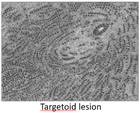
- Single file pattern – concentric circles around ducts (targetoid pattern)
- Invasive with predominant in situ component
- 10% of symptomatic breast ca
- Pathology:
- Rubbery
- Associated with E-cadherin inactivation
- Difficult to detect on mammogram
- 10% have co-existing ductal component
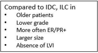
- More often multifocal; 10-20% bilateral
- Better prognosis cf ductal
- 70% ER+, 40% PR+
- 5 main subtypes:
- i. Classical
- small monotonous cells arranged in single file (Indian filing)
- Characteristic can make them clinically occult
- Targetoid lesions – files arranged around duct
- ii. Solid – sheet like, small & monotonous cells
- iii. Alveolar – globular aggregates, small & monotonous cells
- iv. Pleomorphic – diffuse infiltrate; nuclear pleomorphism
- v. Mixed
- i. Classical
- More likely to spread to ovaries, GIT, peritoneum
Other Subtypes
- Undifferentiated carcinoma
- Adenoid cystic
- Squamous cell
- Phyllodes
- Primary Lymphoma
- Metastatic – Melanoma
- Sarcoma
- From interlobular connective tissue
- Angio, rhabo, lipo, leiomyo, chondro, osteo
- Nodal mets rare, spread haematogenous
- Angiosarcoma
- May be as complication of Radiotherapy; 0.3-4% risk within 5-10 years
- Stewart-Treves Syndrome:
- Multiple subcutaneous haemorrhagic nodules develops in chronic lymphoedema (e.g. post-mastectomy)
- Very poor prognosis
- Rx: Mastectomy ± Radiotherapy
- Chemo useless
- High risk of recurrence
- Median time to recurrence = 8 months
- Median survival = 2 years
Tumour Size
- One of the most powerful predictors of prognosis in Breast Ca
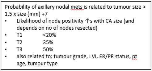
- Nodal mets if tumour < 10mm = 10-20%
- If node negative and <10mm, DFS at 10yrs > 90%
- Nodal mets if tumour <15mm = 15-20%
- If tumour > 15mm = 40%
- Nodal mets if tumour < 10mm = 10-20%
- Association between tumour size and increasing number of positive LNs and worse outcome
- Main aim of screening is to detect smaller tumours
- Smaller tumours have better long-term survival than larger tumours
- Small tumours (< 15mm) sometimes called “minimally invasive carcinomas”
- Good prognosis
Tumour Grade
- Important irrespective of morphological type
- Nottingham Method
- Assessment of grade considers three tumour characteristics (TNM):
- Tubule formation
- Nuclear pleomorphism
- Mitotic counts
- A numerical scoring system of 1-3 is used for each factor individually, the three scores added together to produce scores of 3-9
- Grade 1 – well differentiated = score 3-5 (10-year survival 85%).
- Grade 2 - moderately differentiated = score 6-7
- Grade 3 - poorly differentiated = 8-9 (10-year survival 45%).
- Assessment of grade considers three tumour characteristics (TNM):
- TNM staging system does not include grade
- Highly significant correlation with long-term prognosis
- Both Breast Ca specific survival and disease-free survival
- NB: Effect also shown for ILC
- Both Breast Ca specific survival and disease-free survival
Lymph Node Stage
- One of the most important prognostic indicators
- Clinical assessment of nodal status is unreliable
- Enlarged palpable nodes may be reactive
- Small impalpable nodes involved
- Histologically confirmed positive LN’s confers a significantly poorer prognosis.
- Overall survival decreases as the number of involved nodes increases
- 10-year survival reduced from 85% to 40% with multiple nodes positive
- Overall survival decreases as the number of involved nodes increases
- Sentinel lymph node sampling (SLN) is validated by level 1 evidence in the ALMANAC trial (Mansel 2006)
- Four-node sampling is associated with low rate of lymphoedema (0.04%)
- Assessment of the sentinel LN can involve paraffin histology, intraoperative frozen section, immunohistochemistry, serial sectioning, reverse transcription-PCR
- SLN biopsy false -ve rate varies between 0-11%
- Frozen section has a false negative rate of between 10-30%
- Best applied to selected cases; abnormal LN can be confirmed histologically and further axillary dissection performed immediately
- Intraoperative imprint cytology may reduce false negatives to around 2-5% but not all of been able to achieve this
- Frozen section has a false negative rate of between 10-30%
- Reverse Transcription-PCR
- More sensitive than immunohistochemistry at detecting metastatic tumour
- However, no single genetic defect in all Breast Ca
- Panel of markers may improve this
- PCR has a potentially high false +ve rate
- A major unresolved question about PCR is the prognostic significance of PCR only detected carcinoma compared to HE stained positive histological micromets
- More sensitive than immunohistochemistry at detecting metastatic tumour
- Classification
- Isolated tumour cells = <0.2mm
- Micrometastasis = 0.2mm to 2mm
- Macrometastasis = >2mm
Lymphovascular Invasion (LVI)
- Presence of tumour emboli in vascular and lymphatic spaces
- Important prognostic factor
- Correlates closely with local and regional lymph node involvement
- Can be used as surrogate in cases where nodes not taken
- Correlates with early recurrence
- Independent of grade, size and type for overall survival
Molecular Predictive Factors
- Morphological factors unable to predict response to systemic treatments
- Homogenous features vary in response to therapy
- Molecular markers may allow tailoring of individual therapy to the genetic makeup of a cancer
- Also used to monitor response & detect relapse
Oestrogen/Progesterone Receptors
- Steroid receptors located within the cell nucleus, regulating cell growth
- Expression of hormone receptors and their regulatory pathways
- Degree of hormone receptor expression can be used to predict an individual’s response to hormonal therapy
- More targeted therapies, reduced side-effects, greater efficacy
- Overall, ~ 30% pts will respond to endocrine therapy
- ER/PR+ tumours have a 78% chance of responding to therapy
- ER/PR- tumours rarely, if ever respond
- Detected by:
- Ligand-binding methods (radiolabelled steroid ligand binds to the receptor) - older
- Immunohistochemistry (antibody recognition of receptor protein) – most common
- Assay interpretation methods are subjective but assessed on all histopathological reports of Breast Ca as a predictor of response to systemic endocrine therapy
- Modified Allred score
- Assessed by both
- Intensity of stain and
- % of cells staining
- Gives you a score of 1-8
- Assessed by both
- ER+ if > 10% of tumour cells within the invasive component stain positive
- PR expression requires presence of intact ER machinery
- Positive PR expression = Evidence of functional ER
Type 1 Growth Factors
- Family of 4 receptors that promote cell proliferation and oppose apoptosis
- Overexpression can lead to malignancy
- Includes
- EGFR (HER 1)
- HER 2
Epidermal Growth Factor Receptor (HER1)
- No consensus as to prognostic value (conflicting evidence of its usefulness, heterogeneous study designs)
- Predictive marker for hormone responsiveness/resistance better defined
- EGFR tumours more resistant to edocrine therapy
- Inverse relationship EGFR and ER
- If EGFR negative, more likely ER + and often more sensitive to endocrine therapy
- Future importance is probably in seeing who will respond to therapies targeted at EGFR
- IHC the most practical testing method
Human Epidermal Growth Factor Receptor 2 (HER2)
- Important target of monoclonal antibody-based therapies and vaccines
- Trastuzumab (Herceptin)
- A humanised recombinant mAb that specifically targets the HER-2 extracellular domain
- Overexpression in 15-20% Breast Ca’s
- Critical role in tumour development
- Prognostic Significance
- HER-2 gene amplification independently predicted OS and DFS
- Now well-established HER-2 overexpression/ amplification has poorer prognosis in patients with nodal metastatic disease
- New research also shows poorer prognosis in node negative patients
- Stage for stage, worse prognosis if HER2 +ve
- Testing
- IHC first, then FISH if IHC equivocal
- IHC: HercepTest - looks for overexpression of HER-2 protein
- Negative = 0-1+
- Equivocal = 2+ (up to 25%) – should be tested with FISH
- Positive = 3+ (Strong, continuous membranous expression of HER-2 in at least 10% of tumour cells)
- FISH: Fluorescence in-situ hybridisation
- Measure directly the number of HER-2 genes (≥2 = +ve)
- IHC: HercepTest - looks for overexpression of HER-2 protein
- IHC first, then FISH if IHC equivocal
- Prediction of response to therapy
- HER-2 positivity may result in acquisition of oestrogen-independent growth
- Insensitive to Tamoxifen
- The Naples GUN trial
- HER-2 over-expression predicted resistance to tamoxifen
- Also worse outcome with tamoxifen treatment!
- However more recent studies have not found this same correlation
- HER-2 positivity may result in acquisition of oestrogen-independent growth
Triple Negative Tumours
- ER, PR and HER-2 negative
- Biologically different subgroup
- Relapse pattern very different from hormone positive cancers
- Risk relapse much higher for first 3-5 years
- After this, drops sharply, to substantially below hormone positive tumours
High-throughput Molecular Techniques
- Genomics: = Study of the human genome
- Transcriptomics = Study of the transcriptome (mRNAs)
- Proteomics = analysis of the protein complement of the genome
- Post-translational modifications (glycosylation, phosphorylation) affect their expression and function
- Research into the above may improve understanding, diagnosis, prognostication and provide new therapeutic targets
- Looks at DNA mutations, chromosomal aberrations, epigenetic modifications and protein interactions
Clinical use of high-throughput molecular techniques:
- Gene expression profiling can guide decisions of the choice of hormonal, chemotherapeutic or targeted agents for each individual
- Current examples in use:
- Her2 expression guiding trastuzumab
- Gene profiling to identify primary tumour in metastatic disease
- Transcription profiling to identify BRCA1/2 germ-line mutations
- Prognostic studies
- Poor-prognosis gene signatures can discriminate between good and poor outcome
- Recurrence score gene signatures define tumours based on the risk of disease relapse
- Prediction studies – includes gene signatures that assess response to treatment
- MammaPrint = 70 gene assays, used as a molecular diagnostic test for breast cancer prognostication and prediction.
- Predictor of distant metastasis
- Oncotype DX uses 21 genes to predict a recurrence score for women with early-stage breast cancer to establish the benefit of tamoxifen +/- chemotherapy
- 21 gene assay
- Determines ‘level of recurrence score’ predicting the likelihood of distant recurrence
- Low recurrence score = No benefit from chemo
- High recurrence score = 30% reduction in 10 yr recurrence
- MammaPrint = 70 gene assays, used as a molecular diagnostic test for breast cancer prognostication and prediction.
Molecular Profiling Classifications
- Luminal
- Luminal A
- Less aggressive, ER/PR +ve, HER2 –ve, Low Ki67 (< 14%)
- Luminal B
- More aggressive ER+/PR+ or -ve, HER2 + or –ve, High Ki67
- Luminal A
- Basal-like
- Triple negative but ‘basal’ +ve
- ER/PR –ve, HER2 –ve,
- Expression of basal markers – CK5/6, CK4, CK17 + EGFR
- NB: Not all triple negatives are basal type and not all basal type are triple negative but there is a significant cross-over
- Triple negative but ‘basal’ +ve
- HER-2 Overexpression
- ER/PR –ve, HER2 +Ve, Low Ki67
Clinical Use of Prognostic Factors
- Prognosis depends on the presence of spread of disease and the aggressiveness of the tumour
- The latter dependent upon:
- Morphological features
- Growth rate
- Hormone responsiveness
- Accurate individual prognostication can only be achieved by the use of a prognostic index that includes both time-dependent and biological factors
Nottingham Prognostic Index (NPI)
- Pathological tumour size
- cm x 0.2
- LN stage
- None = 1
- 1-3 LN involved = 2
-
3 LN = 3 +
- Histological grade
- Scored 1-3
Divided into 6 prognostic groups:
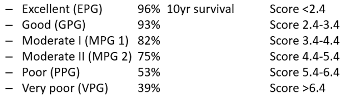
- Predicts 5 and 10-year survival for EPG = 96% and for VPG = 39%
- Verified prospectively in 2 large multicentre studies
Adjuvant Online (AO)
- Available on the internet, assesses risk of developing Breast Cancer metastasis
- Age, size, grade, LN status, hormone receptor status
- Advantage over the NPI in that it integrates treatment and so is the most commonly used by oncologists
St Gallen’s Criteria
- Algorithm for selection of adjuvant systemic therapy for early breast cancer
- Includes: size, grade, nodal status, menopausal status, vessel invasion, endocrine status and HER-2 status
PREDICT
- UK based model to predict 5-10 yr survival and benefit from adjuvant treatment
- Age, tumour size, tumour grade, # of nodes, ER status, HER2 status, Ki67 status, Mode of detection
Advanced Breast Cancer Index (ABC)
- Grade, ER status, site of initial metastasis, disease-free interval
- Can predict survival outcomes for each group
Staging
T Stage
-
TX = Primary tumour cannot be assessed
-
T0 = No evidence of primary tumour
-
Tis = Carcinoma in situ
- Tis (DCIS) = Ductal Carcinoma in situ
- Tis (LCIS) = Lobular Carcinoma in situ
- Tis (Paget’s) = Paget’s Disease of the nipple not associated with invasive carcinoma and/or carcinoma in situ in the underlying breast parenchyma
-
T1 = Tumour ≤ 20mm in greatest dimension
- T1mi - ≤ 1mm invasion through the BM
- T1a - > 1mm but ≤ 5mm
- T1b - > 5mm but ≤ 10mm
- T1c - > 10mm but ≤ 20mm
-
T2 = Tumour > 20mm but ≤ 50mm
-
T3 = Tumour > 50mm
-
T4 = Tumour of any size with direct extension to the chest wall and/or skin (ulceration or skin nodules)
- T4a – Extension to chest wall, not including only pectoralis muscle adherence/invasion
- T4b – Ulceration and/or ipsilateral satellite nodules and/or oedema (including peau d’orange) of the skin, which do not meet the criteria for inflammatory carcinoma
- T4c – both T4a and T4b
- T4d – Inflammatory Carcinoma
-
Invasion of dermis alone does not quality as T4 – dimpling of skin, nipple retraction and/or other skin changes can occur in T1, T2 or T3 without changing classification
N Stage
Clinical
N1 - movable, level 1 and level 2
N2 - matted or isolated internal mammary nodes
N3 - Level 3 or internal mammary + axilary nodes
Pathological
- NX – Regional lymph nodes cannot be assessed
- N0 – No regional lymph node metastases.
- N0 – No regional lymph node metastasis identified or isolated tumor cells (ITCs) only.
- N0(i+) – Malignant cells in regional lymph node(s) no greater than 0.2 mm (detected by hematoxylin and eosin stain (H&E) or immunohistochemistry (IHC) including ITCs)
- pN0(mol+)– Positive molecular findings (reverse transcription polymerase chain reaction (RT-PCR), but no regional lymph node metastases detected by histology or IHC.
- N1 – Micrometastases, or metastases in one to three axillary lymph nodes
- N1mi – Micrometastases (0.2 mm- 2.0 mm)
- N1a– Metastases in one to three axillary lymph nodes, with at least one metastasis greater than 2.0 mm.
- N1b – Metastases in ipsilateral internal mammary sentinel nodes, excluding ITCs.
- N1c – pN1a and pN1b combined.
- N2 – Metastases in four to nine axillary lymph nodes
- N2a – Metastases in four to nine axillary lymph nodes
- N2b – Metastasis only in clinically detected internal mammary nodes with or without microscopic confirmation; with pathologically negative axillary nodes.
- N3 – Metastases in 10 or more axillary lymph nodes; or in infraclavicular (level III axillary) lymph nodes; or in ipsilateral internal mammary lymph nodes in the presence of one or more positive level I, II axillary lymph nodes
- N3a – Metastases in 10 or more axillary lymph nodes (at least one tumor deposit greater than 2.0 mm); or metastases to the infraclavicular (level III axillary lymph) nodes.
- N3b – pN1a or pN2a in the presence of cN2b (positive internal mammary nodes by imaging); or N2a in the presence of pN1b.
- N3c – Metastases in ipsilateral supraclavicular lymph nodes.
N1 - 1-3 LN or internal mammary N2 - 4-9 LN N3 - 10 or more
M Stage
M0 – No clinical or radiographic evidence
- cM0(i+) – No clinical or radiographic evidence of distant metastases, but deposits of molecularly or microscopically detected tumor cells that are no larger than 0.2 mm are present in circulating blood, bone marrow, or other nonregional nodal tissue in a patient without symptoms or signs of metastases.
- M1 – Distant detectable metastases as determined by classic clinical and radiographic means and/or histologically proven metastases larger than 0.2 mm.
TNM (AJCC) - anatomic stage group
| T | N | M | Stage |
|---|---|---|---|
| Tis | N0 | M0 | 0 |
| T1 | N0 | M0 | IA |
| T0 | N1mi | M0 | IB |
| T1 | N1mi | M0 | IB |
| T0 | N1 | M0 | IIA |
| T1 | N1 | M0 | IIA |
| T2 | N0 | M0 | IIA |
| T2 | N1 | M0 | IIB |
| T3 | N0 | M0 | IIB |
| T0 | N2 | M0 | IIIA |
| T1 | N2 | M0 | IIIA |
| T2 | N2 | M0 | IIIA |
| T3 | N1 | M0 | IIIA |
| T3 | N2 | M0 | IIIA |
| T4 | N0 | M0 | IIIB |
| T4 | N1 | M0 | IIIB |
| T4 | N2 | M0 | IIIB |
| Any T | N3 | M0 | IIIC |
| Any T | Any N | M1 | IV |
Staging shortened
TMN (AJCC) – T
- TX = Primary tumour cannot be assessed
- T0 = No evidence of primary tumour
- Tis = Carcinoma in situ
- T1 = Tumour ≤ 20mm in greatest dimension
- T2 = Tumour > 20mm but ≤ 50mm
- T3 = Tumour > 50mm
- T4 = Tumour of any size with direct extension to the chest wall and/or skin (ulceration or skin nodules)
TMN (AJCC) – N (pathological)
- NX – Regional lymph nodes cannot be assessed
- N0 – No regional lymph node metastases.
- N1 – 1-3 positive +/- ipsilateral internal mammary
- N2 – 4-9 positive
- N3 – 10+ or level III, or in ipsilateral internal mammary lymph nodes in the presence of one or more positive level I, II axillary lymph nodes or supraclavicular lymph nodes
TMN (AJCC) – M and Staging
- M1 – Distant metastases
TNM (AJCC) - anatomic stage group
- Stage I
- T1, N0 (can have N1mi IB)
- Stage II
- T1-2, N0-N1
- T3, N0
- Stage III
- T1-T2, N2
- T3, N1-N2
- T4, AnyN
- AnyT, N3
- Stage IV
- AnyT, AnyN, M1
Clinical staging
T- Same N - different M- Same
Clinical N stage
N1 - Level I and II N2 - Fixed or matted level I and II or internal mammary nodes without positive axilla N3 - Level III, IM nodes + axilla, supraclavicular nodes