Section: Breast Curriculum: Curriculum, page 10
Definition
- Ductal carcinoma in situ (DCIS) is a neoplastic proliferation of mammary ductal epithelial cells confined to the ductal-lobular system without evidence of invasion through the basement membrane into the surrounding stroma
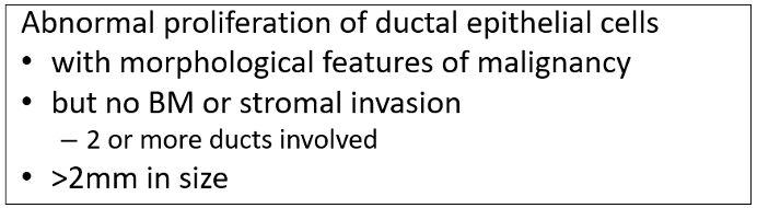
- The above allows differentiation from Atypical ductal hyperplasia
Heterogenous disease, probably two distinct entities
- Slow transition ADH → LG DCIS → LG IDC
- Associated with ER +ve
- Fast transition HG DCIS → HG IDC
- Associated with C-erbB2
- Often larger area at time of diagnosis, younger patients
Epidemiology
- Accounts for 20% of malignant lesions picked up during screening
- Probably about 10% of all (screening + symptomatic)
- Post-mortem studies: 10%
- Can occur at any age, but declines with age
- 60% detected by breast screen
- Progression
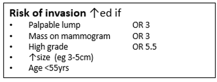
- 60% LG DCIS will become invasive by 40 years
- 50% HG DCIS will become invasive by 7 years
Genetics
- Abnormalities on chromosome 16q (low grade) and 17q (high grade)
Risk Factors
- Same risk factors as invasive cancer - see Breast cancer work up
AJCC pathological staging
- TMN (AJCC) – T
- Tis (DCIS) = Ductal Carcinoma in situ
Clinical
- Most (90%) asymptomatic; microcalcifications on mammogram = Only manifestation in 75-95%
- Minority (10%) present with symptoms
- Mass
- Nipple discharge
- Paget’s Disease of the Nipple
Histology
Progression
- Starts as normal duct → Mild ductal hyperplasia = increased number of cells lining duct and remains uniform
- Progress to Atypical ductal hyperplasia where both layers and cells become heterogeneous - bigger nuclei, more cell layer that arent uniform
- DCIS defined >2 ducts or >2mm
- IDC when these cells invade through the myoepithelial basement membrane
- Note that nodal involvement → rare
Classification
- Comedo (presence of luminal necrosis filling at least one duct with large pleomorphic nuclei and abnormal mitosis)
- Non-comedo (all other subtypes)
- Cribriform – fenestrated/sieve-like appearance
- Solid - ducts filled with malignant cells, considered high-grade
- Micro-papillary – tufts of cells project into duct lumen perpendicular to BM
- Papillary - projecting tufts larger than micropapillary, and have fibrovascular core
- Clinging (flat) – variable columnar alterations along duct margins
- ? Is this truly pre-malignant
- ? Should be categorised as atypical hyperplasia
Grade
- Low Grade (Non-comedo necrosis)
- Evenly spaced cells with central small nuclei, few mitosis and nucleoli not easily seen
- ER +ve, low Ki67, low apoptosis and rarely HER2 +ve
- Intermediate Grade
- Between high- and low-grade features
- High Grade (Comedo necrosis)
- Pleomorphic cells with large irregular nuclei (3x size of RBC), prominent nucleoli and frequent mitosis
- Often solid with comedo necrosis and calcification
- Often HER2 +ve, or ERGF +ve, Rarely ER +ve, High Ki67, high apoptosis
- Multicentric = Separate foci of tumour found in more than one breast quadrant or more than 5cm away from initial primary
- Most DCIS is unicentric, only 1% DCIS multicentric
- Multifocal = Separate foci in same quadrant that are close to each other
- More commonly reported if high grade, poorly differentiated lesion
Histology - DCIS and Malignancy
Microinvasive Breast Cancer
- Foci of tumour cells < 0.1cm in diameter invading the stroma
- If becomes invasive – retains original morphology & grade
- Staged as T1mi
- Invariably encountered in the setting of ductal carcinoma in situ (DCIS)
- Invasion is present in 25% of pts with DCIS
- Likely should have SLNB although axilar usually negative
- Nodal mets 1-10%; long term survival between DCIS & Stage 1
Extensive intraductal component (EIC)
- DCIS surrounding an Infiltrating Ductal Ca (or within)
- Definition = DCIS involving > 25% of the area of the invasive tumour, and also present at edge of tumour
- Can be predicted by area of microcalcifications ≥ 30mm (+ mass) on pre-op mammogram
- More common in tumours in younger women
- EIC is possibly predictor of local recurrence
- x 3-4 if EIC in tumour, but studies did not take positive margins into account
- If clear margins, no increased rate of local recurrence
- Tumours with EIC more likely to have residual tumour burden after WLE
- Nodal involvement is rare
- 1-2% rate of +ve nodes (from a missed microinvasive focus)
DCIS Investigations
Mammogram
- Often underestimates extent of disease (esp. low grade micropapillary)
- Calcifications (Seen in 75-95%)
- NB: Only 25% of biopsies for calcifications yield carcinoma
- Fine calcifications – more likely low-grade DCIS
- Coarse granular/ casting calcifications – more likely high-grade DCIS
- Soft tissue abnormality (seen in 10%)
MRI
- esp. good for pts with no calcifications
- DCIS MRI Sensitivity 90%
- Compared to mammography sensitivity 75%
- Good for assessing extent and distribution
- NB: Substantial false +ve rate & cost ↑
FNA
- Remember invasive malignancy cannot be excluded
- Comedo (reported as malignant)
- High cellularity, high grade nuclei, cellular dissociation, background necrosis, foamy macrophages
- Non-comedo (reported as suspicious)
- High cellularity, architectural features of papillary, low grade nuclei, palisading, monolayer sheets
Core Biopsy
- Method of choice
- Large core, preferably vacuum assisted (mammotome) → X-ray of biopsy to check calcifications
- NB: 10-20% of DCIS on biopsy will have undiagnosed invasive component
- Agreement with surgical Bx: 70-99%
- Under-estimation on core: < 14G = 31%; Mammotome = 2%
- Need to do ER/PR/HER-2 – gives prognostic information and Tamoxifen indicated as treatment
DCIS Management
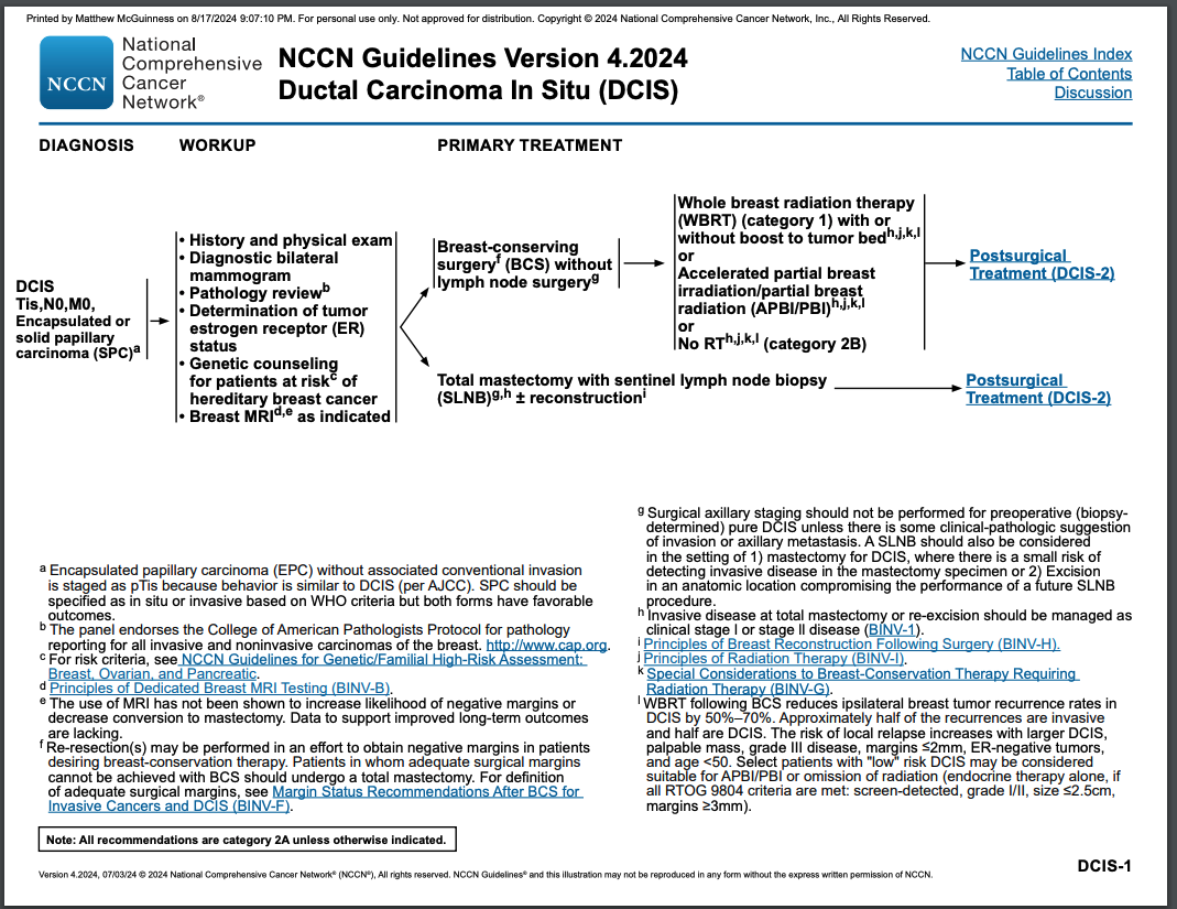
Hook-wire WLE
- WLE with hook-wire guidance preferable (Breast Conserving)
- Aim for 1cm margins intra-op
- Orientate specimen
- Cavity should be marked with clips
- X-ray specimen to determine if calcifications/mass present
- Need to consider
- Size of area to be excised vs size of breast and location
- If patient cannot have radiotherapy, then cannot have conservative surgery
- 2mm histological margins acceptable
- Involved margins - Re-excise
- Close margins – (< 2mm but not involved)
- Consider – age, size, grade, presence of comedo necrosis, which margin (deep/ superficial), extent approaching margin
- Recurrence higher with:
- Close margins = Strongest predictor of recurrence
- High grade DCIS (22% recurrence) or DCIS with comedo necrosis (18% recurrence)
- Poorly Differentiated tumours
- Young age (< 40 yrs)
- Size – Not really a factor if you exclude > 4cm lesions which should be tx with mastectomy
- Can use University of Southern California/Van Nuys Prognostic index (USC/VNPI) which looks at size, margins, pathological classification and patient age to give a score between 4-12 on likelihood of recurrence over next 5 years. 4-6 low risk, 7-9 inter, 10-12 high risk
Mastectomy (+/- Reconstruction)
- Consider if:
- Large + poor cosmetic outcome anticipated
- Multifocal
- Inability to undergo radiotherapy + High grade
Sentinel lymph node biopsy
- Indications not clear
- General consensus: SLNBx if mastectomy
- Mastectomy ± reconstruction make future SNB impossible
- But may consider in certain patients:
- Large volume (> 4.5cm)
- High-grade
- Suspicion of invasive disease
- Mass palpable or suggested on imaging
Axillary Dissection
- Not indicated, < 2% chance of LN involvement
Radiation
- All women undergoing breast conserving surgery should be considered for radiotherapy and discussed at MDM level - NZGG/NICE
- NSABP B17
- RT vs no RT, included occult invasive cancers & +ve margins
- Increase event free survival
- Decreased ipsilateral breast cancer
- Decreased recurrent DCIS and invasive disease by 50%.
- No improvement of overall survival!
- RT vs no RT, included occult invasive cancers & +ve margins
- NSABP B17
Van Nuys Prognostic Index
Scoring system used to predict outcomes
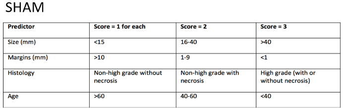
- Score ranges from 4 to 12
- Score 4-6 = Excision only
- Score 7-9 = Excision + Radiotherapy
- Score 10-12 = Mastectomy
Adjuvant Therapy
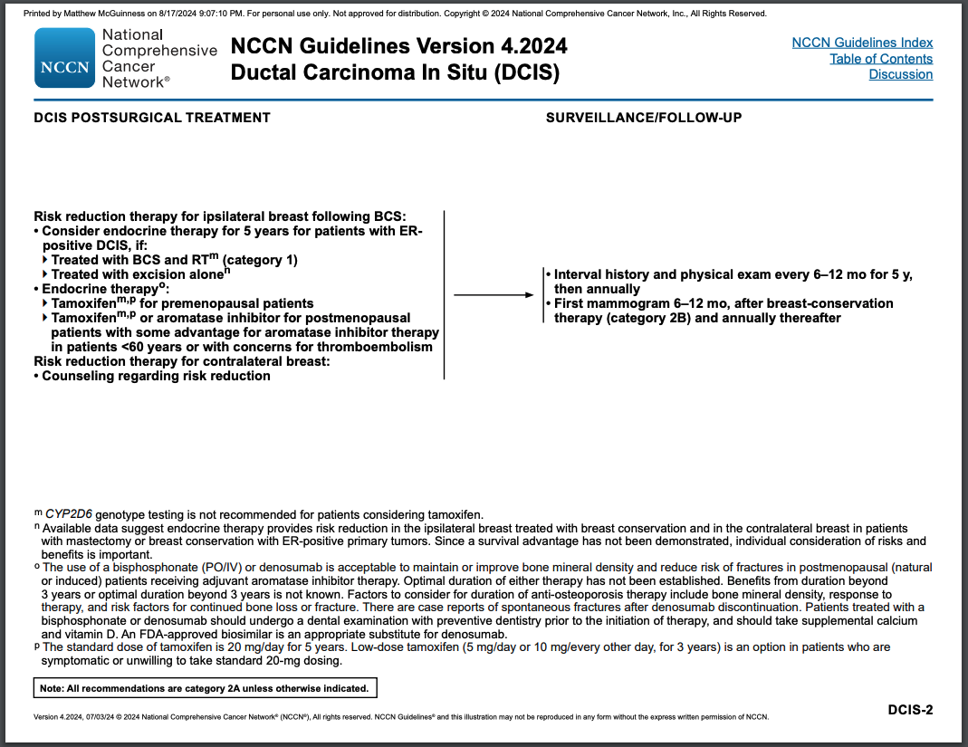
Tamoxifen
- NZGG says “consider” after WLE if
- ER +ve & high risk
- e.g. Age < 45yrs, involved margin, Comedo necrosis
- ER +ve & high risk
- NICE says only if no radiation
- Conflicting reports
- NSABP-B24
- RCT - Tamoxifen after WLE + XRT
- ↑ Disease Free Survival
- ↓ Local recurrence (5% absolute)
- 2% absolute ↓ ipsilateral & contralateral breast CA
- NO overall survival advantage
- RCT - Tamoxifen after WLE + XRT
Chemotherapy
- No significant benefit from chemotherapy
- Survival is already very good and risk of metastatic disease very low
Prognosis/Natural Hx
- Natural Hx unknown for sure
- 10-yr risk of ipsilateral Ductal Ca in same quadrant is 30-50%
- Long-term F/U untreated low-grade DCIS
- 40% develop invasive Ca
- 2/3 of these within 10yrs
- Study of Pts with incorrectly diagnosed benign disease
- By selection bias will tend to be low grade non-comedo
- Comedo has greater chance of microinvasion
- Grows faster & has a higher rate of local recurrence compared to non-comedo
- Poor prognostic features:
-
5cm, high grade, “casting” (neoductogenesis)
-
Recurrence rates following surgery
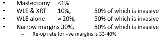
- If invasive Ca is present, then pts with extensive in-situ component (EIC) more likely to have close/involved margins
- No reduction in overall survival, as long as clear margins are obtained (& radiotherapy given if WLE)
- Risk of metachronous Ca ≈ 1% per year
Follow-up
- Annual bilateral Mamogram for 5yrs
- But no diff in DFS or OS if f/up mammo biannual
- Clinical surveillance – role not clearly supported in the literature
Management of Recurrence
In-situ Recurrence
- If initially had breast conserving therapy alone
- Could offer repeat WLE + radiotherapy
- If has previously had radiotherapy
- Offer completion mastectomy
Invasive Recurrence
- Also dependent on initial therapy for DCIS
- If no previous radiotherapy
- Could offer WLE + radiotherapy, with axillary staging
- If WLE not an option
- Offer mastectomy and axillary staging as per usual Ca
NCCN Guidelines