- Radiation exposure of mammogram
- ≈ Background radiation on a flight LA → Auckland
- Less radiation than CXR
- Views
- MLO - med-lateral oblique (can see some of pectoralis major)
- CC - cranio-caudal (NB: name/markers usually placed laterally)
- If abnormalities, can add
- Magnification views (esp. for calcifications)
- Compression views (to further assess mass characteristics)
- Describe
- Density
- Calcifications
- Architectural distortion
- Masses/spicules, circumscribed or not?
- Mammogram can’t often differentiate between cystic & solid
- Cysts typically seen as very round dense (white) lesions
- Skin changes
- Asymmetry
- NB: Should be a ‘fat plane’ against the chest wall = No densities here
- Calcifications on Mammogram:
- Benign
- Rounded
- Homogeneous in size & density
- Regional or scattered distribution
- Malignant
- Increased number
- Heterogeneous density / shape / size
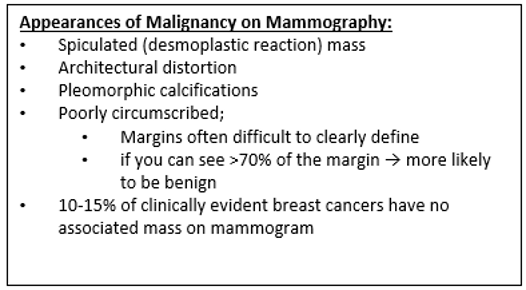
- Clustering, linear / vermicular forms, casting / branching → PPV of Ca is 88%
- Mostly dystrophic calcifications secondary to necrotic tumour cells
- If none of the above are present → PPV of Ca is 7%
- Require stereotactic biopsy (as often not palpable)
- If Bx is non-diagnostic / suspicious / malignant → Needs excision with hook-wire
- NB: Only 25% of Bx for calcifications yield carcinoma
- Benign
Interpretation
Normal
- A normal breast is composed of a honeycomb supporting fibrous structure made up of Cooper ligaments that houses fatty tissue, which in turn supports the glandular elements of the breast
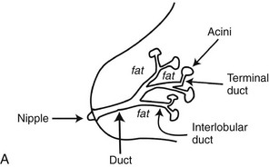
- The glandular elements are composed of lactiferous ducts leading from the nipple and branching into excretory ducts, interlobular ducts, and terminal ducts leading to the acini that produce milk.
- The ducts and glandular tissue extend posteriorly in a fanlike distribution consisting of 15 to 20 lobes draining each of the lactiferous ducts, with most of the dense tissue found in the upper outer quadrant.
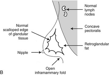
- Posterior to the glandular tissue is retroglandular fat, described by Dr. Laszlo Tabar as a “no man’s land,” in which no glandular tissue should be seen. The pectoralis muscle lies behind the fat on top of the chest wall.
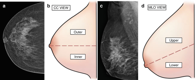
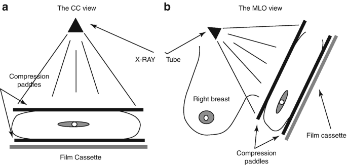
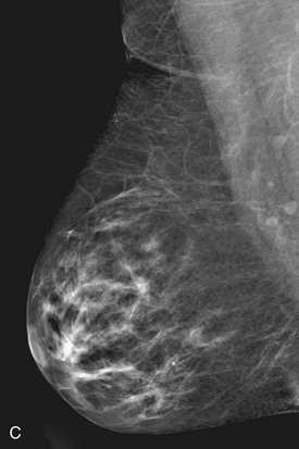
Mediolateral oblique view (MLO)
- Pectoralis major
- Concave structure posterior to the retroglandular fat near the chest wall
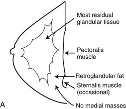
Craniocaudal project (CC)
- Pectoralis major
- Produces a half-moon–shaped density near the chest wall
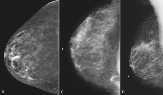
- Fat lies anterior to the muscle, and the white glandular tissue lies anterior to the fat. In older women, most of the glandular tissue in the medial breast undergoes fatty involution, and therefore most of the residual dense glandular tissue exists in the upper outer breast.
- There should be only fatty tissue in the medial breast near the chest wall. The only normal exception is the sternalis muscle, a muscular density near the medial aspect of the chest wall that should not be mistaken for a mass
- Seen in C below
- Seen in C below
General features
- Glandular tissue
- White
- Fat
- Black
- Dense
- Mostly white
- Fatty replaced
- Mostly black
- Breast cancer
- Mostly white
BI-RADS® for breast density
-
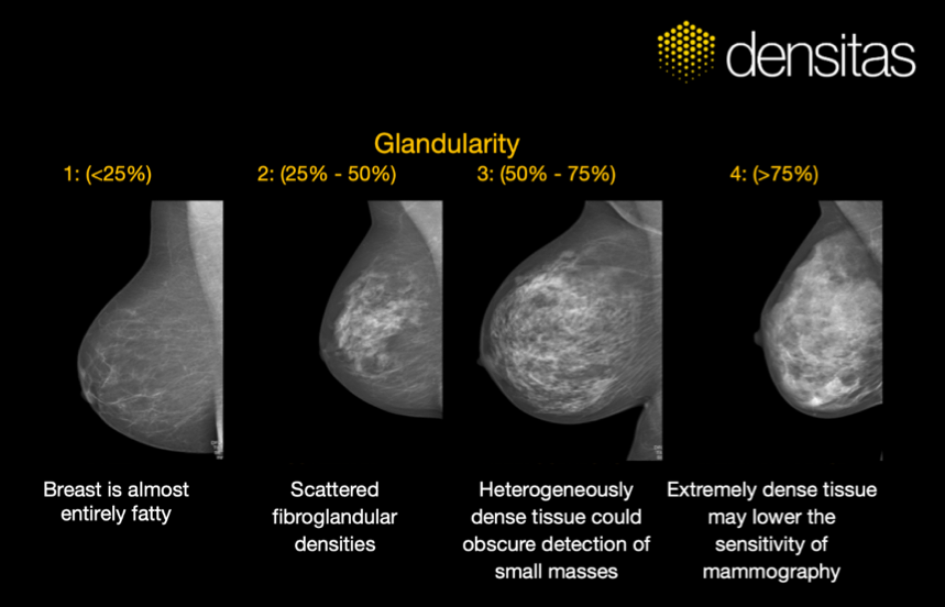
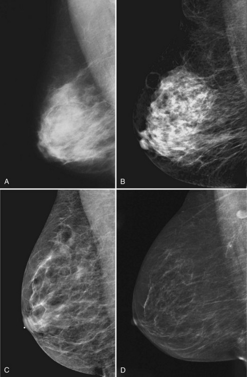
The breast is almost entirely fat (<25% glandular).
-
There are scattered fibroglandular densities (approximately 25–50% glandular).
-
The breast tissue is heterogeneously dense, which could obscure detection of small masses (50–75% glandular)
-
The breast tissue is extremely dense (>75% glandular).
-
As women age, the fibroglandular tissue involutes into fat, which is black
- the amount remaining depends on genetics, parity, and exogenous hormone replacement therapy.
-
It is important to know about the relative decrease in breast tissue and breast density over time.
- Increases in breast density in normal women occur only in:
- Pregnant and lactating women
- Women starting exogenous hormone replacement therapy.
- Unexplained generalized increases in breast density may indicate breast edema or inflammatory cancer
- Increases in breast density in normal women occur only in:
BI-RADS assessment categories
| Category 0: Incomplete – Need additional imaging evaluation and/or prior mammograms for comparison | Recall for additional imaging and/or comparison with prior examination(s) | N/A |
| Category 1: Negative | Routine mammography screening | Essentially 0% likelihood of malignancy |
| Category 2: Benign | Routine mammography screening | Essentially 0% likelihood of malignancy |
| Category 3: Probably benign | Short-interval (6-month) follow-up or continued surveillance mammography | >0 but ≤2% likelihood of malignancy |
| Category 4: Suspicious | Tissue diagnosis* | >2 but <95% likelihood of malignancy |
| Category 4A: Low suspicion for malignancy | Tissue diagnosis* | >2 to ≤10% likelihood of malignancy |
| Category 4B: Moderate suspicion for malignancy | >10 to ≤50% likelihood of malignancy | |
| Category 4C: High suspicion for malignancy | >50 to <95% likelihood of malignancy | |
| Category 5: Highly suggestive of malignancy | ≥95% likelihood of malignancy | |
| Category 6: Known biopsy-proven malignancy | Surgical excision when clinically appropriate | N/A |
Approach
- Side by side
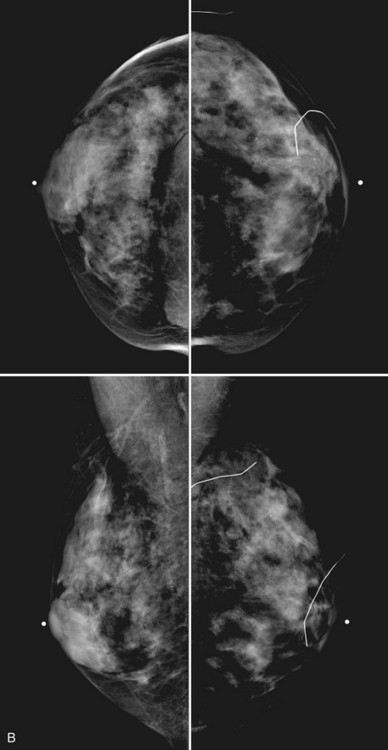
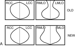
- Breast tissue is usually symmetric, or “mirror image,” when comparing left to right mammograms, although 3% of women have normal asymmetric glandular tissue
- Breast tissue is usually symmetric, or “mirror image,” when comparing left to right mammograms, although 3% of women have normal asymmetric glandular tissue
Findings suggestive of cancer
| Finding | Differential Diagnosis |
|---|---|
| Pleomorphic calcifications | Cancer (most common), benign disease, fat necrosis |
| Spiculated mass | Cancer, postsurgical scar, radial scar, fat necrosis |
| Round mass | Cyst, fibroadenoma, cancer, papilloma, metastasis |
| Architectural distortion | Postsurgical scarring, cancer |
| Developing density | Cancer, hormone effect, focal fibrosis |
| Asymmetry: focal or global | Normal asymmetric tissue (3%), cancer (suspicious: new, palpable, a mass containing suspicious calcifications or spiculation) |
| Breast edema | Unilateral: mastitis, postradiation therapy, inflammatory cancer Bilateral:systemic disease (liver disease, renal failure, congestive heart failure) |
| Lymphadenopathy | Unilateral: mastitis, cancer Bilateral: systemic disease (collagen vascular disease, lymphoma, leukemia, infection, adenocarcinoma of unknown primary) |
| Single dilated duct | Normal variant, papilloma, cancer |
| Mass with calcifications | Cancer, fibroadenoma, papilloma; exclude calcifying oil cyst |
| Nothing | 10% of all cancers are false-negative on mammography |
Localisation
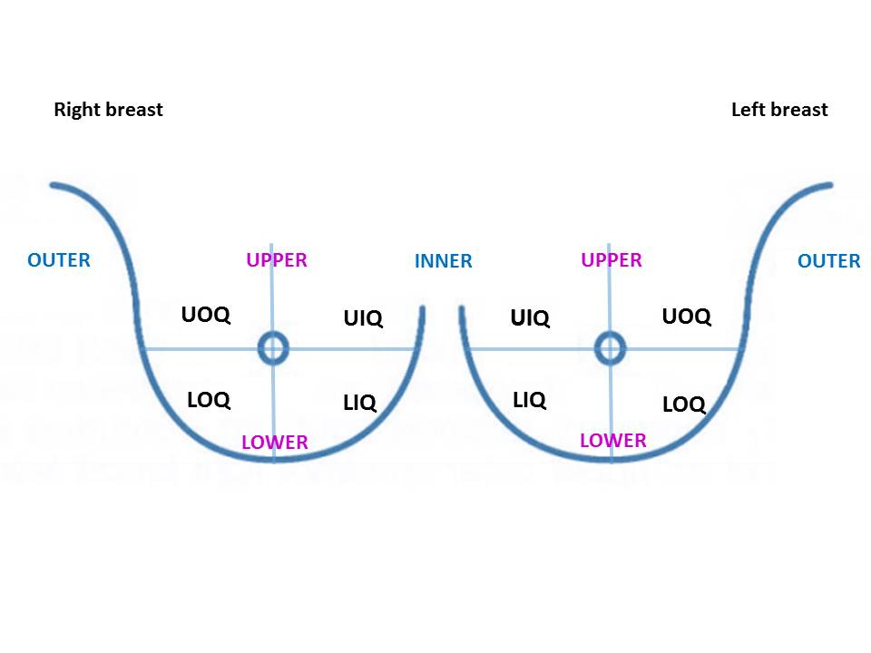
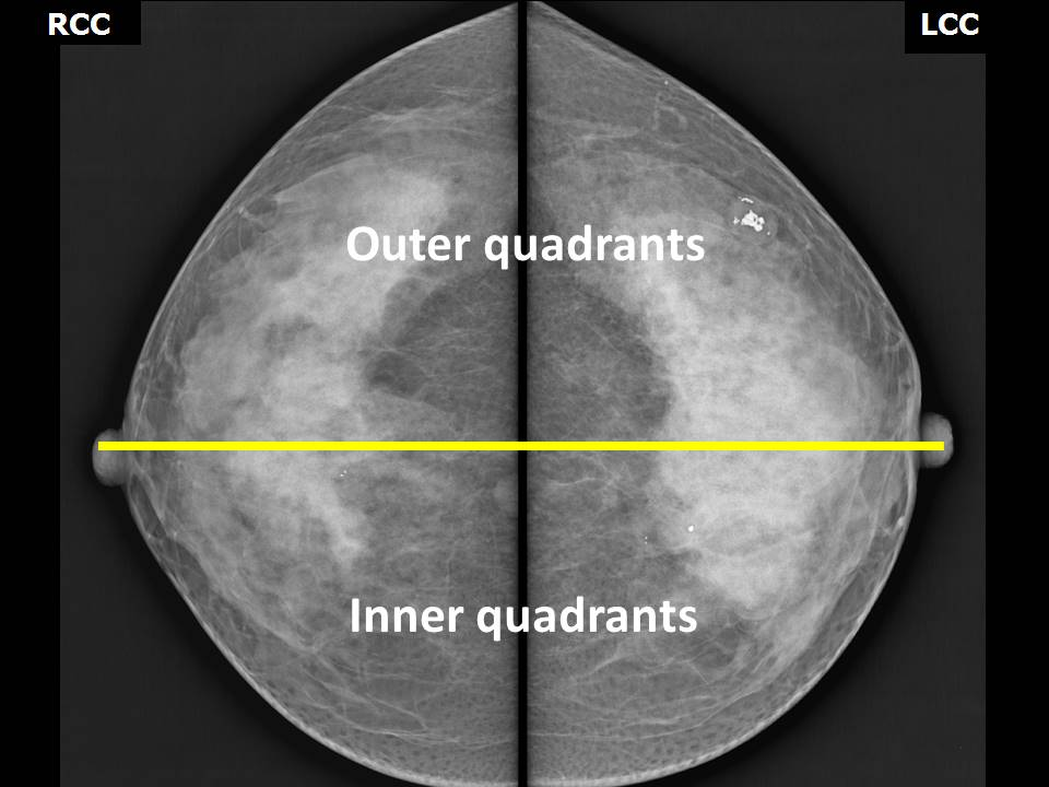
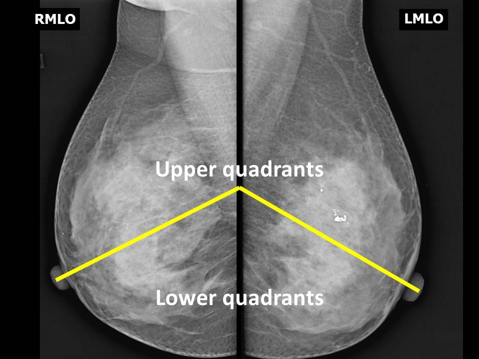
Examples
Case 1
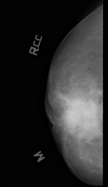
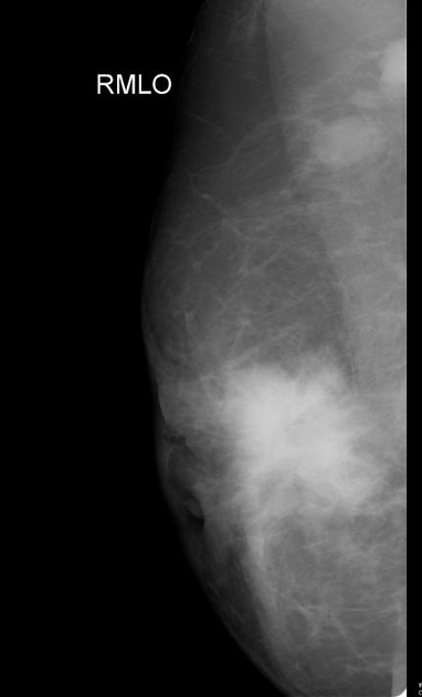
- Large spiculated multilobulated mass almost in all four quadrants with skin thickening and nipple retraction.
- Multiple axillary nodes are also seen with no distinct fatty hilum.
Case 2
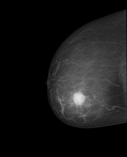 Mammography demonstrates a fairly well defined dense lesion with microlobulated borders. No spiculation, architectural distortion or underlying microcalcification.
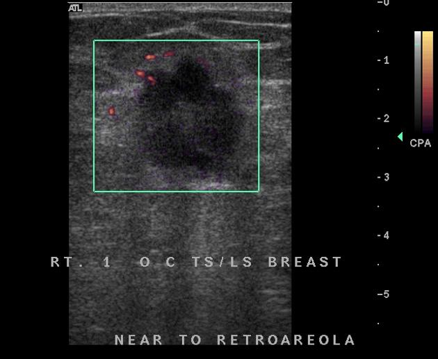 Targeted breast US demonstrates a round, solid mass with homogeneous echogenicity and microlobulated margins.
- Diagnosis: metaplastic carcinoma of the breast (path proven).
Case 3
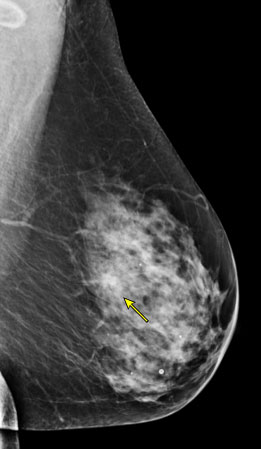 The arrow identifies a suspicious mass in the left inner upper breast in a 54-year-old woman with heterogeneously dense breast tissue. Biopsy revealed invasive carcinoma.
Cass 4
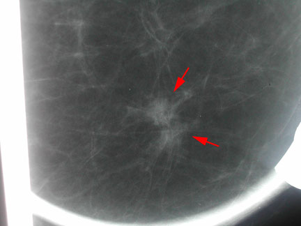 Spot magnification view of a mammogram showing 2 small adjacent interconnected spiculated masses (red arrows). Pathology revealed tubular carcinoma. Tubular carcinoma characteristically appears spiculated on mammogram and is often associated with satellite lesions.
Case 5
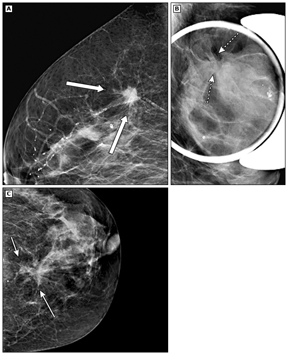 Spiculation from invasive ductal carcinoma in 3 different cases. In Panel A, an irregular mass with spiculated margins is seen (thick arrows). In Panel B, the cancer is not seen as a mass, but extensive distortion (dashed arrows) is seen. In Panel C, an indistinct mass is seen with spiculations extending far beyond the mass (arrows).
Case 6
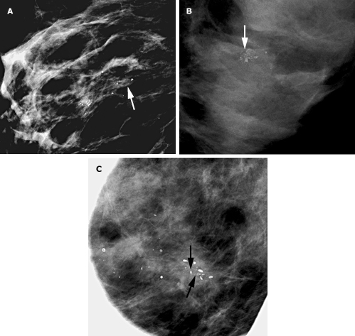 Grouped microcalcifications in 3 different cases. A loose group of coarse heterogenous calcifications is seen in Panel A (arrow). A small cluster of amorphous calcifications is seen in a background of dense breast parenchyma in Panel B (arrow). Fine pleomorphic calcifications are seen in between coarse larger calcifications in Panel C (arrows).
Case 7
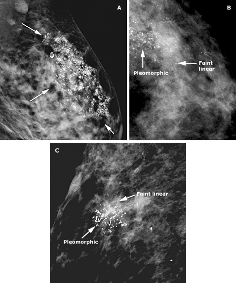 3 different cases of suspicious calcifications (arrows): segmental coarse heterogenous distribution (Panel A); and faint, with some grouped linear branching and fine pleomorphic calcifications (Panels B and C).
Compare this to benign patterns
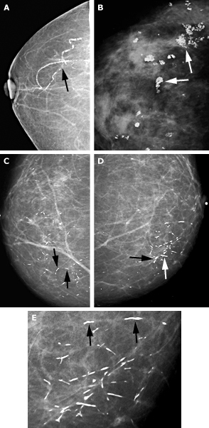 Benign patterns of calcifications (arrows) in 3 patients. Vascular calcifications are seen in the lateral aspect of the breast on a CC mammogram (Panel A). Multiple coarse “popcorn” calcifications are characteristic features of calcifying fibroadenomas (Panel B). Bilateral, benign, large, rod-like secretory calcifications are seen in CC views of both breasts (Panels C and D). The rod shape is better seen on the magnification view (Panel E).
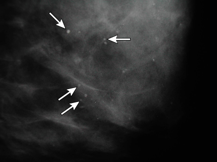 Magnified medial lateral oblique mammogram showing scattered, well-defined, round calcifications (arrows) that can be characterized as benign. These findings are benign and would be described as BI-RADS 2.
Case 8
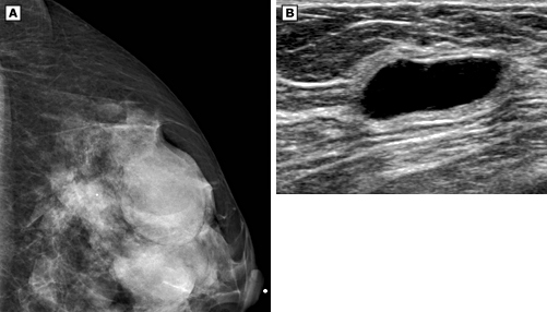 Multiple circumscribed oval and round masses seen on CC mammogram (A). Simple cyst is seen on ultrasound (B), confirming the benign nature of the mammographic mass.