Axilla – General Points
- 95% of breast lymph drained by axilla
- 50% symptomatic patients and 10-20% screened cancers are node positive
- Axillary LN status one of the most important prognostic actors in Breast Ca
- Average of 20 nodes

Axilla – Pre-op Evaluation
- Palpable node
- Proceed to USS guided FNA, then core biopsy if negative
- If decline biopsy → ALND
- Abnormal node on imaging
- Pre-op USS is effective way to screen axillary LNs
- – Sensitivity 50%
- If NAD → SLNB
- If Abnormal node → FNA/Core →
- If +ve FNA →
- Neoadjuvent treatment (HER+ve/TNCB),
- ALND if undergoing primary surgery
- if luminal A/B → ?overtreatment, role of neoadjv? (ask breast surgeon)
- If -ve FNA → SNB
- If +ve FNA →
- Pre-op USS is effective way to screen axillary LNs
Sentinel Lymph Node Biopsy
Indications
- All patients with clinically negative nodes
- DCIS if mastectomy performed
- Or consider if suspicious features
Advantages
- Axillary dissection does not improve survival but provides local control and staging
- Adequate staging can be gained with SLNB
- Adequate local control can often be gained avoiding ALND with radiotherapy
- Decreased lymphoedema, pain, numbness, movement restriction
- Intra-op assessment can be done by frozen section → False negative rate of up to 20%!
- Touch prep cytology has sens of 90%
- Equivalent staging to level II clearance, with less morbidity
- Predicts status of remaining axillary LNs in > 95% of pts
- False negative rate 5-10 %
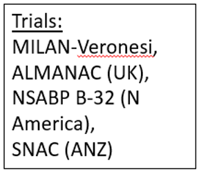
Five randomized clinical trials have been performed to evaluate the efficacy and safety of SLNB in early breast cancer patients
- The NSABP B32 trial (Krag et al., 2007)
- Randomized 2807 patients into the ALND group and 2804 patients into the SLNB group.
- SLN identification rate was 97.2%, and the FNR was 9.8% (Krag et al., 2007).
- DFS, regional control and OS were equivalent between SLNB and ALND groups in breast cancer patients with clinically negative ALN at a median follow-up 95.6 months (range 70.1-126.7)
- Milan trial (Veronesi et al., 2006b)
- Compared with patients underwent SLNB, patients underwent ALND had substantially more arm swelling, arm movement restrictions, paresthesias and pain on the operated side
- Those difference in mobidities persisted 2–3 years after surgery, which significantly impaired patients’ QoL in ALND group
- SNAC trial - Sentinel Node Biopsy versus Axillary Clearance (Gill, 2009)
- GIVOM trial (Zavagno et al., 2008)
- ALMANAC Trial - Axillary Lymphatic Mapping Against Nodal Axillary Clearance trial (Goyal et al., 2006)
- The ALMANAC trials demonstrated that combination of blue dye and radioisotope (dual mapping method) permitted an improved SLN identification (combined 96% versus blue dye 85.6% versus radioisotope 85.6%) and positive SLN identification (combined 93.5% versus blue dye 90.9% versus radioisotope 89.1%)
SNB Technique
-
Technetium99 colloid
- t1/2 6hrs; injected on am of surgery, or on pm prior to am list
- SLN by radioisotope defined as containing ≥ 10 x background count
-
± 2-4 mL Patent Blue V dye after induction
- cf Methylene blue: faster & can lead to breast tissue necrosis
- Anaphylaxis 1:2000
- Intradermal / subareolar injections → Track to axilla in 15mins
- Peritumoral injection → Track to axilla ± internal mammary nodes
-
Combination of patent blue and radioactive colloid results in significantly higher success rates and lower false negative rates
- Compared to patent blue alone
-
Ideally get 1-3 LNs
- 1 x LN → Sensitivity 87%,
- 2 x LN → Sensitivity 97% (Sens 91% and False negative 4-10%)
- No advantage if > 3 LNs
-
In 4% (in SNAC trial) SLN not found → Axillary dissection required
-
False -ve ≈ 5%
- 8% in SNAC trial up to May 05
- ≈ 13% for tumours >3cm
- Axillary recurrence rate in SNB –ve 2.7%
- 8% in SNAC trial up to May 05
-
50% with +ve SNB have no other +ve nodes
Pathologic Analysis of Nodal Mets
- Subgroup based on size of largest contiguous tumour deposit in sentinel node (on H&E)
- Isolated Tumour Cells
- < 0.2mm tumour deposit in SLN
- pN0(i+)
- Prognostically = LN -ve pts
- Isolated Tumour cells give 5% risk of another node being +ve
- Micrometastases
- 0.2-2mm tumour deposit in SLN
- pN1mi
- Slightly worse prognosis vs. LN -ve pts
- Do not predict recurrence
- ACOSOG-20010 & European IBCSG 23-01 trials
- No survival benefit or recurrence benefit if axillary dissection done for Micrometastasis
- Micromets unlikely to have additional nodes involved
- 97.3% 5y survival and only 1.5% local recurrence
- Macrometastases
-
2mm tumour deposit in SLN
- Worse prognosis the greater the nodal involvement
-
- Extranodal Extension
- Invasive tumour cells or clusters outside of LN capsule
- Generally tumour present both subcapsular and adjacent extranodal fat
- More significant if extensive
- NB: ITCs in perinodal axillary channels are not classified as extranodal extension
- Occult Metastatic Disease
- Nodal disease not seen on H&E stain but picked up if doing IHC
- No significance in terms of surgical management and patient outcomes, still classed as pN0
- IHC not routinely performed
- Missed node
- Risk of a +ve (non-sentinel) node being missed is dependent on
- Size of micrometastases
- Extra-capsular spread
- ≈ 8-20% overall
- Risk of a +ve (non-sentinel) node being missed is dependent on
- Isolated Tumour Cells
Additional considerations
- If no Axillary dissection done after positive SLN
- 1.4 % (vs. 0.8%) recurrence rate
- Alternative if can’t do Axillary dissection - offer adjuvant RTx to Axilla (NICE 2009)
- Z11 – No survival difference or recurrence difference
- But only T1/T2 tumours and pts did actually get axillary radiotherapy
- Z11 – No survival difference or recurrence difference
SNB after Neoadjuvant Therapy
- Clinically node negative
- Negative - no further treatment
- Shows evidence of cPR - radiotherapy
- Postive - ALND
- Clinically node positive prior
- If remains positive
- ALND
- If clinically negative post treatment
- Target axillary dissection (TAD)
- ACOSOG Z1071
- Aim: to determine the false positive rate in SLN after neoadjuvent chemotherapy in patients with node positive disease
- Inclusion: T1-4, N1-2, M0
- SLNB alone
- False negative rate 12.6% (39 patients who had an ALND after negative SLNB)
- SLNB + clip removed in specimen
- False negative 6.8%
- Improved Axillary Evaluation Following Neoadjuvant Therapy for Patients With Node-Positive Breast Cancer Using Selective Evaluation of Clipped Nodes: Implementation of Targeted Axillary Dissection. Caudle AS et al
- Target axillary LND (TAD) has a false negative rate of 2% compared with SLNB FNR 10.1%
- Among patients shown to be N+ prior to preoperative systemic therapy, SLNB has a >10% false-negative rate when performed after preoperative systemic therapy, which can be improved by marking and removing the most suspicious biopsied node, using dual tracers, and by obtaining ≥3 sentinel nodes (targeted axillary lymph node dissection). (Caudle AS, et al. J Clin Oncol 2016;34:1072-1078.)
- ACOSOG Z1071
- Target axillary dissection (TAD)
- If negative - radiotherapy
- If positive - ALND
- Residual cancer = Isolated Tumour Cells, Micromets, Macromets
- If remains positive
- Uptodate authors recommend ALND in all patients with pretreatment N2-N3 independent of clinical response
Frozen Section During SNB
- Melanoma
- Sensitivity 50%, risk of positive LN = 20%
- Therefore 10% whom have frozen section will need ALND
- Breast
- Sensitivity 75%, Risk of positive LN = 40%
- Therefore – 30% whom have frozen section will need ALND
- Frozen section more useful in breast cancer than melanoma
- NB: False negative rate up to 20%
- Touch imprint cytology
- 30-100% sensitivity (poor for lobular)
- False-negative rates for intra-op assessment are up to 38%
- Path protocol:
- 2mm slices, H&E & CAM 5.2 stains: single slice thru each LN
Management after SNB
- No nodes positive, ITC, or occult mets → No ALND
- No diff in regional control, overall survival, or disease free survival
- Positive nodes
- 1-2 nodes
- Breast conserving surgery
- Z11 Eligible → No ALND
- Clinically negative nodes, T1 or T2, < 3 positive nodes on SNB
- Although in practice now criteria is expanding including from SENOMAC trial
- Undergoing breast conserving therapy followed by whole breast radiation
- Similar LR, DFS and OS
- Clinically negative nodes, T1 or T2, < 3 positive nodes on SNB
- Z11 Eligible → No ALND
- Mastectomy
- AMAROS criteria → axillary radiotherapy
- T1-T2, unifocal, invasive breast cancer without palpable axillary lymphadenopathy
- Also included patients with mastectomy 17.4% (and BCS, still the majority) which showed axillary radiotherapy is a safe alternative to ALND
- Similar LR, DFS and OS
- AMAROS criteria → axillary radiotherapy
- Breast conserving surgery
- 3+ nodes
- ALND
- 1-2 nodes
Landmark trials
- IBCSG 23-01 trial - The International Breast Cancer Study Group 23-01 trial (Galimberti et al., 2013).
- Designed to determine whether ALND was warranted in patients with one or more micrometastatic (≤ 2 mm) SLNs and tumor ≤ 5 cm.
- 934 patients were randomly allocated to receive either ALND (N = 465) or no ALND (N= 469).
- Patients receiving either mastectomy or breast conserving surgery were eligible in this study.
- Median follow-up of five (Interquartile range IQR 3.6–7.3) years, the 5-year DFS (ALND 84.4% versus no ALND 87.8%, log-rank P = 0.16) and 5-year OS (ALND 97.6% versus no ALND 97.5%, log-rank P = 0.73) were comparable between two groups
- ACOSOG Z0011 trial- The American College of Surgeons Oncology Group ACOSOG Z0011 trial (Giuliano et al., 2011)
- Designed to determine whether no ALND is non-inferior to ALND in patients with clinical T1-T2 N0 invasive breast cancer, and 1–2 SLNs containing micrometastases (≤2 mm) or macrometastases (> 2 mm).
- Local recurrance similar
- At a median follow-up of 9.25 years, the 10-year cumulative locoregional recurrence incidence was not significantly different between two groups (ALND 6.2% versus SLNB 5.3%, P = 0.36)
- Similar disease free and overall survival
- In an earlier report of data at a median follow-up of 6.3 years, non-inferiority for DFS (ALND 82.2% versus SLNB 83.9%) and OS (ALND 91.8% versus SLNB 92.5%) in the SLNB group was observed .
- Critics have argued that patients enrolled in Z0011 trial were highly selected population with favorable outcome (e.g. hormonal receptor positive rate 83% in SLNB group) and considered that the Z0011 findings may not be applicable to all patients undergoing breast conserving surgery.
- It is very important to know that the majority of patients in Z0011 trial (ALND 96% and SLNB 97%) received systemic therapies, which could eliminate the residual disease in ALNs.
- These results demonstrated that omission of ALND had no apparent negative effect on locoregional recurrence and survival in patients with limited SLN involvement who are treated with breastconserving surgery, whole-breast radiation therapy and adjuvant systemic therapy.
- AMAROS - After Mapping of the Axilla Radiotherapy or Surgery trial
- Non-inferiority trial, patients with clinical T1-T2 breast cancer and a positive SLN were randomly assigned to receive either ALND (N = 744) or axillary radiotherapy (N = 681).
- All patients underwent either mastectomy or breast-conserving surgery.
- Similar local recurrence
- At a median follow-up of 6.1 (IQR 4.1-8.0) years in the intention-to-treat population, the 5-year axillary recurrence was non-inferior (Hazard ratio 0.00-5.27 with a non-inferiority margin of 2) in the axillary radiotherapy group to the ALND group (Axillary radiotherapy 1.19% versus ALND 0.43%).
- Similar DFS and OS
- Fiveyear DFS (Axillary radiotherapy 82.7% versus ALND 86.9%, P = 0.18) and 5-year OS (Axillary radiotherapy 92.5% versus ALND 93.3%, P = 0.34) between two groups were also comparable (Donker et al., 2014).
- Several limitations of this trial hamper the extrapolation of its results to clinical practice. First, due to fewer axillary recurrence events than anticipated, the study was under powered to address the primary endpoint, which was non-inferiority of 5-year axillary recurrence. Second, 82% of the patients enrolled in the AMAROS trial received breast-conserving surgery, 95% had one or two positive SLN and 40% had only micrometastatic disease or ITCs.
- According to the results from Z0011 and IBCSG 23-01 trials, most of the patients enrolled in the AMAROS trial would not have needed ALND or axillary radiotherapy. It is inappropriate to extrapolate the AMAROS results to patients with three or more positive SLNs, since there were only 5% of the patients with three or more positive SLNs in the trial (Boughey, 2014).
- For a small proportion of patients who do not meet the criteria of the Z0011 trial, e.g. patients with clinical T1-T2 N0 invasive breast cancer, and 1–2 SLNs containing macrometastases who are scheduled for mastectomy, the results from AMAROS trial suggest that axillary radiotherapy could be an alternative to ALND. It should be noted that the extensive radiotherapy used in the AMAROS trial, which includes level 1, 2 and 3 nodes, and the supraclavicular nodes, might be viewed as overtreatment by current standards. It might have been sufficient to treat only the level 1 and level 2 nodes.
- Non-inferiority trial, patients with clinical T1-T2 breast cancer and a positive SLN were randomly assigned to receive either ALND (N = 744) or axillary radiotherapy (N = 681).
- SENOMAC trial
- T1-T3 cancer with 1 or 2 positive nodes on SLNB
- ALND vs omission (most patients in both groups received radiotherapy including nodal target volumes)
- Included patients with extra-nodal extension and undergoing mastectomy
- This trail expands the indications to include T3
- T1-T3 cancer with 1 or 2 positive nodes on SLNB
- The results of the IBCSG 23-01, Z0011, MSKCC and AMAROS studies suggest that the aggressive ALND can be safely omitted in a proportion of patients to avoid morbidities and improve patients’ QoL. Thus, omission of axillary surgery in patients with a low risk of ALN metastasis has gained more research interest.
Axillary Lymph Node Dissection
Boundaries of the axilla
- Axillary vein superiorly.
- Serratus anterior muscle and chest wall medially
- Subscapularis and teres minor posteriorly
- Latissimus dorsi posteriorly
- Medial aspect of the humerus laterally
- Pectoralis minor and major muscles anteriorly.
Lymph node levels
- Level I – lateral to pec minor.
- Level II – posteriorly to pec minor.
- Level III – medially to pec minor
- Rotter’s node – in between pec minor and pec major.
Clinical considerations
- Axillary node clearance should strive to preserve the thoracodorsal pedicle, long thoracic nerve, medial pectoral nerve, and intercostobrachial nerve.
- The thoracodorsal supplies the LD, it is vital to preserve this if you want to perform a LD reconstruction.
- The thoracodorsal nerve supplies motor function to LD - injury to this effects shoulder extension and rotation.
- The long thoracic nerve supplies the serratus anterior and injury to this results in winged scapula.
Surgical access
- If the patient is undergoing a modified radical mastectomy then the lateral extension of the mastectomy incision is used.
- Otherwise, and oblique incision at the inferior margin of the axillary hairline extending from the lateral edge of pectoralis muscle to the medial edge of latissimus dorsi is used.
- Skin flaps should be raised – superiorly to the level of the axillary vein and inferiorly to the level of the 4th-5th rib.
Operative steps
- The clavipectoral fascia should be identified and opened.
- Blunt dissection through the axillary fat pad superiorly to identify the axillary vein should occur. The vein will course horizontally in the dissection field and be anterior and inferior to the axillary artery.
- Once the axillary vein is identified, the lateral edge of pectoralis muscle should be identified. Dissection should continue laterally from this lateral edge until the lateral thoracic vein (LTV), approximately 2-3cm from the lateral chest wall is identified.
- The LTV drains the lateral portion of the breast and can be ligated and divided if needed, but only after the thoracodorsal bundle has been identified.
- Located deep and slightly lateral to the LTV is the TDB, which includes the nerve, artery, and vein running to latissimus dorsi muscle.
- The nerve is located laterally, artery in the middle, and vein medially (same as the femoral canal!)
- The TDB should be identified and preserved along its entire course to its insertion into the medial edge of LD.
- Approximately 2cm inferior to the axillary vein is the intercostal brachial sensory nerve. This runs from the second intercostal rib space and transverses across the axilla to provide sensory innervation to the medial portion of the upper inner arm.
- It courses anterior to the TDB. An attempt to preserve it should be made.
- The long thoracic nerve runs parallel to the TDB along the lateral deep surface of the serratus anterior muscle fascia and provides motor function to the muscle.
- The medial pectoral nerve wraps around the lateral edge of pec major and supplies both pectoral muscles.
- Once all the axillary anatomic strictures have been identified, resection of the level I and II nodes can be performed. Currently, there is little indication to remove level III nodes unless there is gross nodal involvement.
- Superior dissection behinds under the pectoralis minor muscle to remove level II lymphatics.
- Lymphatic channels and blood vessels should be clipped or ligated.
- After a superior dissection is complete, the specimen can be retracted inferiorly and the remained of the lymphatic tissue removed.
Rationale:
- Stages disease & dictates need for adjuvant treatment
- Provides local control of axilla - local recurrence < 1% (0.4%)
- Possible survival implications – unknown if ↑s survival
Procedure
- Level II dissection is operation of choice
- Level III if macroscopic evidence of tumour at level II
- No improved DFS/OS after routine level III AD
- Up to 25% will have level III involvement if Level I involved
- Aim to remove 20 nodes for optimal staging
Complications
- Lymphoedema
- 15%
- Physical therapy, arm strengthening, and stretching; compression garments
- Limitations in postoperative shoulder and arm mobility
- Prolonged postoperative pain
- Cording
- Cording of the axilla and upper arm, is a self-limited, harmless condition
- Believed to be caused by thrombosis in subcutaneous veins and ligation of veins after axillary dissection
- Paraesthesia
- intercostal bracial nerve
- Upper inner arm
- intercostal bracial nerve
- Winged scapula - LTN
- Weakness of the arm with respect to extension, adduction and medial rotation - TDB/Lats