Arterial exam
Expose: ARMS + LEGS + GROIN + ABDO
HANDS:
- Nicotine staining
- Nails-splinter haemorrhage
- Endocarditis
- SLE
- RA
- Vasculitis
- Radial Pulse
- Rate + Rhythm
NECK:
- Carotid pulse-AF
- Aucultate Bruits
- Scar- Carotid Endart
ABDOMEN:
- AAA (N=<2.5cm)
- Renal Bruits
- Laparotomy Scar
OTHERS:
- DOPPLER
- ABPI
- N=0.9-1.2
- <0.7=claudication
- <0.4=rest pain
- NEURO
- Sensation
- motor
DIABETIC FOOT:
- clawed toes
- flattened arch
- Decreased sensation (glove + stocking)
- dry + cracked skin
- atrophic
- infection
LEGS/GROIN: INSPECTION:
- Colour a. Haemosidoren b. Erythema c. Purple d. white
- ulcers a. location b. depth c. granulation
- trophic changes a. hair loss b. tissue loss c. dry
- scars
- varicose veins
- guttering of veins (suggests decreased art flow)
- between toes + back of foot
PALPATION:
- Capillary refill
- Temperature (bck of hand)
- Pitting oedema
- Pulses a. DP/PT/popliteal/femoral
AUSCULTATE:
- Aorta + femoral + popliteal for BRUITS
BEURGERS TEST/ANGLE
- Angle to which leg must be raised before becomes white.
- N = > 90 dgrs AND toes remain PINK
- < 90 dgrs ischaemic leg
- < 20 dgrs severe ischaemia
- (angle is proportional to pressure in foot small vessels)
ISCHAEMIC ULCERS:
- tips of toes / pressure areas / punch out / deep/surrounding tissue cold
NEURPATHIC/DIABETIC ULCERS:
- Painless / punched out +/- surrounding tissue healthy
Venous exam
2 FEATURES OF VENOUS CIRCULATION:
- Adequate CROSS-SECTIONAL area to allow normal OUTFLOW from limb
- If VALVES ensure ONE-WAY flow
INSPECTION STANDING:
- Varicosities- (collapsed when lying)
- Venous stars
- Skin changes: a. Pigmentation b. Eczema
- ‘inverted champagne’ bottle look
- Venous ulcers / healed ulcers (medial gutter area) a. Painful b. Shallow c. Sloping edge d. Purple/blue
- Oedema
SAPHENA VARIX COUGH TEST:
- 2-4 cm inferolateral to pubic tubercle
- ask patient to cough
- N= no impulse palpable
- +ve test = smooth palpable thrill
- indicates incompetent valve at SFJ
PALPATION (lying):
- Temperature
- Tenderness
- Induration
- Thrombosis
- Pulses (DP/PT/Pop)
TRENDELENBURG TEST: • Used to find how far up the incompetent valves are • Pt lying • Raise leg (to empty veins) • Place torniquet below SFJ (remove after 60sec) • Stand Patient • N = vein fills > 35 sec • If incompetent valves, the superficial veins fill IMMEDIATELY o If fills from ABOVE, then SFJ incomp. o If fills from below, then incomp below torniquet.
PERTHES TEST: • Used for assessing PATENCY of DEEP VENOUS system • Pt STANDS → veins fill → torniquet placed (mid thigh) → walk 5 mins • N = long saphenous veins collapse o Indicates deep veins competent + communicating veins patent • If unchanged = SFJ + communicating veins incompetence • If veins increase (and pain) = deep veins occluded
More detail Arterial
Examination
- Inspection:
- Trophic changes: Loss of hair over calf/toes/foot, Thickening of toenails (onychomycosis), muscle wasting, Thin skin, Pale legs, Ulceration/ gangrene
- Feel:
- Coolness in feet, cap refill
- Feel all pulses – rate, rhythm, quality
- Examine aorta
- Listen to heart
- Check BP bilaterally - 5% PAD pts have supra-aortic occlusive disease with diff arm pressures (Use higher of two pressures to calculate ABPI)
- Special tests
- ABPI
- Calculate pedal pressure with cuff at ankle (use doppler)
- Compare to brachial (again use doppler)
- Values
- Normal = 1-1.2
- < 0.5 = Critical Ischaemia
- ABPI = Pedal BP/Brachial BP
-
0.9 Normal
- 0.6-0.9 Claudication
- 0.3-0.6 Rest pain
- < 0.3 Critical ischaemia
-
- DM leads to falsely high ABI due to arteriosclerosis
- Wall calcification (in elderly / diabetics) should be suspected if ABI > 1.1
- Usually triphasic, biphasic – 50% stenosis, monophasic 70%
- Actual ankle pressure can be useful
- < 50mmHg – severe disease
- Toe pressures
- Useful in when have non-compressible tibial artery (DM) - digital vessels spared & hence compressible
- Small pneumatic cuff around toe & photoplethysmographic sensor
- Values
- Normal ≥ 0.7
- Claudication 0.4
- Rest pain, gangrene 0.1
- If > 30mmHg – indicator that there is healing potential for ulcers
- ABPI
- Buerger’s Test
- Elevate the limb
- Normally toes should stay pink at 90 degrees
- In PVD: Leg becomes waxy/cadaveric, white (esp. on sole) ± Venous guttering appears if a finger is run along a vein
- When the limb is lowered, the time it takes for the vein to refill is a good indicator of the degree of ischaemia (normal < 15secs)
- Buerger’s angle = Angle to which the leg is raised to become white (or doppler pulses disappear) (< 20-30 degrees = severe PVD, often with rest pain)
- Dependent rubor - When the limb is hung down there is reactive hyperaemia because the ischaemia produced by elevation result in maximum cutaneous vasodilation - Dark red cyanosis (blood in foot relatively stagnant, oxygen extraction high)
- Also: evaluate pt for associated cerebrovascular/cardiovascular disease
- Listen for carotid bruit
More detail Venous
-
Look
- Areas
- Examine in standing position with groin and leg exposed
- Externally rotate each leg then turn around
- As with the post-thrombotic limb, an incompetent GSV disrupts the capillary circulation around the ankle, which may lead to venous hypertensive changes in gaiter area
- Distribution and extent of varicosities – esp. Saphenovarix
- Presence of skin changes of chronic venous stasis
- Varicose eczema
- Lipodermatosclerosis = Fibrosis of s/c tissues with fat necrosis & chronic inflammatory changes → Hard tissue
- Champagne leg
- Hemosiderin pigmentation / hyperpigmentation
- Induration
- Ulceration (esp. at med ankle) ± surrounding white scarred areas (atrophie blanche)
- Corona phlebectatica = Cluster of telangiectasia over the medial ankle (distended subdermal & intradermal venules)
- Look for thrombophlebitis – 40% associated with DVT
- Areas
-
Feel:
- Pulses
- Check arterial status – distal pulses
- If not palpable – ABPI
- Other
- Feel for temperature differences
- Check for pitting oedema
- Look for scars from previous repairs/abdominal surgery
- Veins tense on palpation & cough impulse may be elicited at SFJ
- Saphena varix - soft compressible dilatation of GSV adjacent to SFJ
- Pulses
-
Special Tests
- Cruveihier’s Sign
- Thrill over saphena varix on coughing
- Chevrier’s Sign
- Impulse felt over SFJ if saphenous vein is distally percussed
- Trendelenburg Test
- Direct digital pressure over the SFJ preventing retrograde filling of the GSV
- Brodie-Trendelenburg Test
- Tourniquet test to identify the sites of reflux from deep to superficial vein systems
- With pt lying:
- Tourniquet applied to elevated limb just below groin
- Patient stands up
- Normal: vein fills within 35secs from below with tourniquet in situ
- Rapid filling of veins below the tourniquet indicates short saphenous vein incompetence or an incompetent perforator distally
- Tourniquet removed after 60 secs
- If rapid filling from above, it is due to incompetence above the tourniquet (i.e. SFJ)
- Tourniquet can then be repositioned distally to find the level of incompetence or two tourniquet can be used to define an incompetent communicating vein
- Trendelenburg test becoming obsolete
- Sensitivity for GSV incompetence = 55%, SSV incompetence = 35%
- Tourniquet test to identify the sites of reflux from deep to superficial vein systems
- Perthes Test
- Assesses patency of deep veins
- Pt standing and veins filled
- Tourniquet is applied to mid-thigh
- Pt walks for 5 minutes
- If Saphenous Veins collapse below the tourniquet = Deep veins are patent and the communicating veins are competent
- If unchanged = Both saphenous and communicating veins are incompetent
- If the veins ↑ in prominence and pain occurs = Deep veins are occluded
- Assesses patency of deep veins
- Hand-held Doppler Test:
- Traditional tests have poor diagnostic value
- Using hand-held doppler the examiner ask pt to wt bear on the contralateral leg
- Locate femoral pulse then move medially – confirm vein by squeezing and releasing the calf to hear antegrade flow. Move inf to locate SFJ – squeeze calf to get antegrade flow and listen for reflux – can test SSV also
- Pressure Changes
- Normal
- P drops from 90mmHg to < 20mmHg on 10s exercise
- P returns to 90mmHg over > 20sec rest
- Primary Varicose Veins
- P drops from 90 mmHg to 20-40mmHg on 10s exercise
- P returns in about 10s
- Post thrombotic syndrome
- P drop if present is minor (perhaps down to 60-70)
- P returns in about <10s
-
60 – risk of ulceration > 50%
- Normal
- Incompetent perforators
- May be found clinically as gaps in the fascia → Aneurysm-like distension can be palpated as a soft compressible lump
- INDIRECT – to a muscular deep vein
- DIRECT – to one of the main deep veins
- “Horny dogs breed chaos”
- Hunterian – middle 1/3 of thigh (level of adductor canal)
- Dodd – distal 1/3 of thigh
- Boyd – around knee & upper 1/3 of medial calf – MOST COMMON
- Cockett (I-III)– distal calf – connects to Posterior Arch Vein
- Inframalleolar
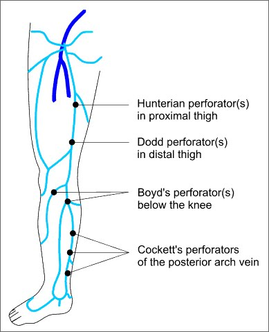
- May be found clinically as gaps in the fascia → Aneurysm-like distension can be palpated as a soft compressible lump
- Cruveihier’s Sign
-
Percussion impuls
- Can palpate for a transmitted percussion impulse from above downwards → Implies absent / incompetent valves
-
NB: Relying on examination alone
- Inappropriate surgery in 12-25%
- Recurrence & re-op rate of 10% after 2 yrs (cf 2% if scanned)
- Most pts with venous ulcers have incompetent communicating veins
Evaluating a diabetic foot
History
- Duration of diabetes, overall glycaemia control, presence of micro/macrovascular disease
- History of foot injury
- Claudication symptoms
- Seldom get claudication as disease mostly infragenicular
- Smoking
- History consistent with foot neuropathy - burning pain in feet, wakes at night, worst at night, relived by walking around.
Examination
- Inspect
- Lesions between toes, bunions, macerated areas.
- Claw-foot deformity
- Look for signs of infection
- Assess gait
- Feel
- Evaluation of pedal pulses
- Test sensation - Semmes-Weinstein monofilament - 12 sites of the foot. Test vibration at dorsum of first toe just proximal to nail bed.
- ABPI - normal is 0.9-1.3
- < 0.9 = PVD
- < 0.4 = 0.4 critical limb ischaemia
- PVD examination
- Ulcer
- Neuropathic ulcers: occur @ MTP heads or dorsum of toe, foot is typically warm with bounding pulses and distended veins
- Neuro-ischaemic: occur @ tip of toes, heel and medial aspect of 1st metatarsal
- No overlying callus, rim of ischaemic tissue around and often necrosis
- Pain will depend on underlying neuropathy
- Gangrene – dry (not infected) can be left and will autoamputate eventually
- Wet gangrene (infected) will require debridement and often amputation
- Infection: Cellulitis &/or osteomyelitis
- Systemic signs of infection may be absent
- Probe to bone – if +ve – PPV for osteomyelitis 50-65%