Section: Vascular Sub-section: Curriculum, page 99
Definition
- Peripheral arterial insufficiency is caused by Atherosclerosis which causes progressive narrowing of the arteries leading to claudication, ulcers or rest pain and eventually ischaemia. ABPI <0.9
- Encompasses
- Atherosclerosis
- Arteriosclerosis obliterans
- Thromboangiitis obliterans (Buerger’s)
- Systemic vasculitis affecting medium sized arteries and veins
- Associated with smoking – esp. middle eastern ethnicity
- Strongly associated with smoking, recurrent inflammation & thrombosis of medium & small arteries/ veins
Incidence
- 20% > 70yrs
- Incidence of claudication 5-7%
- Incidence of severe leg ischemia (rest pain or tissue loss) 75/ 100,000/ year
- M:F = 2-5:1
Aetiology
- Atheroma
- Most common
- Risk Factors (Modifiable)
- Smoking – 4x RR (ex-smoker 2x RR), associated with 80% of claudication
- Hypertension
- Hyperlipidaemia
- Obesity
- Glycaemic control
- Risk Factors Non-Modifiable:
- ↑ Age
- Male
- FHx
- Ethnicity
- Pre-existing Disease:
- CVD
- CVA/TIA
- PAD
- DM – 5 x RR, often presents with unreconstructible disease
- Emboli
- AF / post-MI / Popliteal or Iliac aneurysm
- Thrombosis
- Risk factors = Hypercoagulable states
- Malignancy
- Polycythaemia
- Factor V Leiden mutation
- Protein C / S deficiency
- Lupus anticoagulant
- Risk factors = Hypercoagulable states
- Vasculitides
- Buerger’s, SLE, Takayasu’s
- Congenital Anomalies:
- Coarctation of Aorta, Popliteal Entrapment, Fibromuscular dysplasia
- DDx = Spinal Stenosis or Lumbar Nerve root irritation that pain comes on with STANDING and is not relieved by resting pts need to bend or sit down. Exacerbated by straight leg raise
Differential Diagnosis
- For Intermittent Claudication
- Chronic Compartment Syndrome
- Usually seen in young athletes, with regular exercise
- Symptoms often bilateral
- Cystic Adventitial Disease
- 80% male, average age 42yr
- Popliteal Artery most commonly affected
- Almost always unilateral
- Popliteal cystic disease/ cystic degeneration of popliteal artery
- Cystic abnormality of adventitia of Popliteal Artery
- Contents resemble ganglion
- Pedal pulses may disappear on knee flexion
- Adventitial mucoid cysts of unknown aetiology, occur at level of knee joint leading to occlusion of artery, angiogram often shows a curved stenosis/ hour-glass “Scimitar sign”
- US/CT often diagnostic
- Rx - Cystectomy/ evacuation (occasional vein bypass)
- External Iliac Endofibrosis
- Seen in 20-30 year old long-distance cyclists; buttock, thigh claudication
- Popliteal Artery Entrapment/ Compression
- Congenital anomaly – runs medial to Medial Head of Gastrocnemius OR deep to Popliteus
- 85% male, average age 33
- Pedal pulses disappear with plantar flexion
- Often MRI useful
- Rx: Return popliteal artery to normal course
- Persistent Sciatic Artery
- Rare Congenital anomaly
- Absent femoral pulse, but popliteal pulse present
- Embryological Sciatic Artery persists and continues with popliteal artery, often ‘held up’ at sciatic foramen
- Sx develop if femoral artery not well developed
- Venous Claudication
- Occurs in > 40% of those with Iliofemoral DVT
- Clinically: Stigmata of chronic venous insufficiency
- Unlike arterial claudicant, pts require 15-20mins rest, often with leg elevation for pain to resolve
- Spinal Stenosis
- 75% male, average age 50-60yrs,
- Due to degenerative lumbosacral disease
- May occur on standing – paraesthesia and weakness
- Normal pulses & ABI
- Chronic Compartment Syndrome
Pathology
- Atherosclerosis related lesions
- Early: Confined to intima
- Advanced: Intima & media are involved (adventitia spared)
- Plaques usually develop in areas of low shear stress such as arterial bifurcations and posterior wall of aortoiliac segment
- Aortoiliac
- Confined to aorta & common iliacs 10%
- Extends into external iliacs 25%
- Multi-segmental & infrainguinal 65%
- Femoral artery often affected where the vessel passes through adductor hiatus
- Aortoiliac
- Patterns of disease
- Smokers < 55ys → Accelerated disease involving aortoiliac segments
- Diabetics → Tibioperoneal Artery disease more likely
- Lesions often symmetrically distributed
- Relationship of Flow & Radius
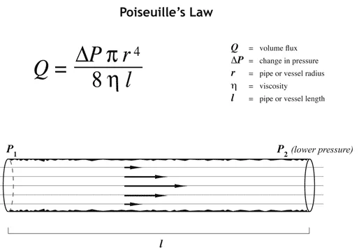
- Flow & resistance are inversely related to the 4th power of the radius
- Small changes in radius → Big changes in flow
Clinical
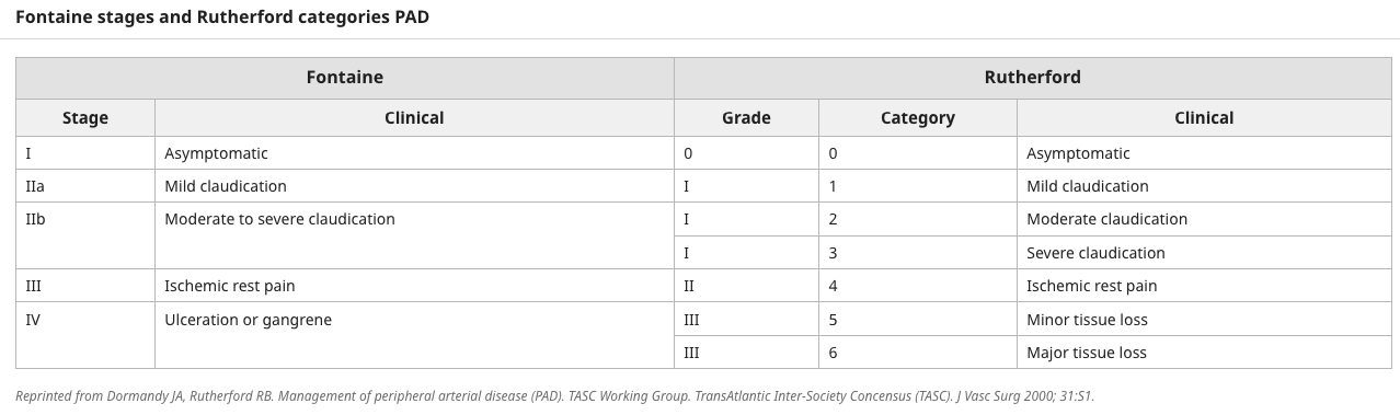
- Fontaine Stages
- 1 Asymptomatic
- 2 Intermittent claudication
- 2a >200m
- 2b <200m
- 3 Rest pain (often worse at night)
- 4 Tissue loss (Ulceration/Gangrene)
- Rutherford
- Stage 0 – Asymptomatic
- Stage 1 – Mild claudication
- Stage 2 – Moderate claudication
- Stage 3 – Severe claudication
- Stage 4 – Reѕt paiո
- Stage 5 – Minor tissue loss with ischemic nonhealing ulcer or focal gangrene with diffuse pedal ischemia
- Stage 6 – Major tissue loss – Extending above transmetatarsal level, functional foot no longer salvageable
-
WIfI (Wound, Ischemia, foot Infection)
- Wound
- Grade 0 – Reѕt рaiո; no wound, no ulcer, no gangrene.
- Grade 1 – Small shallow ulcer(s) on distal lеg or foot, any exposed bone is only limited to distal phalanx; no gangrene, or gangrene limited to distal toe (ie, minor tissue loss: limb salvage possible with no more than simple digital amputation, or skin coverage)
- Grade 2 – Deeper ulcer on distal leg or foot with exposed bone, joint, or tendon, or shallow heel ulcer without involvement of the calcaneus; gangrenous changes confined to the digits (ie, major tissue loss: salvageable with ≥3 digital amputations or standard transmetatarsal amputation plus skin coverage)
- Grade 3 – Extensive deep ulcer of the forefoot and/or midfoot, or full-thickness heel ulcer with or without involvement of the calcaneus (ie, extensive tissue loss: salvageable only with complex foot reconstruction or nontraditional TMA
- Ischemia (note that toe systolic pressures are preferred in patients with diabetes):
- Grade 0 – ABI ≥0.8, ankle systolic pressure >100 mmHg, toe pressure (TP)/transcutaneous oxygen (TcPO2) ≥60.
- Grade 1 – ABI 0.6 to 0.79, ankle systolic pressure 70 to 100 mmHg, TP/TcPO2 40 to 59.
- Grade 2 – ABI 0.4 to 0.59, ankle systolic pressure 50 to 70 mmHg, TP/TcPO2 30 to 39.
- Grade 3 – ABI ≤0.39, ankle systolic pressure <50 mmHg, TP/TcPO2 <30.
- Foot infection
- Grade 0 – No symptoms or signs of infection.
- Grade 1 – Infection is present and at least two of the following are present: local swelling or induration, erythema >0.5 to ≤2 cm around ulcer, local tenderness or раiո, local warmth, or purulent discharge. Other causes of an inflammatory response of the skin have been excluded (eg, gout, fracture).
- Grade 2 – Local infection is present as defined for Grade 1 but extends >2 cm around ulcer or involves structures deeper than the skin and subcutaneous tissues (eg, abscess, osteomyelitis, septic arthritis, fasciitis). No clinical signs of systemic inflammatory response.
- Grade 3 – Local infection is present as defined for Grade 2, but clinical signs of systemic inflammatory response are present as manifested by two or more of the following: temperature >38°C or <36°C; heart rate >90 beats per minute, respiratory rate >20 breaths per minute or PaCO2 <32 mmHg; white blood cell count >12,000 or <4000 (cu/mm) or >10 percent immature band forms present.
- Wound
-
Claudication
- Calf (± thigh/gluteal) pain is typical of SFA (± iliac) disease
- Calf most commonly involved due to high workload of calf muscles with normal walking
- Buttock (cf thigh) claudication may signify Internal Iliac disease
- 25% of males with Aortoiliac occlusion suffer Leriche’s Syndrome
- Triad: Buttock/thigh pain/ Claudication + Impotence + Absence of Femoral Pulses
- 25% of males with Aortoiliac occlusion suffer Leriche’s Syndrome
- Buttock (cf thigh) claudication may signify Internal Iliac disease
- Gradual onset over years
- Claudication distance → Reproducible
- Resting in upright position rapidly relieves pain, within minutes (2-10), (Venous = 20mins)
- Usually worse uphill
- DDx
- Spinal claudication / spinal stenosis – rest doesn’t relieve, worse in am, need to sit/lie down to relieve pain
- Disc herniation – worse with lordosis, standing, walking
-
Rest pain
- (± exacerbated by warmth) suggests critical ischaemia
- Distal - more in the feet, often forefoot & toes
- Pt may sit in bed with knee bent, holding the foot still to relieve pain
- Night → Leg-hanging (often worsens oedema & hence worsens tissue damage)
-
Ulcers – Pressure areas
- Acute Ischaemia
- Paraplegia – can knock off Arteria Radicularis Magna of Adamkiewicz (dominate artery which arises from somewhere within T8-L1 that supplies distal spinal cord)
-
Upper limb
- Claudication
- Rest pain, ulcer – rare as are extensive collaterals
- Digital gangrene usually embolic
- Most commonly in subclavian proximal to vertebral (supraclavicular bruit)
- Can get vertebral steal
- Non-hemispheric symptoms
- If cerebral symptoms usually concomitant ICA disease
- Claudication
History
- Claudication
- Hx of Risk Factors (associated conditions, current medications and family hx)
- Previous treatments
- Hx of CVD/CVA/angina
- Post-prandial pain (Mesenteric ischaemia)
- ***Fitness for major surgery esp. weight, resp fx – smoking risk of COPD
Examination
- Inspection:
- Trophic changes: Loss of hair over calf/toes/foot, Thickening of toenails (onychomycosis), muscle wasting, Thin skin, Pale legs, Ulceration/ gangrene
- Feel:
- Coolness in feet, cap refill
- Feel all pulses – rate, rhythm, quality
- Examine aorta
- Listen to heart
- Check BP bilaterally - 5% PAD pts have supra-aortic occlusive disease with diff arm pressures (Use higher of two pressures to calculate ABPI)
- Special tests
- ABPI
- Calculate pedal pressure with cuff at ankle (use doppler)
- Compare to brachial (again use doppler)
- Values
- Normal = 1-1.2
- < 0.5 = Critical Ischaemia
- ABPI = Pedal BP/Brachial BP
-
0.9 Normal
- 0.6-0.9 Claudication
- 0.3-0.6 Rest pain
- < 0.3 Critical ischaemia
-
- DM leads to falsely high ABI due to arteriosclerosis
- Wall calcification (in elderly / diabetics) should be suspected if ABI > 1.1
- Usually triphasic, biphasic – 50% stenosis, monophasic 70%
- Actual ankle pressure can be useful
- < 50mmHg – severe disease
- Toe pressures
- Useful in when have non-compressible tibial artery (DM) - digital vessels spared & hence compressible
- Small pneumatic cuff around toe & photoplethysmographic sensor
- Values
- Normal ≥ 0.7
- Claudication 0.4
- Rest pain, gangrene 0.1
- If > 30mmHg – indicator that there is healing potential for ulcers
- ABPI
- Buerger’s Test
- Elevate the limb
- Normally toes should stay pink at 90 degrees
- In PVD: Leg becomes waxy/cadaveric, white (esp. on sole) ± Venous guttering appears if a finger is run along a vein
- When the limb is lowered, the time it takes for the vein to refill is a good indicator of the degree of ischaemia (normal < 15secs)
- Buerger’s angle = Angle to which the leg is raised to become white (or doppler pulses disappear) (< 20-30 degrees = severe PVD, often with rest pain)
- Dependent rubor - When the limb is hung down there is reactive hyperaemia because the ischaemia produced by elevation result in maximum cutaneous vasodilation - Dark red cyanosis (blood in foot relatively stagnant, oxygen extraction high)
- Also: evaluate pt for associated cerebrovascular/cardiovascular disease
- Listen for carotid bruit
Investigation
- Bloods
- FBC, BSL, UEC, Lipids, ESR
- Young pts < 50 yrs
- Thrombophilia screen (Lupus, Protein C& S, Antithrombin III, Anticardiolipin, Fibrinogen, Plasminogen, Homocysteine)
- Urinalysis
- Glycosuria, proteinuria
- Exercise testing
- ABI after exercise – Useful for pts with normal pulses but good hx of claudication
- Duplex USS
- Operator dependent – can identify areas of stenosis and degree of stenosis
- Velocity measurements are used to infer degree of stenosis
- Velocities >400-500 m/sec suggest Clinically sig stenosis
- 2 x increased velocity ~ 50% stenosis
- Can check ‘pulse waveform’
- Normally triphasic wave form due to normal elasticity of vessels
- Moderate stenosis = Biphasic
- Severe stenosis = Monophasic
- NB: Difficult to assess suprainguinal vessels but if good views attained then high accuracy
- Velocities >400-500 m/sec suggest Clinically sig stenosis
- Contrast Enhanced MRA
- Preferred 1st line imaging for chronic ischaemia
- Non-ionizing radiation and no iodine based contrast
- However, long study duration ⇒ Images ‘contaminated’ by venous architecture – now compensated by faster image processing
- CI – Pacemaker, metal cardiac valves, aneurysmal clips, claustrophobia
- NB: Gadolinium now recognised to cause Nephrogenic Systemic Fibrosis (NSF) in up to 2% of high risk pts Attributed to gadolinium based contrast agents esp. in setting of renal impairment
- CT-angiography
- Excellent for acute ischaemia or bleeding
- Enables 3-D reconstruction
- Good for surgical planning and graft selection
- Calcification in arteries can limit use in chronic setting
- Requires high volumes of contrast (nephrotoxic)
- Arteriography / DSA = Digital Subtraction Angiography
- No longer considered as diagnostic test – replaced by CTA/MRA
- Invasive procedure with complication risks
- Transfemoral catheterization
- Can use vasodilators (tolazoline, nitroglycerin) at same time to assess hemodynamic significance of lesions
- 50% reduction in arterial diameter → 75% stenosis of cross-sectional area → Enough resistance to ↓ downstream flow & pressure
- Technical complications:
- Haematomas (3%)
- AV fistulas
- False aneurysms
- Arterial dissection
- Distal emboli/thrombosis (0.5%-2%)
- Contrast reaction
- Renal effects (↓ by pre-hydration ± N-acetyl cysteine & stopping NSAIDs)
Non-operative Management
- Always first!
- Goals of treatment: REDUCE CVS risk and prevent progression of PAD
- Lifestyle modification:
- Smoking cessation
- Increased exercise (ideally with supervised exercise programme)
- Improves walking distant and reduces CV risk
- Dietary changes
- Risk reduction medications:
- Statin
- Antihypertensive
- Antiplatelet therapy – Aspirin or Clopidogrel/Dipyridamole
- Good glycaemic control
- Symptom improving medications for intermittent claudication
- Vasoactive drugs
- Naftidrofuryl (5-hydroxytryptamine-2-receptor antagonist)
- Cilostazol (phosphodiesterase inhibitor that suppresses platelet aggregation)
- Antiplatelets
- Vasoactive drugs
Operative Management
- Treatment options depend upon:
- Age/comorbidities of pt
- Sx and impact on QOL
- Underlying disease/anatomy and reconstruction options
- Duration of procedure benefit
- Best medical tx and supervised exercise results in same improvement in walking distance than angioplasty – however pts perceive better quality of life after angioplasty. Therefore BMC and exercise should ALWAYS be first line tx and intervention reserved for those with claudication with major impact on life and no improvement with exercise
- Pts with critical ischemia require some form of intervention
- Broken down
- Aortoiliac occlusive disease
- Infra-inguinal occlusive disease
- Indications for Intervention
- Acute occlusion
- Critical ischaemia
- Rest pain or tissue loss
- Intermittent claudication if
- Inadequate response to non-operative after 6 months
- Lifestyle-limiting symptoms
- Severe worsening disability
- Subclavian Steal Syndrome – occlusion at proximal subclavian results in blood from contralateral side to flow ‘down’ vertebral artery or ‘UP’ in thoracic to supply arm
- Neurological sx from reduction of cerebral flow
Endoluminal Procedures
- Basic steps
- Obtain arterial access
- Perform initial аrtеriоgrарhy and determine whether to proceed with the intervention.
- Anticoagulate the patient and monitor to maintain an adequate level.
- Identify target lesions and place a sheath through which wires, catheters, and other devices are delivered.
- Then, cross stenoses and occlusions and treat the diseased segment to achieve a patent lumen, followed by completion аrtеriοgraрhy.
- For multilevel disease, the goal of treatment is to establish in-line flow to the foot in a patient with tissue loss, and typically, the lesions are treated from proximal to distal in sequence.
- Remove devices and manage the access site.
- Options
- Balloon angioplasty
- Stents
- Athrectomy
- Embolectomy/lysis
- Beneficial in acute ischaemia due to emboli (e.g. AF)
- Complications
- Puncture site problems: Bleeding / haematoma (3%), pseudoaneurysm, AV fistula, nerve damage, vascular stenosis / occlusion
- Bleeding and pseudoaneurysm:
- Digital pressure usually effective
- Occasionally thrombin injection, stent or surgery
- Arterial dissection/arterial perforation
- Dx by arteriogram after angioplasty/ stenting.
- Tx with tamponade with balloon then covered stent
- Embolisation and thrombosis
- More common in occlusive disease and CLI
- Depends on nature of emboli – thrombus responds to thrombolytic agents, plaque needs suction thrombectomy ~ occasionally require surgery
- Stent occlusion/infection
- Generally tx with surgery
- Contrast problems: allergy, renal impairment
- Fracture or loss of guidewire
Surgical Procedures
- Work up
- Vascular evaluation
- Vein mapping for autogenous conduit
- Arteriography - procedure planning
- Vascular evaluation
- Graft options
- Vein graft - typically GSV
- Prosthetic - typically ePTFE for lower extremity and Dacron for the Aorta
- Options
- Endarterectomy
- Bypass
- Amputation
- Sympathectomy
- Endarterectomy
- Bypass steps
- Arterial exposure and control
- Vein exposure/harvest
- Systemic аոtiϲοаgulatiοո
- Proximal anastomosis
- Graft tunneling
- Distal anastomosis
- Completion imaging
- Options
- Aortoiliac disease
- Aorto-bifemoral Bypass
- Expose both femoral vessels first and control them, if profundaplasty is needed do it now, then form retroperitoneal tunnel on anterior surface of external iliacs under inguinal ligament
- Aorta – transperitoneal versus lateral retroperitoneal
- Transperitoneal – right visceral rotation – mobilise SB, incise ligament of Treitz, mobilise duodenum, mobilise up to renal vein level, down to IMA
- Potential to damage ANS
- Complete retroperitoneal tunnels
- Anticoagulate – 5000 IU heparin
- Clamps; infra-renal, supra-IMA
- End-to-end anastomosis (dividing distal aorta) – 3/0 Prolene
- (can do end-to-side if some patent flow distally to IMA/internal iliacs)
- Re-site clamp to end of graft
- Anastomose to CFA
- Can close retroperitoneum over an end-to-end
- Extra-anatomical bypass
- No danger to nerves but prone to late occlusion & infection
- Indications
- Re-operation for aortobifemoral occlusion
- Contraindication to transabdominal reconstructive approach (co-morbidities or intra-abdominal pathology)
- Aorta is no good in first place
- Axillofemoral Bypass
- Find axillary artery with least evidence of disease (if equal use right as has less risk of developing subclavian occlusive disease
- Axillary artery exposed via transverse incision over deltopectoral groove – deep to vein, inferior to brachial plexus, occasionally dividing pectoralis minor tendon helps
- Expose femoral arteries
- Use tunnelling device – midaxillary line, superficial to EOA, medial to ASIS to prevent kinking, make Extrafascial suprapubic tunnel between the two sides
- Anticoagulate
- Use 6 or 8mm externally supported PTFE graft
- Anastomosis
- Axilla
- Ipsilateral femoral
- Contralateral femoral
- Femorofemoral bypass
- Requires 1 functioning iliac
- Tunnel as above
- Unilateral Iliofemoral Bypass
- Indication:
- Ext disease in EIA with failure to respond to endovascular tx or disease extends beyond CFA
- Often have to do combined CFA endarterectomy and patch
- Retroperitoneal approach to CIA
- Tunnel created above/below inguinal ligament – with care to avoid injuring femoral vein
- Indication:
- Aorto-bifemoral Bypass
- Common femoral
- Femoral endarterectomy with patch angioplasty ensuring adequate flow into the deep femoral artery can be performed concomitantly with an inflow procedure (eg, iliac stenting),
- Femoropopliteal bypass
- Femoral-tibial bypass
- Femoropopliteal artery disease
- Distal Popliteal/ Posterior Tibial or mid-peroneal
- Aortoiliac disease
- Issues
- Beware reperfusion injury - metabolic acidosis, hyperkalaemia, myoglobulinemia & pulmonary dysfunction (respiratory failure due to activated white cells)
- Always do a 6 week USS to assess graft and determine if ongoing graft surveillance required. If shows low flow rate may salvage with long term anticoagulants.
- NB: Prevalence of thrombophilia in PVD pts (≈27%) is higher than general population
- Pts can be tested pre-op & considered for post-op warfarin, if requiring high-risk bypass / anastomosis
- Poorly controlled glucose perioperative in DM pts → Poorer outcomes after bypass
- Sympathectomy
- Lumbar Chemical Sympathectomy – Phenol injected under radiological guidance
- Side Effects: Lateral thigh pain - can last 2 -6 weeks (Post-sympathectomy Neuralgia)
- Complications: 1/5000 risk of motor/sensory deficit
- Useful in pts not suitable for surgery (multilevel disease / no remaining surgical options) and Buerger’s
- Not effective in Mx of gangrene of toes/feet and doesn’t usually prevent amputation
- Main indication is for complex regional pain disorder (pain, swelling and vasomotor dysfunction)
- Other Possible Treatments
- Hyperbaric oxygen therapy
- Intermittent pneumatic compression – 3x 2hr sessions per day required long-term
Prognosis / Natural History
- Non-disabling intermittent claudication
- 1/3 improve; 1/3 stay the same;
- 1/3 worsen
- Of which 20% get intervention
- 5% get gangrene in first year
- 2% ulcerate
- 5-7% come to amputation after 5 yrs
- Low ABI is one of the strongest risk factors for all-cause mortality
- IC has a RR 3 for mortality
- 85% MR at 5 years (50% cardiac, 25% CVA or AAA, 25% other)
- Critical ischaemia
- 25% die within 1yr; 50% 5yrs
- Surgical site infection rate 10-20%
- Early aggressive treatment associated with limb salvage rates of 85-90% and mortality rates of 10-15%
- Prognosis after bypass is worse in diabetics → ↑ amputation rates
- Amputees have a poor prognosis for survival
- 40% die within 2 yrs; 50% 3 yrs
- Major amputation of other leg is required in 30% of patients
- 25% of non-diabetics and 50% of diabetics within 5 yrs
- Full mobility is achieved in only 50% of BKA & 25% of AKA
Lower limb amputations
- Indications
- Acute non-salvageable limb
- Chronic non-reconstructable disease
- Necrosis of significant weight-bearing areas
- Fixed, irremediable flexion contractures
- Terminal illness or very limited quality of life
- Gangrene in diabetic
- Contraindications to Prosthetic Rehabilitation
- Extreme frailty
- Severe Dementia
- Severe cardiorespiratory disease
- Gross fixed flexion deformities (> 15°)
- Severe arthritis
Pathology
- Ischaemia - 75%
- Diabetic infection - 17%
- Osteomyelitis - 3-5%
- Trauma - 2-5%
- Other: frostbite, tumours - 5-10%
Types:
- Digit amputations
- If through a joint, you must nibble away the avascular cartilage
- Especially for DM – use “tennis racquet” or V-shape incision anteriorly to be able to excise tendons proximal
- Transmetatarsal = Digit + part of metatarsal
- Ray amputation = Digit + metatarsal
- Syme Amputation = Ankle disarticulation
- Below knee
- Above knee
- Others (rare)
- Lisfranc’s amputation = Through the tarsometatarsal joint
- Chopart’s amputation = Partial foot amputation = Transtarsal amputation of the forefoot through the talonavicular-calcaneo-cuboid joint
- Piroff’s amputation = Removal of the talus and rotation of the calcaneus
- Knee-bearing amputation = Knee disarticulation
- Through knee amputation useful if very FAST amputation is required or if there is femoral metal
- Van-ness rotation / rotationoplasty = Foot being turned around and reattached to allow the ankle joint to be used as a knee
- Hip disarticulation
- Hemipelvectomy / hindquarter
- Arm amputations: digits / metacarpal / wrist disarticulation / forearm (transradial) / elbow disarticulation / above-elbow (transhumeral) / shoulder disarticulation / forequarter amputation / Krukenberg procedure (converts a forearm stump into a pincer by separating the radius & ulna to provide a pincer like grasp powered by pronator teres)
- Hemicorporectomy
Level of Amputation Determined by:
- Blood supply
- In general, presence of a palpable pulse in major artery immediately above the amputation site (e.g. popliteal for BKA) indicates a high probability of amputation primary healing
- Rehab potential / Pt overall condition
- Prosthesis:
- BKA: 25-40% extra energy expenditure
- AKA: 60-100%
- NB: Digit/ray amputations should be carried out through shafts of bones as the joint cartilage will secrete fluid into the wound and slow healing
- Tendons are avascular and should be divided as proximally as possible
Management
- Use compressive garment / Stump shrinker – to control oedema
- Massage techniques – to soften scar and ↓ sensitivity
- Orthotics – for diabetics & anyone with anything more than a digit amputation
Prognosis
- 45% suitable for prosthesis
- 15-50% of diabetic amputees will lose a second leg within 2-5 yrs (M > F)
- 20% of pts undergoing major amputation (BKA or AKA) will be dead within 2yrs
- 50% 5YS (cf 85% for age-matched controls); less in diabetic pts