Section: Vascular Sub-section: Curriculum, page 99
Definition
- Sudden occlusion of a previously patent artery
- Due to embolus, thrombosis, trauma or dissection
- Characterized by the abrupt onset of
- Severe pain
- Coldness
- Numbness
- Motor weakness
- Absent pulses in the involved extremity
Aetiology
- Embolism
- Thrombosis
- Hypercoagulable state/Sepsis
- Trauma
- Endothelial injury causes aggregation of platelets & components of the clotting cascade
- Dissection
- Air
- IVDU
- Non-Occlusive mechanisms
- Hypotension
- Vasospasm
Embolism
- Thrombotic Emboli
- Heart source in 80-90%
- 5-10% source unknown
- Recent MI (within approximately 6/52)
- 1/3 (ventricular mural thrombus)
- AF 2/3 (atrial mural thrombus)
- Prosthetic heart valves/ RHD/ Endocarditis with emboli forming in the dilated left atrium
- Popliteal /Iliac aneurysms or (rarely) Aorta
- Paradoxical Emboli
- Venous thrombi migrating through a patent foramen ovale
- Heart source in 80-90%
- Atheroembolism
- Proximal atherosclerotic plaques (thoracic or abdo aorta) disrupted by trauma/intervention or spontaneously
- Dislodge platelet rich thrombus AND cholesterol plaque
- Often small and large debris that will occlude small and large vessels (Trash foot)
- Far more difficult to treat
Thrombosis
- Most common cause of Acute limb ischaemia
- Stenosing (ulcerated) atherosclerotic plaque or flow arrest in critical stenosis
- Thrombosed aneurysm
- Results in laminar thrombus that can occlude popliteal artery or distal embolism that occlude tibial vessels
- Graft occlusion
- Early = technical (inadequate run-off)
- < 1 yr – Hyperplasia at anastomosis
-
1 yr – Atherosclerotic plaque
- Suggested by history of claudication, bilateral disease
- Risk factors for PVD
- Usually a degree of collateralization as is chronic
- Most common = SFA
- Also Popliteal Artery Aneurysms are predisposed
Hypercoagulable State/Sepsis
- Spontaneous occlusion of native high flow vessel
- Any inflammatory process
- Herediatry/Congenital causes
- Hyperhomocysteinaemia
- Antithrombin III deficiency
- Lupus anticoagulant (antiphospholipid antibody)
- Protein C deficiency
- Factor V Leiden with associated increased Protein C resistance (usually associated with venous thrombosis)
- Heparin exposure if have antibodies
Aortic dissection
- Severe sudden onset migratory pain
- Stanford Classification
- Type A – Ascending, 60%,
- Elastic tissue degeneration (Marfans, Ehlers Danlos), younger
- Rx: Surgical – Aortic replacement
- MR 50% in first 24 hours, 90% 3/12
- Type B – Descending
- Smooth muscle degeneration in media, older patients
- Rx – Medical (ß-blocker), stent, surgical
- Type A – Ascending, 60%,
- DeBakey Classification
- I Whole length
- II Ascending aorta – stops at Brachiocephalic
- III Descending aorta – starts distal to Left Subclavian
- IV enters descending to aortic valve
IVDU
- E.g. Cocaine
- Often partial obstruction, arteritis, thrombosis, spasm
- Clinical – Pain & burning sensation characteristic
- Rx; Heparinize, angiogram, vasodilators (papaverine, maybe dexamethasone), thrombolysis
Classification
- Acute Limb Ischaemia
- Rutherford Rutherford
- Class 1 – Viable limb
- Capillary return intact, no paralysis, no sensory loss
- Dopplers present
- Class 2a – Marginally threatened
- Capillary return intact/slow, no paralysis, partial sensory loss
- Arterial doppler absent, venous present
- Class 2b – Immediately threatened
- Capillary return slow/absent, partial paralysis, partial/complete sensory loss
- Arterial doppler absent, venous present
- Class 3 – Irreversible (needs amputation)
- Absent staining, tense paralysis, complete sensory loss
- Both arterial and venous dopplers absent
- Class 1 – Viable limb
- Rutherford Rutherford
Pathology
- Acute Phase
- Insufficient substrate delivery: Glucose and O2
- Anaerobic metabolism → Lactic Acidosis
- Failure of ATP pumps:
- Cellular damage
- K+ release
- Cytokine release
- Oedema (due to ↑ membrane permeability)
- ± 2° Bacterial Infections
- Reperfusion injury
- Generation of oxygen free radicals → Attach to FAs in phospholipids membrane ⇒ Mechanical & functional derangements
- Release of ischaemic metabolites from muscle ischaemia leads to SIRS
- Proteins (myoglobin), enzymes & K+ that are released from damaged cells are washed out to the circulation
- ↑ Myoglobin → Acute renal impairment
- ↑ K+ ⇒ Possible Arrhythmias
- Compartment Syndrome
- Pressure in the compartment becomes greater than the perfusion pressure
- → ↑ Ischaemia
- → ↑ Swelling > Secondary ischaemia
Clinical Presentation
-
Emboli
- Usually large so lodge high
- When it comes from atherosclerosis proximally it is usually smaller & more likely to present with a clinical syndrome similar to blue foot syndrome & be more difficult to fix
- 70-90% lower limb
- Main spot = Femoral Bifurcation
- 10% upper limb (usually Brachial Artery)
- Cerebral 10%
- Visceral circulation 5%
- Usually large so lodge high
-
6 P’s
- Pain
- Sudden severe pain with emboli
- Pallor
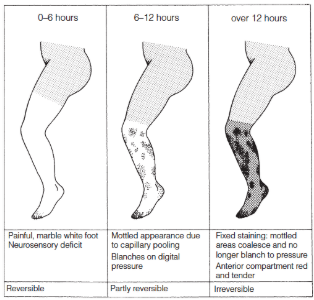
- Initially reversible but with prolonged ischaemia there is hypoxic vasodilation resulting in rubor in dependent position (partly reversible)
- With ongoing ischaemia there is thrombosis in capillaries/arterioles and rupture resulting in fixed blue staining and a mottled appearance (irreversible))
- Initially reversible but with prolonged ischaemia there is hypoxic vasodilation resulting in rubor in dependent position (partly reversible)
- Paraesthesia
- Important to determine if sensitivity to light touch maintained
- These fibres are most susceptible to ischaemia and dysfunction heralds the beginning of irreversible ischaemic changes
- Paralysis
- Onset of motor paralysis implies impending gangrene
- Swelling with ++ tenderness of a muscle belly (calf after femoral artery occlusion) usually denotes irreversible muscle ischaemia/infarction
- Pulseless
- Pulse immediately above may be particularly prominent – water-hammer
- If contralateral pulses present be suspect of emboli
- Polikothermia = ↓ Temp
- Pain
-
Microemboli
- Most significant when microemboli occlude a digital artery
- Pain, cyanosis, coldness, numbness in affected digit
- Usually improve over several days
- Sudden onset differentiates embolism from other causes of blue toes
- Vasculitis, Thromboangitis obliterans, trauma, chronic ischaemia
- Multiple emboli → Trash foot / blue toe syndrome
- Proximal pulses are intact
- Most significant when microemboli occlude a digital artery
-
Thrombosis
- More likely to result in reversible ischaemia
- Acute on chronic limb ischemia
- New onset rest pain
- Usually with hx of claudication
- Less ‘acute’ as more likely to have collaterals well established
- Should always get pre-intervention imaging
- CTA or MRA
- Ideally management with
- Thrombolysis +/- Angioplasty +/- Stent
- If unsuccessful may need bypass
- Always need good run-off vessels for bypass
Investigations
- Bloods
- FBC, U+E, Coags, G+H
- ECG -
- CXR
- CT Angiogram
- Imaging of choice in the acute setting
- MRA is alternative
- Transoesophageal ECHO to demonstrate thrombus
- Often do this post-op, Can flag if in AF
- ABPI – limited value in acute ischaemia
- Except in acute on chronic ischaemia
Management
Emergency Management
- Analgesia
- O2
- IV fluid
- Heparinize 5000IU bolus then infusion. Stop 4-6 hours pre-angio
- Aim: Prevent thrombosis resulting from stagnant flow beyond 1° occlusion
- Would continue past intervention if had a source which wasn’t corrected
- Stay on anticoagulation re-embolization rate 7%
- No anticoagulation re-embolization rate 21%
- Risk of heparin induced thrombocytopenia 5%
- Assess reversibility and manage accordingly
- Use Rutherford Classification
- Lower extremity may be salvaged after up to 5-6 hours of profound ischaemia
- Class 1 – Viable limb
- Class 2a – Marginally threatened
- Class 2b – Immediately threatened
- Class 3 – Irreversible (needs amputation)
| Viable | Marginally threatened | Immediately threatened | Nonviable | |
|---|---|---|---|---|
| Pain | Mild | Moderate | Severe | Variable |
| Capillary refill | Intact | Delayed | Delayed | Absent |
| Motor deficit | None | None | Partial | Complete, paralysis (rigor) |
| Sensory deficit | None | None or minimal (toes) | More than toes | Complete, anesthetic |
| Arterial Doppler | Audible | Inaudible | Inaudible | Inaudible |
| Venous Doppler | Audible | Audible | Audible | Inaudible |
| Treatment | Urgent evaluation | Urgent revascularization | Emergency revascularization | Amputation |
Threatened but Reversible Ischaemia
- Rutherford category I or IIa:
- Usually presents as new onset rest pain
- Not paralysis and no sensory change
- Usually due to thrombosis rather than embolus
- Time available to plan intervention
- CTA or MRA
- Management more complex
- Often have diffuse disease at multiple levels
- Variable collateralization Unpredictable run-off
- No neurological deficit
- Thrombolysis
- Neurological deficit is present
- Reconstruction (if run-off is OK)
Partly Reversible Ischaemia
- Rutherford category IIb: Immediately threatened
- White limb with sensory/motor deficit
- Embolus most likely, esp. if
- Rapid onset
- No prev hx of claudication
- Normal contralateral pulses
- Probable embolic source (AF, cardiomyopathy, AAA)
- Surgery most appropriate - Embolectomy
- CT-A helpful but “should NOT delay surgery” although in this day and age it is unlikely that a CTA is not able to be performed/not done
- Thrombolysis not appropriate
- Prepare both groins and lower limbs
- If embolectomy not effective can do Fem-Fem Cross-over bypass
Irreversible Ischaemia
- Rutherford Category III:
- Muscle paralysis, tense swollen fascial compartments, fixed staining
- 10% of pts
- Consider pt’s condition and level of ischaemia
- Revascularisation inappropriate
- If high level and moribund patient
- Palliative care
- If level treatable by amputation and pt appropriate for amputation
- Resuscitate/stabilize and amputate limb
- Amputation considered as primary management if QOL / mobility poor
- or if disease is unreconstructible or if there is extensive tissue loss
- Even for partly reversible disease
- or if disease is unreconstructible or if there is extensive tissue loss
Non-Operative Management
- Sometimes considered for emboli to major arteries in the upper extremities where collateral circulation is outstanding
- Or for rare event where signs & symptoms spontaneously resolve after an initial ischaemic episode
- Can consider Iloprost
- Prostacyclin
- ↓ Platelet aggregation
- Dilatation of arterioles & venules
- Inhibition of leukocyte adhesion
- Indications: Buerger’s disease (when revascularization is not indicated/possible) or Raynaud’s
- Prostacyclin
Post-Intervention Cares
- Consider fasciotomy after prolonged ischaemia
- Anticipate renal insufficiency from myoglobin release after reperfusion of ischaemic muscle
- Consider ECHO/CT-A if not done before to determine site of embolus if patient not in AF
- Will help guide ongoing anticoagulation therapy and may require treatment
Thrombolysis
Indications
- For acute thrombosis
- Achieved by stimulating conversion of plasminogen to plasmin
- A protease that degrades fibrin
- Results in thrombus dissolution
- Must be injected via angiography
- AT THE SITE of thrombus
- Otherwise ineffective and high risk of complications
- Achieved by stimulating conversion of plasminogen to plasmin
- Short segment thrombotic occlusions
- Catheter directed followed by balloon angioplasty to underlying offending lesion
- Ischaemic interval short
- Can be used up to 1-2/52 post initial event
Contraindications
- 5 Absolute (active or high risk of active bleeding)
- CVA within 2 months (or TIA)
- Neurosurgery within 3 months
- Intracranial trauma within 3 months
- Active bleeding or bleeding diathesis
- Recent GI haemorrhage (<10 days)
- 5 Relative
- CPR within 10 days
- Uncontrolled hypertension
- Major trauma (10 days) or Major surgery within 2 weeks
- Puncture of incompressible vessel
- Intracranial tumour
- Minor
- Liver failure
- SBE
- Pregnancy
Agents Used
- rTPA (Recombinant Tissue Plasminogen Activator)
- Streptokinase
- Urokinase (probably superior)
- Heparin given concurrently
- Prevents clot formation around catheter, prevents early re-thrombosis & for 48 hours after
- Consider life-long Warfarin
- or Aspirin + Clopidogrel if warfarin contraindicated
Technique
- Need to deliver to site of thrombus
- Intra-thrombus method
- Initial bolus dose followed by infusion for up to 3 days
- Duration is dictated by reassessment
- Occasionally can be supplemented with percutaneous thrombectomy
- Suction/aspiration or disruption and net catchment
- Not really recommended for distal vessels and can cause further arterial damage
Results
- Recanalization rate 80-90%
- Allows demonstration of underlying anatomy
- Better planning subsequent surgery if required
Complications
- 1% MR (5-10% MR in 30 days – mostly MI or CVA)
- CVA – 3%
- GI bleed rare
- Bleeding at catheter site up to 40%
- Haemorrhage requiring transfusion 5%
- Usually at groin but can be retroperitoneal or intra-abdominal
- Manage with Aprotinin (plasmin inhibitor), RBC (or whole blood), FFP + Fibrinogen
- Distal embolus – 2-3%
- Allergic reaction
Compartment Syndrome
Definition
- Compartment syndrome occurs when increased pressure within a compartment compromises the circulation and function of the tissues within that space
- Pressures increase beyond capillary perfusion pressure (30mmHg)
- Due to muscular swelling within the confines of an unyielding osseofascial bound space
Aetiology
- Orthopaedic
- Fractures and fracture surgery
- Vascular
- Arterial and venous injuries
- Reperfusion injury
- Haemorrhage
- Phlegmasia caerulea dolens
- Soft tissue
- Crush injury
- Burns
- Iatrogenic
- Prolonged limb compression
- Puncture in anticoagulated patients
- Use of a pneumatic antishock garment
- Casts and circular dressings
- Pulsatile irrigation
- Occasional
- Snakebite
- Overuse of muscles
- Chronic exertional compartment syndrome in runners
Clinical Presentation
- Severe out of proportion pain on examining compartment and passive movement
- Paraesthesia often first sign
- Check by compartment
- Pallor, decreased capillary refill, perishing cold
- Doppler can be intact until late
Investigations
- Measure compartment pressures with needle catheter attached to pressure transducer
- Normal = 10-12 (> 30mmHg)
- Or if Diastolic pressure – Compartment pressure < 30mmHg
- Zero at position where transducer will sit during measurement
- Do within a 5cm radius of injury
- Measure all four compartments
- Take highest reading
- https://www.youtube.com/watch?v=kFIyFTKim3U&ab_channel=MedStarEmergencyPhysicians
See: Fasciotomy
Embolectomy
Principles
- Prepare the whole limb and lower abdomen and contralateral side to mid-thigh
- Because you don’t know exactly where problem is
- You can evaluate your success (foot in transparent plastic bag)
- Embolectomy via Fogarty balloon catheter placed through a proximal arteriotomy
- Sizes 2-7 available (Fogarty)
- Check balloon with saline first
- Catheter passed in 10cm increments
- Gentle inflation
- Resistance free withdrawal
- Repeat until no more clot, and good back flow
- Back flow not a reliable sign though
- Heparinize distally 5000 IU in 250mL
- Can use intraoperative direct arterial infusion of urokinase (50,000-250,000 units) in settings where cleared proximal disease but might be left with some stagnant thrombosis which can’t clear
- For an aortic bifurcation saddle embolus
- Will often do bilateral femoral cut-downs, clamp both sides and clear
- To avoid forcing trash down one side
- Always do arteriogram on completion
Femoral Embolectomy
- Setup and Positioning
- Fogarty size 3 or 4 for Femoral
- Sizes 2-7 available – 2 – smallest
- Check the balloon
- Prepare both limbs and lower abdomen completely (can wrap feet in transparent plastic bags)
- E.g. If you identify an aortic bifurcation saddle embolus will need to do bilateral femoral cut downs, clamping both sides before starting to clear so trash doesn’t get thrown down one side
- Thigh abducted, knee on pillow
- NB: Mid-inguinal point – ASIS Pubic Symphysis
- Femoral Artery
- NB: Mid-point of the inguinal ligament; ASIS to Pubic Tubercle
- Femoral Nerve
- NB: Mid-inguinal point – ASIS Pubic Symphysis
- Fogarty size 3 or 4 for Femoral
- Incision and Control
- Longitudinal skin incision at mid-inguinal point
- Divide skin, subcutaneous tissue
- Superficial & deep fascia, vascular sheath
- May come across superficial branches of GSV/ Femoral Artery
- Control Common femoral artery, superficial femoral artery, PFA individually with vascular loops
- Encircle vessels by working medial to lateral under vessels with right angles
- Avoid damage to Femoral Vein
- Identify PFA as the one with the branches
- Control Common femoral artery, superficial femoral artery, PFA individually with vascular loops
- Before opening – 5000 IU heparin
- Take into account body weight, renal function
- Embolectomy
- Before opening – 5000 IU heparin (take into account body weight, renal function)
- Just above bifurcation make
- Transverse cut if artery normal & unlikely to need to re-construct
- Longitudinal if diseased, will often need to close with patch to prevent stenosis
- Transverse arteriotomies are more predisposed to flaps & you are limited with the size of the cut you can make (therefore problematic in small vessels)
- Longitudinal arteriotomies: More prone to stricturing
- Will accept up to 50% stenosis
- Use size (3) 4 catheter mostly
- Do proximal (common), then SFA then PFA
- Catheter passed in 10cm increments, gentle inflation, resistance free withdrawal
- Repeat until no more clot, good back flow
- Check distal pulses
- Check other side pulses
- From here if placed distally
- Generally can enter peroneal artery
- If want ATA or PTA, make separate distal (below knee) incision
- Closure and Considerations
- Once finished
- Flush with heparinized saline
- Arteriogram can be done if facilities/ experience available but not if unexperienced in technique
- Transverse arteriotomy closed with 4/0 or 5/0 Prolene
- Consider about fasciotomy
Popliteal Embolectomy
- Leg abducted, hip & knee flexed, externally rotated
- Standard medial incision
- Parallel to long axis of leg,
- 1 finger breadth posterior to medial border of Tibia
- Approximately 10cm long, preserve LSV
- Fat, fascia & deep fascia divided
- Medial head of gastrocnemius mobilized & retracted posteromedially
- Soleus taken down off tibia to expose Tibioperoneal Trunk
- Get individual control (sometimes take ATV to aid dissection)
- Distal popliteal arteriotomy (usually longitudinal)
- Use size (2) 3 catheter for embolectomy
- Close with graft or vein patch
Brachial Embolectomy
- Longitudinal incision just above elbow
- Exposed at Medial border of Biceps
- In the groove between Biceps & Triceps
- Deep fascia is incised & the groove opened up to display the neurovascular bundle embedded in connective tissue
- Size 3 catheter for embolectomy
.7-.9 - long distant .5-.7 - short distant .3-.5 - ischaemic rest pain <.3 - tissue loss, gangrene
Toe pressure
- ABPI is inaccurate in diabetes
- Normal toe pressure >70
- If >40 then an amputation will heal