Due to muscular swelling within the confines of an unyielding osseofascial bound space
Pathophysiology
Insult
Leads to accumulation of fluid, blood, or oedema within a fixed volume
In a contained osteo-fascial comparment
Cascade
Venous outflow obstruction
- Venous return is first impeded → increased hydrostatic pressure
- Leads to further interstitial fluid accumulation and swelling
Arterial inflow impairment**
As pressure exceeds arterial capillary pressure, oxygen delivery to tissues is impaired
Cellular hypoxia
Within 4–6 hours:
Myocytes undergo necrosis
Peripheral nerves undergo demyelination and axonal injury
Cell death releases potassium, myoglobin, lactate, worsening local and systemic
Reperfusion injury
Aetiology
Orthopaedic
Fractures and fracture surgery
Vascular
Arterial and venous injuries
Reperfusion injury
Haemorrhage
Phlegmasia caerulea dolens
Soft tissue
Crush injury
Burns
Iatrogenic
Prolonged limb compression
Puncture in anticoagulated patients
Use of a pneumatic antishock garment
Casts and circular dressings
Pulsatile irrigation
Occasional
Snakebite
Overuse of muscles
Chronic exertional compartment syndrome in runners
Clinical Presentation
Severe out of proportion pain on examining compartment and passive movement
Anterior > Lateral > Deep Posterior > Superficial Posterior
Anterior most susceptible as is tightest
Compartments
Anterior compartment syndrome
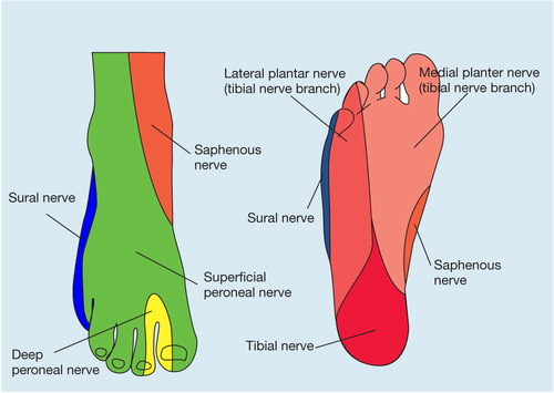
Numbness of first web space
Deep peroneal nerve, also weakness of great toe dorsiflexion
Web space between 1st & 2nd toes, dorsiflex toes
Nerve then skeletal muscle has most tenuous blood supply
Lateral compartment
Superficial Peroneal Nerve
Sensation to dorsum of foot except 1st web space
Deep Posterior compartment
Posterior Tibial Nerve
Sensation to plantar surface, flex toes
Superficial Posterior compartment
Sural Nerve – lateral foot border sensation
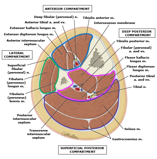
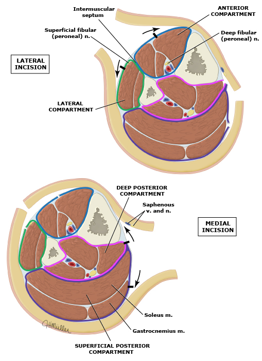
Anterior compartment (blue): Tibialis anterior, extensor muscles of the foot, and fibularis (peroneus) tertius muscles. The anterior tibial artery and deep fibular (peroneal) nerve supply the anterior compartment.
Lateral compartment (green): Fibularis (peroneus) longus and fibularis (peroneus) brevis muscles. The superficial fibular (peroneal) nerve and branches from the anterior tibial artery supply these muscles.
Superficial posterior compartment (purple): Gastrocnemius, soleus, and plantaris muscles. Tibial nerve branches supply these muscles. The arteries that supply these muscles descend from the popliteal artery. The sural arteries (medial, lateral) supply the gastrocnemius. The soleus is variably supplied by the popliteal artery, posterior tibial artery, and fibular (peroneal) artery.
Deep posterior compartment (pink): Tibialis posterior, flexor muscles of the foot, and popliteus muscles. The deep posterior compartment is innervated by the tibial nerve and supplied by the posterior tibial and fibular (peroneal) arteries.
Procedure
Two-incision, four compartment, knee to ankle fasciotomy
Easier, faster, safer
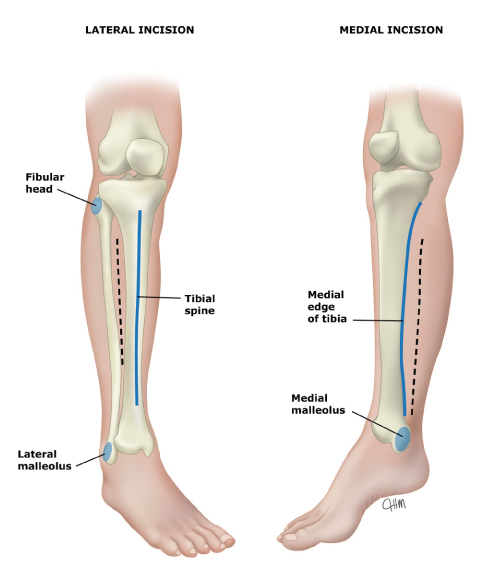
Posteromedial longitudinal incision 2cm posterior to posterior medial palpable edge of tibia
Carried down through fascia into superficial posterior space
Soleus incised longitudinally near its tibial insertion to reach deep fascia
Deep fascia incised longitudinally to decompress deep
2 fingerbreadths posterior to medial tibial condyle & Medial Malleolus
Avoids LSV/ Saphenous Nerve
Anterolateral longitudinal cut
Through fascia to anterior compartment
Second longitudinal fascia cut over lateral compartment (via same skin incision
2 fingerbreadths lateral to anterior border of Tibia – avoids Peroneal Nerve
Make sure divide Superior Extensor Retinaculum above ankle.
Excise any necrotic tissue
Splint limb
Close 3-5 days later – may rarely need SSG
Thigh
Three compartments
Lateral incision
Two parallel incisions in fascia lata with intervening bridge of 4-5cm
Through this decompress the anterior and posterior compartment
Medial incision
Adductor compartment
Because the medial compartment of the thigh is less prone to acute compartment syndrome, the compartment pressure should be measured prior to undertaking medial compartment fаѕϲiotοmy. A second incision overlying the adductor muscle group is used for medial thigh compartment decompression.
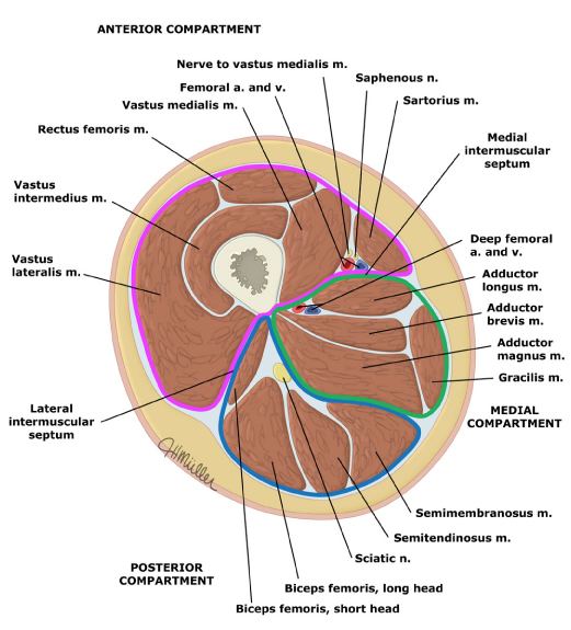
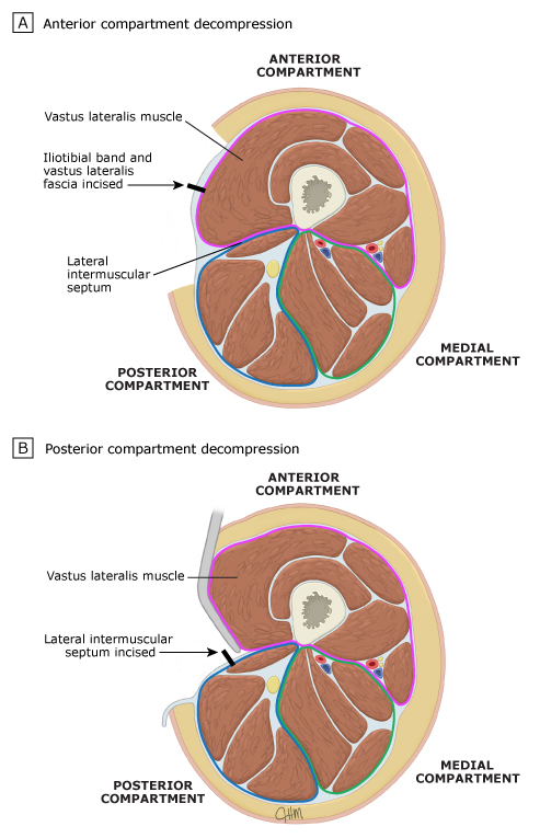
The thigh has three muscle compartments:
Anterior compartment (pink) – Sartorius and quadriceps muscles (rectus femoris, vastus lateralis, vastus intermedius, vastus medialis). The femoral nerve and superficial femoral artery supply these muscles.
Medial compartment (green) – Pectineus, obturator externus, gracilis, and adductor muscles (longus, brevis, magnus, minimus). The obturator nerve innervates the medial compartment.
Posterior compartment (blue) – Biceps femoris, semimembranous, and semitendinous muscles. The sciatic nerve innervates the posterior compartment. The deep femoral artery supplies the posterior compartment.
For thigh fasciotomy, a single, generous lateral incision is made that originates from a point just distal to the intertrochanteric line and extends to the lateral epicondyle of the femur. Through this incision, the iliotibial band and fascia of the vastus lateralis are incised the full length of the skin incision to decompress the anterior compartment. The posterior compartment is decompressed by reflecting the vastus lateralis muscle medially to expose the lateral intermuscular septum, which is incised the length of the skin incision.
Upper Limb
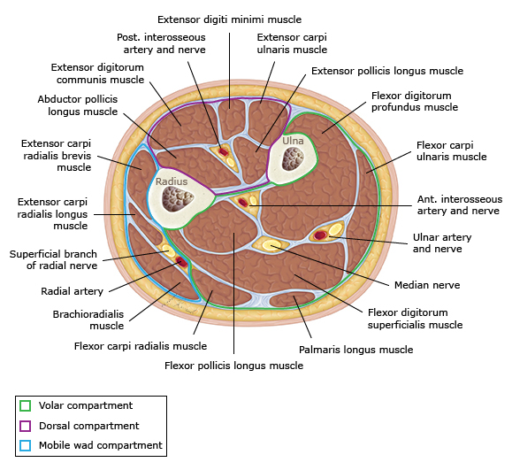
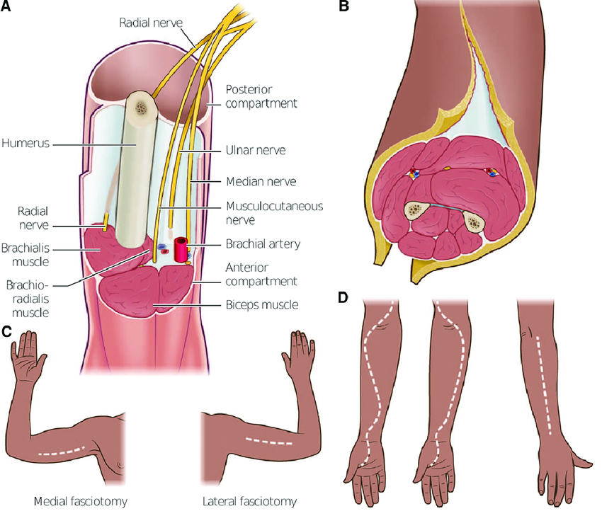
Anterior or volar forearm compartment > Dorsal forearm, hand, upper arm
 For thigh fasciotomy, a single, generous lateral incision is made that originates from a point just distal to the intertrochanteric line and extends to the lateral epicondyle of the femur. Through this incision, the iliotibial band and fascia of the vastus lateralis are incised the full length of the skin incision to decompress the anterior compartment. The posterior compartment is decompressed by reflecting the vastus lateralis muscle medially to expose the lateral intermuscular septum, which is incised the length of the skin incision.