- Indications
- Do prophylactically if had
- Severe (warm) ischaemia for > 6 hours
- Ligation of major veins in popliteal region or distal thigh
- Crush injuries
- Do as treatment in
- Clinical compartment syndrome
- Pressure > 30
- Perfusion Pressure < 30
- NB: Abdo is 20/60
- Rhabdomyolysis from localized breakdown of muscle
- Hyperkalaemia, Myoglobinuria
- Reperfusion injury
- Clinical compartment syndrome
- Do prophylactically if had
Foot
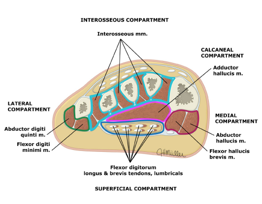
- Compartments of the foot:
- Interosseous (turqoise) — Interosseus muscles, each in its own compartment.
- Calcaneal (pink) — Flexor digitorum brevis, quadratus plantae and adductor hallucis.
- Lateral (green) — Flexor digiti minimi and abductor digiti quinti.
- Medial (red) — Abductor hallucis and flexor hallucis brevis muscles.
- Superficial (blue) — Flexor digitorum brevis, lumbricals, flexor digitorum longus tendons.
Leg
- Anterior > Lateral > Deep Posterior > Superficial Posterior
- Anterior most susceptible as is tightest
- Compartments
- Anterior compartment syndrome
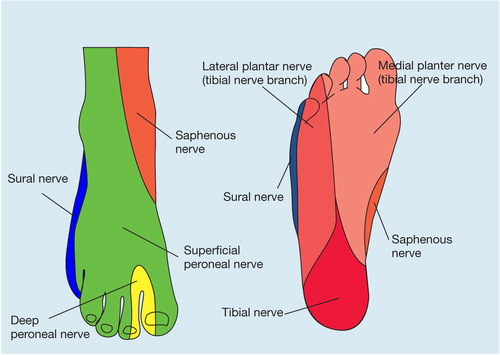
- Numbness of first web space
- Deep peroneal nerve, also weakness of great toe dorsiflexion
- Web space between 1st & 2nd toes, dorsiflex toes
- Nerve then skeletal muscle has most tenuous blood supply
- Deep peroneal nerve, also weakness of great toe dorsiflexion
- Numbness of first web space
- Lateral compartment
- Superficial Peroneal Nerve
- Sensation to dorsum of foot except 1st web space
- Deep Posterior compartment
- Tibial Nerve
- Sensation to plantar surface, flex toes
- Superficial Posterior compartment
- Sural Nerve – lateral foot border sensation
- Anterior compartment syndrome
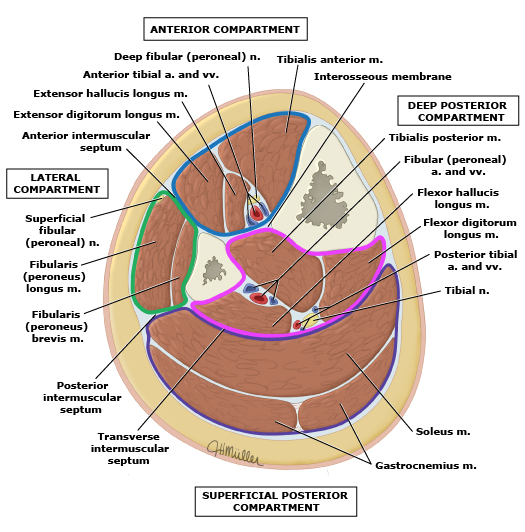
- Anterior compartment (blue): Tibialis anterior, extensor muscles of the foot, and fibularis (peroneus) tertius muscles. The anterior tibial artery and deep fibular (peroneal) nerve supply the anterior compartment.
- Lateral compartment (green): Fibularis (peroneus) longus and fibularis (peroneus) brevis muscles. The superficial fibular (peroneal) nerve and branches from the anterior tibial artery supply these muscles.
- Superficial posterior compartment (purple): Gastrocnemius, soleus, and plantaris muscles. Tibial nerve branches supply these muscles. The arteries that supply these muscles descend from the popliteal artery. The sural arteries (medial, lateral) supply the gastrocnemius. The soleus is variably supplied by the popliteal artery, posterior tibial artery, and fibular (peroneal) artery.
- Deep posterior compartment (pink): Tibialis posterior, flexor muscles of the foot, and popliteus muscles. The deep posterior compartment is innervated by the tibial nerve and supplied by the posterior tibial and fibular (peroneal) arteries.
- Procedure
- Two-incision, four compartment, knee to ankle fasciotomy
- Easier, faster, safer
- Posteromedial longitudinal incision 2cm posterior to posterior medial palpable edge of tibia
- Carried down through fascia into superficial posterior space
- Soleus incised longitudinally near its tibial insertion to reach deep fascia
- Deep fascia incised longitudinally to decompress deep
- 2 fingerbreadths posterior to medial tibial condyle & Medial Malleolus
- Avoids LSV/ Saphenous Nerve
- Anterolateral longitudinal cut, incision 2cm below fibula head to 2cm above lateral malleolus
- Through skin
- Vertical incision to identify anterior and lateral compartment
- Convert to an H incision with two longitudinal fascial incisions
- Second longitudinal fascia cut over lateral compartment (via same skin incision
- Avoids Peroneal Nerve which perforated the fascia of the lateral compartment
- Make sure divide Superior Extensor Retinaculum above ankle.
- Excise any necrotic tissue
- Splint limb
- Close 3-5 days later – may rarely need SSG
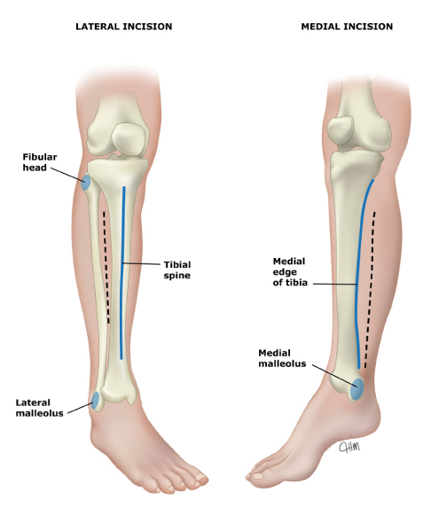
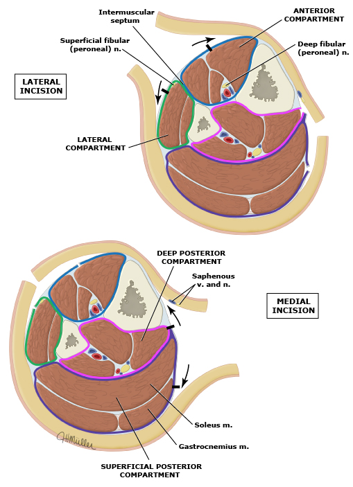
- Two-incision, four compartment, knee to ankle fasciotomy
https://youtu.be/WNyKp1hlZKc?si=e2ZQjQVe5zXqQgmX
https://www.youtube.com/watch?v=AYlcxypkZ4w&ab_channel=HoustonMethodistDeBakeyCVEducation
Thigh
- Three compartments
- Lateral incision
- Incisions in fascia lata
- Through this decompress the anterior and posterior compartment
- Medial incision
- Adductor compartment
- Because the medial compartment of the thigh is less prone to acute compartment syndrome, the compartment pressure should be measured prior to undertaking medial compartment fаѕϲiotοmy. A second incision overlying the adductor muscle group is used for medial thigh compartment decompression.
- Adductor compartment
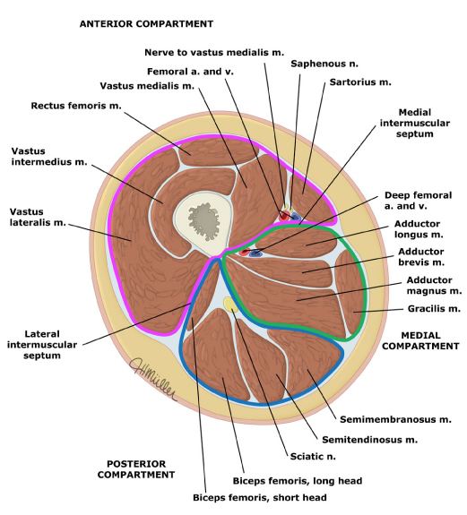
- The thigh has three muscle compartments:
- Anterior compartment (pink) – Sartorius and quadriceps muscles (rectus femoris, vastus lateralis, vastus intermedius, vastus medialis). The femoral nerve and superficial femoral artery supply these muscles.
- Medial compartment (green) – Pectineus, obturator externus, gracilis, and adductor muscles (longus, brevis, magnus, minimus). The obturator nerve innervates the medial compartment.
- Posterior compartment (blue) – Biceps femoris, semimembranous, and semitendinous muscles. The sciatic nerve innervates the posterior compartment. The deep femoral artery supplies the posterior compartment.
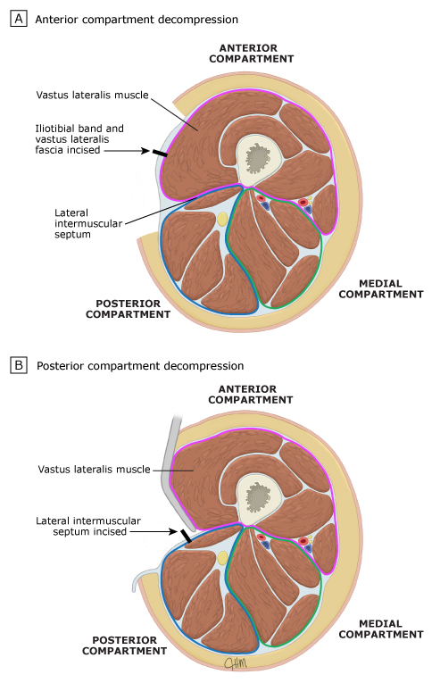 For thigh fasciotomy, a single, generous lateral incision is made that originates from a point just distal to the intertrochanteric line and extends to the lateral epicondyle of the femur. Through this incision, the iliotibial band and fascia of the vastus lateralis are incised the full length of the skin incision to decompress the anterior compartment. The posterior compartment is decompressed by reflecting the vastus lateralis muscle medially to expose the lateral intermuscular septum, which is incised the length of the skin incision.
https://www.youtube.com/watch?v=DtBFLQ8kHGQ&ab_channel=HoustonMethodistDeBakeyCVEducation
Upper Limb
- Anterior or volar forearm compartment > Dorsal forearm, hand, upper arm
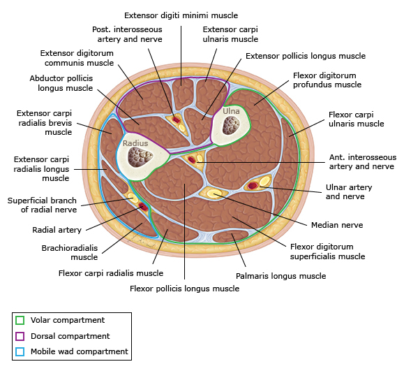
- 3 compartments of the forearm
- Volar
- Mobile wad compartment - brachioradialis. extensor carpi radialis brevis. extensor carpi radialis longus
- Dorsal
- Usually release
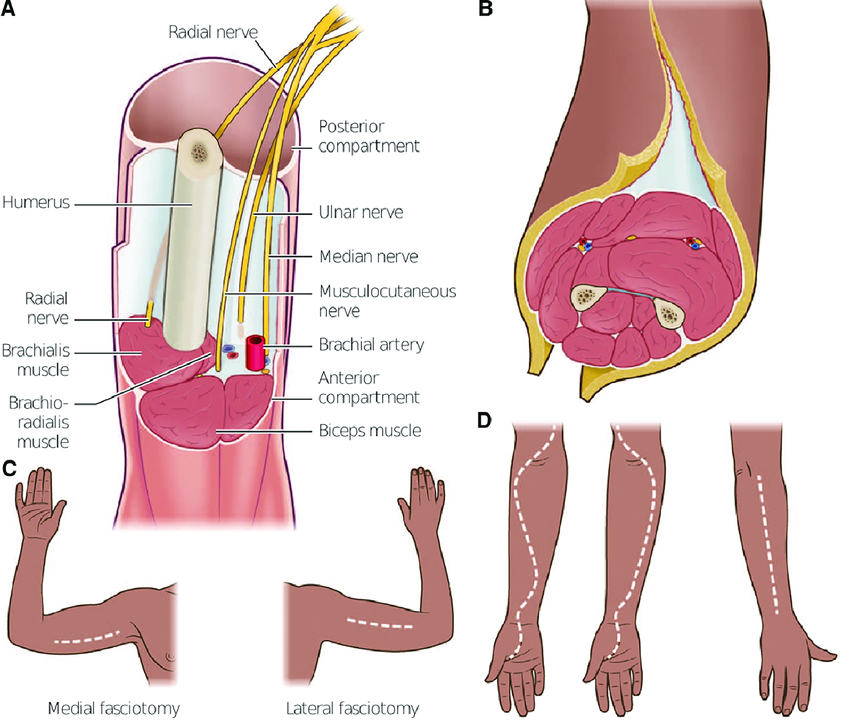
- Volar – Curvilinear and open carpal tunnel
- Mobile wad compartment - accessed adjacent to volar compartment
- Dorsal – Straight
- Volar – Curvilinear and open carpal tunnel
- Gelberman Technique
https://www.youtube.com/watch?v=GYpjFfTQjLc&ab_channel=Mojo