Relative anatomy
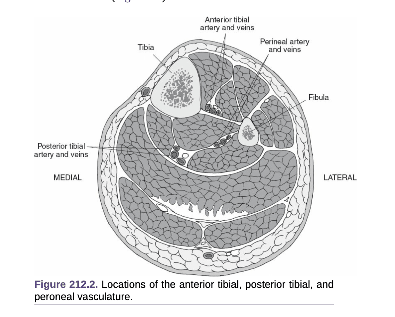
- Anterior = Extensor compartment
- Tibialis anterior, EHL, EDL
- Deep peroneal nerve
- Lies on interosseous membrane lateral to ATA
- Supplies the muscles above + a small area of skin at 1st web-space
- Anterior tibial artery
- From popliteal bifurcation, via interosseous membrane at neck of fibula
- Has two companion veins on either side
- Lateral
- Peroneus longus, peroneus brevis
- Superficial peroneal nerve - Supplies those two muscles + sensation to dorsum of
- foot
- Posterior Superficial
- Gastrocnemius , soleus (soleus deep)
- Posterior Deep
- Tibialis posterior, FHL, FDL
- Tibial nerve
- Deep to soleus, PTA crosses from lateral to medial
- Supplies all the muscles of flexor compartment
- Posterior tibial artery
- Runs down on Tibialis posterior between the two flexor
- Also has paired veins
- Gives of the peroneal branch 2.5cm distal to popliteus
Key principles
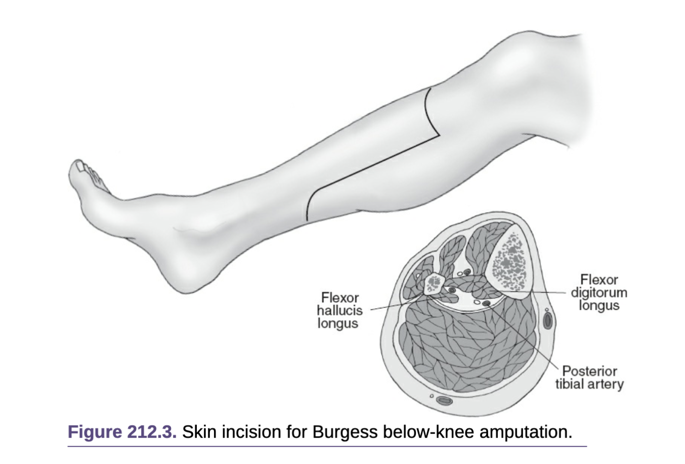
- This describes the Long Posterior Myocutaneous Flap (Burgess Technique)
- Longer posterior flap than Above knee amputation
- Alternative is the Skew Flap
- Skin – thick, no tension
- Muscle – good bone cover
- Nerves – cut under tension
- Bones – bevel & smooth
- Optimal tibial section is 1/3 of its length
Specific preoperative preparation
- Carefully assess viability of the soft tissues of the lower leg
- Only consider in ambulant patient, otherwise perform AKA
Operation Details
- GA/Spinal
- Prep/ drape – leg free draped, foot wrapped
- Incision
- Anterior skin incision 12cm below tuberosity – 1/3 calf circumference then
- Down vertical axis for about 1.5x the length of transverse incision
- Posterior flap the created with a transversely posterior incision
- Incise down through deep fascia ligating LSV
- Anterior and lateral compartment
- Scalpel dissection to expose tibia and fibula at the same level as the anterior incision
- Anterior
- Anterior tibial artery and vein identified and ligated centrally in the anterior compartment
- Deep peroneal nerve transected under tension
- Lateral
- Superficial perineal nerve
- Anterior
- Scalpel dissection to expose tibia and fibula at the same level as the anterior incision
- Bone
- Elevate periosteum
- Divide fibula as high as possible with bone cutters
- Take fibula first – if take tibia first the fibula can shatter
- Divide tibial– bevel & smooth end
- Posterior compartment
- Posterior compartment muscles are then divided obliquely to create a posterior muscle flap
- Ligate posterior tibial artery & vein
- Ligate & divide posterior tibial nerve under tension
- Ligate the peroneal artery
- Flap
- Fashion posterior flap on fascia overlying gastrocnemius, debulk soleus medially and laterally
- Suture gastrocnemius fascia to pretibial fascia with 1/0 vicryl
- Drain
- Subcuticular monocryl
- Softban & crepe bandage
https://www.youtube.com/watch?v=7CR-j-3o27E&ab_channel=HoustonMethodistDeBakeyCVEducation