Background
- Transposition and reimplantation of the superior mesenteric artery (SMA) into a healthy area of the aortic wall is a therapeutic alternative for revascularization for chronic mesenteric ischemia, as long as the occlusive atheromatous lesions are limited to the most proximal (postostial) portion of the SMA.
- This procedure seems particularly apt in the potentially septic setting of mesenteric ischemia since it allows the avoidance of prosthetic material.
- To be feasible in optimal conditions, the infrarenal aorta must be relatively free from atherosclerosis.
- The quality of the aortic wall is best assessed preoperatively by CT imagery.
Exposure
- Laparotomy
- Lateral approach
- Colon Cranial
- Small bowel to the right
- Division of the ligament of Treitz
- Identify and loop left renal vein
- Dissection of the infrarenal aorta
- With the duodenum retracted to the right, dissection begins along the anterior face of the aorta just below the left renal vein and the aorta is dissected circumferentially over a length of 4–5 cm or until the aorta can be safely cross-clamped.
- The quality of the aortic wall is assessed by palpation. The best area for anastomosis often lies between the renal arteries; this may necessitate upward mobilization of the left renal vein. If the quality of the anterior aortic wall is unsatisfactory, it may be necessary to choose a different method for SMA revascularization.
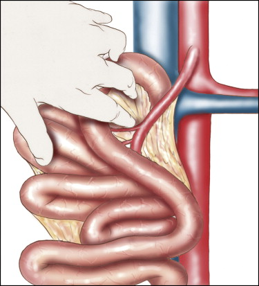
- Choosing the optimal placement for anastomosis to the aorta
- The most critical judgment of this procedure lies in choosing an optimal site for anastomosis. When the patient is upright, the small bowel exerts downward tension on the SMA. To avoid kinking or tension at the anastomosis, the SMA should have adequate length and follow a supple course.
- The rightward traction on the eviscerated or retracted small bowel should also be relaxed somewhat to simulate its final position. The site on the aortic wall chosen for anastomosis should then be marked with a prolene suture (asterisk)
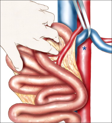
- Preparation of the superior mesenteric artery
- The patient is anticoagulated by intravenous injection of 50 IU/kg of heparin
- The SMA is clamped at its origin and divided at a point where the arterial wall is healthy.
- The stump of the SMA on the aorta is oversewn with a double layer of running 5/0 prolene suture.
- The distal SMA is carefully examined to verify the absence of atheromatous lesions. Its orientation is marked with a guide suture or methylene blue; the retraction of the small intestine is relaxed to ascertain that the position of the anastomosis of the SMA on the aorta is satisfactory.
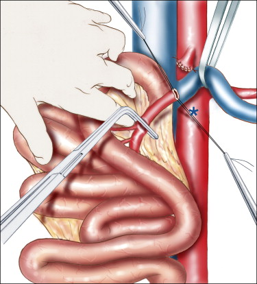
- Aortotomy and first steps of the anastomosis
- The aorta is clamped, preferably with a totally occlusive cross-clamp placement.
- A small circular arteriotomy of a size to match the caliber of the SMA is then made.
- This orifice can also be created with an aortic punch.
- Tension should be avoided during the anastomosis; the root of the mesentery should be maintained in a relaxed position.
- A running suture of 6/0 prolene is placed beginning at the heel of the anastomosis.
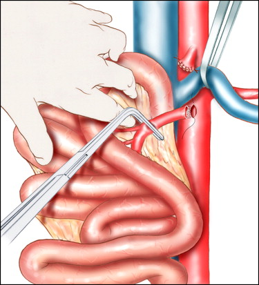
- Completion of the anastomosis
- The aorta and SMA should be cautiously flushed before completion of the running anastomotic suture line. The position and hemostasis of the anastomosis are carefully inspected and the coloration of the revascularized intestine is assessed
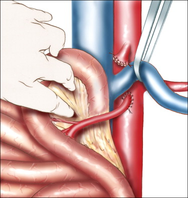
- The aorta and SMA should be cautiously flushed before completion of the running anastomotic suture line. The position and hemostasis of the anastomosis are carefully inspected and the coloration of the revascularized intestine is assessed
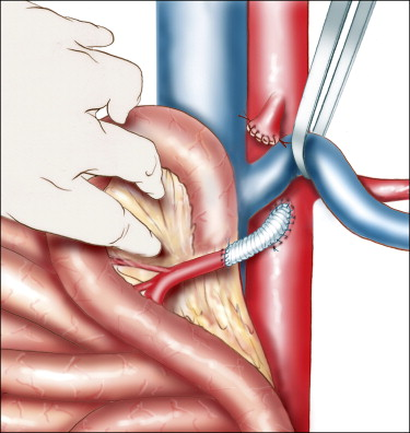
- Prosthetic graft interposition
- Transposition of the SMA may be technically impossible if there is extensive atheromatous involvement of the distal artery or if critical collateral vessels prevent the SMA from being brought down to the aorta without tension.
- In such cases, a short segment of prosthetic arterial graft (7–8 mm diameter) must be interposed. In that case, the prosthetic graft should first be anastomosed to the aorta according to the same principles we have previously described; an end-to-end anastomosis is then made between the prosthetic graft and the SMA. It is then essential to place a pedicle of omentum between the prosthetic graft and the intestines.