Aetiology
- Occlusive
- Embolic (50% of all cases)
- Typically cardiac in origin
- May arise from atherosclerotic plaques or arterial aneurysms
- Typically emboli lodge just past the origin of the Middle Colic
- Thrombotic (20% due to plaque at origin of SMA)
- Iatrogenic
- Traumatic
- Embolic (50% of all cases)
- Nonocclusive mesenteric ischemia (20%)
Risk Factors
- Older
- Smoker
- HTN
- Low output cardiac states
- Cardiac arrhythmias
- Severe valvular disease
- Recent MI
- Intra-abdominal malignancy
Pathophysiology
- Control of Mesenteric Flow
- Intrinsic factors affecting flow
- Arteriole smooth muscle response to adenosine and other metabolites of mucosal ischaemia
- Extrinsic factors affecting flow
- Neural and hormonal mechanisms
- NB: Intestine able to tolerate significant reduction in blood flow for up to 12 hours
- Get rapid vasodilation of collateral vessels
- However with on-going ischaemia and oedema/inflammation in ischaemic bed
- Associated vasoconstriction and increased vascular resistance
- Hence reduction in collateral supply and a rapid reduction in O2 supply
- Increased tissue hypoxia & injury
Stages of Mesenteric Ischaemia
- Stage 1 = Mucosal Ischaemia
- Visceral pain however no clinical signs
- Stage 2 = Disruption of microvascular integrity
- Stages 1 and 2 within 15 mins
- Progressive ischaemia leading to damage to microvascular integrity
- Capillaries in submucosa damaged leading to further mucosal damage
- Loss of mucosal integrity leading to increased permeability
- May leak blood causing GI bleed and bowel wall thickening
- Stage 3 = Progressive mucosal injury
- Leakage of protein/fluid and electrolytes
- Translocation of bacteria and entry of gas forming organisms
- Pneumatosis + Portal venous gas
- Can get full thickness necrosis and perforation
- Stage 4 = Reperfusion injury
- Stages 3-4 over 3-6 hours
- Paradoxical exacerbations of cellular dysfunction and death following restoration of blood flow to previously ischaemic tissues
- Damage due to
- Release of oxygen free radicals
- Release and activation of cytokines, proteases, complement
- Release of toxins/cell breakdown products/bacteria into systemic circulation
- Leads to both local and systemic injury
Presentation
- Severe pain out of proportion to findings
- With minimal signs
- May have benign abdo on examination.
- May have watery diarrhoea
- Marked forced evacuation of bowels
Investigations
- Bloods: no helpful markers:
- CRP, WCC, U&E
- Lactate: 100% sensitive, 42% specific
- LDH / GGT / amylase may all be elevated
- CTA
- Angio – diagnostic identification of point – if will not delay management
- Interventional angiography:
- Allows therapeutic intervention
- Suction thrombectomy
- Clot lysis
- Allows therapeutic intervention
- Duplex: useful screening, especially for chronic ischaemia
Management
- Initial Resuscitation
- Aggressive initial resuscitation prior to OT
- Haemodynamic monitoring and support
- IVF, correction of acidosis
- IV antibiotics
- Nasogastric decompression
- Avoid vasoconstrictors
- Or consider dobutamine if required
- Systemic anticoagulation unless bleeding
- Heparin infusion
Risk Stratification
- Needs decision: Resuscitation vs Palliation
- Stratify risk:
- Age > 80
- Bad lungs: home O2, FEV1 < 80%, DLCO < 50% predicted
- Bad heart: LVEF < 25%, NYHA III/IV, Angina
- MI < 90 days
- Renal Impairment, Cr > 3.0mg/dL
-
4 = 25% 30-day mortality
- 60% dead within 5 years
-
Embolic vs Thrombotic
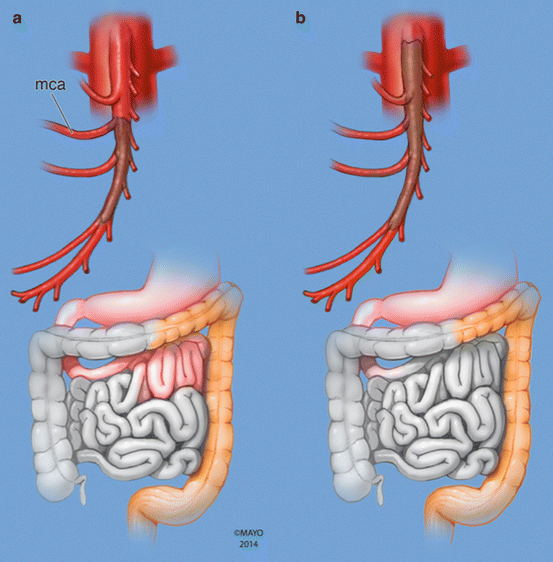 Diagrammatic representation of typical location of embolic (a) and thrombotic (b) occlusion of the SMA with corresponding pattern of extent of bowel ischemia. (a) Embolic occlusion of the SMA at the branching point of the middle colic artery (mca). (b) In situ thrombosis of the SMA starting at the ostium
Open Exploration
- Immediate surgery if suspected intestinal infarction or perforation
- Exploratory laparotomy advisable to assess bowel viability
- Open revascularization:
- Embolic = Has proximal SMA pulse
- Needs a Embolectomy of the Superior Mesenteric Artery
- Mobilise SMA: reflect D3/D4 or follow middle colic down
- Vascular loop to get vascular control above and below
- Transverse arteriotomy, trawl with a 3F Fogarty balloon catheter
- Close 6/0 prolene with interrupted or continuous
- Needs a Embolectomy of the Superior Mesenteric Artery
- Thrombotic = No proximal pulse
- Options
- Reimplantation of the superior mesenteric artery (if vascular surgeon)
- Retrograde bypass an option
- Retrograde iliac to SMA with PTFE (6-8mm)
- Could use autologous graft if sig contamination (GSV)
- Anastomosis with 6/0 prolene SMA, 5/0 prolene iliac
- Retrograde SMA stenting
- Options
- Embolic = Has proximal SMA pulse
- After any procedure - enable time for reperfusion to assess bowel viability
- If not viable will need resection
- If any on-going concern
- Recommend exteriorisation with stoma and relook laparotomy
Endovascular Revascularisation
- Improved mortality from 70 to 40%
- Pre-operative:
- Needs emergency access to angio suite
- May allow treatment prior to surgical exploration
- On-table:
- Thrombolysis
- Suction thrombectomy
- Antegrade/retrograde
- Angioplasty + stent via SMA/Celiac
- Give Papaverine to counter persistent arterial spasm
- Post-procedure:
- To avoid intra-abdominal bypass graft, performed ASAP
- Generally would still do laparoscopy/ laparotomy to asses bowel viability
Prognosis
- Depends upon
- Patients CVS status
- Extent of ischemia
- Speed to surgery
- Even with best treatment mortality remains high
- Patients should continue on:
- Optimal medical therapy for those with atherosclerosis
- Anticoagulation for those with embolic disease