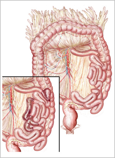
Assess bowel - if non survivable necrosis then close and comfort cares
Exposure
Anterior or lateral approach,
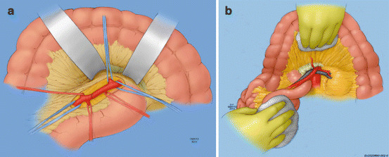
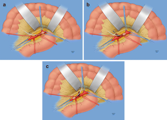
Anterior approach (Figure A)
Transverse colon is retracted cephalad and the small bowel caudal and to the left
Allow for palpation of the vessel at the root of the mesentery, along the inferior margin of the pancreas
Peritoneum overlying it is next incised at the base of the transverse mesocolon, and careful dissection is carried down to the artery.
It can often be difficult to expose due to the lack of pulsatility and significant mesenteric edema.
It lies to the left of the superior mesenteric vein, and multiple small venous tributaries crossing the SMA may require ligation and division to facilitate exposure.
The middle colic artery can also serve as a landmark to identify the SMA.
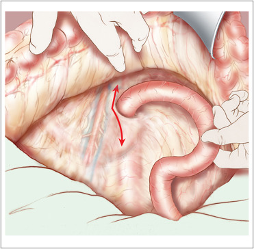
Lateral approach (Figure B)
Take down the ligament of Treitz
Mobilise the entire small bowel to the right side of the abdomen.
Ideally suited for revascularization of the SMA with antegrade bypass from the supraceliac aorta or retrograde bypass from the infrarenal aorta or either iliac artery to treat in situ thrombosis.
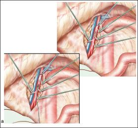
Embolectomy
Proximal and distal control of this vessel is obtained
A transverse arteriotomy
Thrombus is extracted with 3–4 mm Fogarty balloons.
balloon catheter proximally as well as distally down the main trunk and into the branches if necessary
After all thrombus has been removed, appropriate forward and backward bleeding followed by careful flushing with heparinized saline (10 units of heparin per mL) is performed.
The arteriotomy is then closed primarily with interrupted 6–0 Prolene sutures, or with a GSV/bovine pericardium patch, and flow is restored