Sigmoid volvulus
Aetiology:
- Aetiology unknown but thought to be secondary to constipation and colonic lengthening.
Risk factors:
- Elderly, debilitated, neurological conditions, institutionalised or taking psychiatric medications, laxative abuse, pregnancy, congenital anomalies e.g. Hirschsprung disease and gut malrotation, and parasitic – Chagas disease. M=F.
Presentation
- abdominal pain, distension and constipation.
Investigations:
- X-ray – coffee bean sign, apex pointing to right upper quadrant.
- CT – whirl sign
Management:
- Fluid and electrolyte abnormalities
- Acute
- Without perforation or peritonitis
- Rigid or flexible sigmoidoscopy to detort volvulus
- With perforation, peritonitis, ischaemia or inability to endoscopically reduce volvulus.
- Emergency resection
- Segmental resection + primary anastomosis; unless haemodynamically unstable, intraperitoneal contamination.
- Emergency resection
- Without perforation or peritonitis
- Elective
- Recurrent sigmoid volvulus or volvulus that was endoscopically detorted
- Fixation procedures should be avoided. High rate of recurrence
Caecal volvulus
Aetiology:
- Commonly occurs in clockwise orientation when there is lack of retroperitoneal fixation.
Risk factors:
- Younger patient, prior abdominal operation, female, pregnancy and constipation.
Presentation:
- Abdominal pain, distension, nausea and vomiting.
Investigations:
- Xray coffee bean extending across to left upper quadrant
- CT – whirl sign
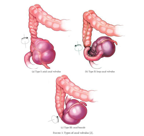
Types:
- Type 1: clockwise axial twisting along long axis
- Type 2: twisting of caecum and terminal ileum with caecum displaced to ectopic location, usually LUQ, and relocated in an inverted orientation. Usually counter-clockwise.
- Type 3: Caecal bascule – type of caecal volvulus from caecum folding anteriorly onto itself without mesenteric twisting.
- Often present with intermittent and recurrent obstructive symptoms.
- CT most useful imaging. Management same for caecal volvulus.
 Management:
- Endoscopic decompression should not be attempted
- Definitive operative management required.
- Once resuscitated, segmental resection and primary anastomosis – if stable
- Resection and ileostomy if unstable and bowel not viable.
- Caecostomy and caecopexy are not recommended; high recurrence rate.