Epidemiology
- Parasite Entamoeba histolytica.
- Four species of intestinal amebae with identical morphologic characteristics:
- E. histolytica
- E. dispar
- E. moshkovskii
- E. bangladeshi.
- Most symptomatic disease is caused by E. histolytica.
- Amebiasis occurs worldwide; the prevalence is disproportionately increased in resource-limited countries because of poor socioeconomic conditions and sanitation levels.
- Areas with high rates of amebic infection include India, Africa, Mexico, and parts of Central and South America.
- In resource-rich countries, amebiasis is generally seen in migrants from and travelers to endemic areas
Clinical presentation
- Subacute - one to three weeks
- Mild diarrhea to severe dysentery, producing abdominal pain, diarrhea, and bloody stools
- Fulminant colitis with bowel necrosis leading to perforation and peritonitis can occur
- Toxic megacolon
- Amoebic liver abscess
Life cycle
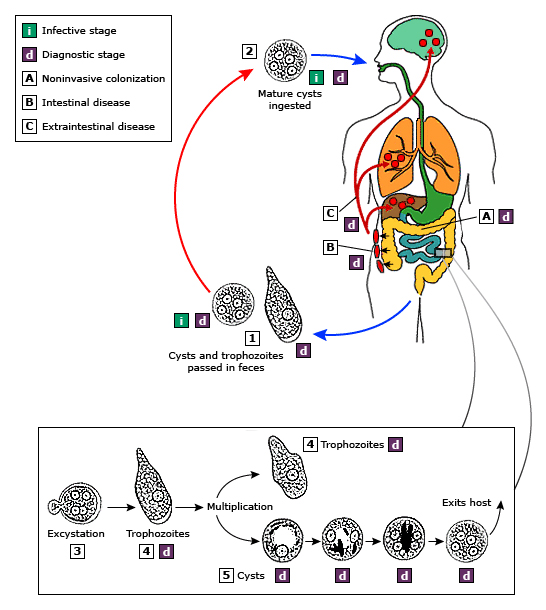 Cysts and trophozoites are passed in feces (1). Cysts are typically found in formed stool, whereas trophozoites are typically found in diarrheal stool. Infection by Entamoeba histolytica occurs by ingestion of mature cysts (2) in fecally contaminated food, water, or hands. Excystation (3) occurs in the small intestine and trophozoites (4) are released, which migrate to the large intestine. The trophozoites multiply by binary fission and produce cysts (5), and both stages are passed in the feces (1). Because of the protection conferred by their walls, the cysts can survive days to weeks in the external environment and are responsible for transmission. Trophozoites passed in the stool are rapidly destroyed once outside the body, and if ingested would not survive exposure to the gastric environment. In many cases, the trophozoites remain confined to the intestinal lumen (A: noninvasive infection) of individuals who are asymptomatic carriers, passing cysts in their stool. In some patients the trophozoites invade the intestinal mucosa (B: intestinal disease), or, through the bloodstream, extraintestinal sites such as the liver, brain, and lungs (C: extraintestinal disease), with resultant pathologic manifestations. It has been established that the invasive and noninvasive forms represent two separate species, respectively E. histolytica and E. dispar. These two species are morphologically indistinguishable unless E. histolytica is observed with ingested red blood cells (erythrophagocystosis). Transmission can also occur through exposure to fecal matter during sexual contact (in which case not only cysts, but also trophozoites could prove infective).
Diagnosis
- Stool specimens
- PCR
Treatment
- All E. histolytica infections should be treated, even in the absence of symptoms, given the potential risk of developing invasive disease and the risk of spread to family members.
- The goals of antibiotic therapy of intestinal amebiasis are to eliminate the invading trophozoites and to eradicate intestinal carriage of the organism.
- Systemic
- Luminal eradication
- Treatment of invasive disease usually consists of oral or intravenous metronidazole as well as a luminal agent to eradicate colonisation of the intestine. Paromomycin is typically used.