Section: Hepatobiliary Sub-section: Liver Curriculum: Curriculum, page 86
Definition
Liver abscess caused by Entamoeba
Epidemiology
- Amebiasis occurs worldwide; the prevalence is disproportionately increased in resource-limited countries because of poor socioeconomic conditions and sanitation levels.
- Areas with high rates of amebic infection include India, Africa, Mexico, and parts of Central and South America.
- In resource-rich countries, amebiasis is generally seen in migrants from and travelers to endemic areas
Aetiology
- Entamoeba histolytica account for 10% of liver abscesses. Found in endemic areas such as tropics
- Only 10% of infected patients experience symptoms – colitis
- Liver abscesses most common extraintestinal manifestation, found in 1% of clinically infected patients.
Pathology
- E. histolytica trophozoites normally remain confined to bowel lumen.
- Some virulent trophozoites migrate through mucosa of bowel and reach liver through portal system.
- Can provoke enzymatic focal necrosis of hepatocytes and multiple micro-abscesses that coalesce to form a single lesion.
- The central cavity contains a homogenous thick liquid, typically reddish-brown and yellow. This material is usually sterile, except when secondary infection has occurred.
- Amoebae can be found at edge of lesion but rarely detected in pus or within abscess cavity itself.
Clinical
- RUQ pain
- High fever
- Hepatomegaly
- Usually more ill than pyogenic abscess. Usually younger and from high-prevalence area or recent traveller
- Jaundice – unusual, unless multiple or very large, associated with poor prognosis.
Investigations
- USS and CT sensitive
- Combination of epidemiology/clinical in conjunction with positive amoebic titres may suggest diagnosis.
- Entamoeba histolytica Antibodies
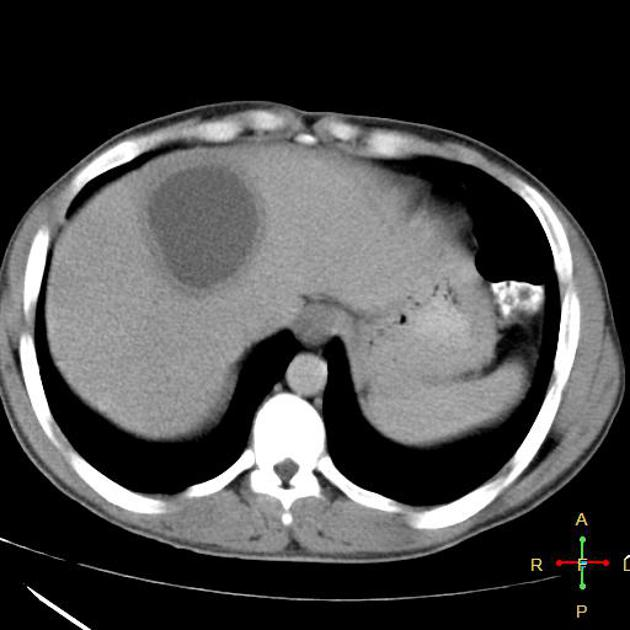
- On contrast CT, usually rounded, well defined, with enhancing wall 3 – 15 mm in thickness, and peripheral zone of oedema. Central cavity may contain multiple septa or fluid debris levels.
- Serum antibodies to Entamoeba species present in > 90% of cases
Management
- Majority with metronidazole alone. + Luminal agent to eliminate intestinal colonisation by E. histolytica (e.g paromomycin)
- 2nd line – chloroquine & iodoquinol
- Therapeutic aspiration controversial. Reserved for uncertain diagnosis, no response to metronidazole and large left lobe abscesses (risk of rupture into pericardium) and severely ill patients