Section: Colorectal Sub-section: Colorectal cancer Curriculum: Curriculum, page 21
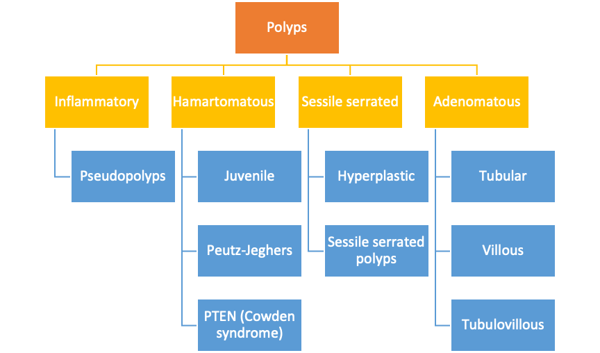
Sessile serrated lesions
- Hyperplastic polyps
- Small rectal HP are not associated with increased risk of cancer.
- Distal HP may be associated with a small risk
- Sessile serrated polyps/adenoma
- Increased risk of cancer and therefore managed like adenomas
- Serrated polyposis syndrome
- May carry a mutation in rare polyposis-associated genes including SMAD4, BMPR1A, PTEN, GREM1, RNF43, and MUTYH, these genes are not altered in the majority of individuals with SPS
- Significantly increased risk of CRC (20%)
Adenomatous polyps
Epidemiology:
- 2/3 of colonic polyps are adenomas Risk factors:
- Age
- Obesity
- M>F Clinical features:
- General asymptomatic, most detected by colon cancer screening tests
- Small adenomas do not typically bleed Endoscopic features:
- Majority are <1 cm
- Adenomas may be: o Sessile – base & top of lesion have same diameter o Pedunculated – base is narrow with mucosal stalk interposed between polyp and wall o Flat o Depressed
- Features suggestive of malignancy include friability, induration, ulceration, adherence to underlying tissue.
Adenoma-carcinoma sequence:
- Observational data supporting this hypothesis: o Early carcinomas are frequently seen within large adenomatous polyps. Areas of adenomatous change are often found surrounding CRC. o Adenomas and carcinomas are found in similar distributions throughout the large bowel o Adenomas are typically observed 10-15 years prior to the onset of cancer in both sporadic and familial cases o Ability to reduce the incidence of CRC through removal of polyps has been shown in controlled trials
- Genetic basis: o Accumulation of mutations (germline or somatic) causes malignant transformation. o Mutations of APC usually occur early, leading to formation of early adenomas, whereas p53 generally occurs late, with transformation of adenoma to carcinoma.
Malignant polyp:
- Definition: Polyp containing adenocarcinoma with breaching the muscularis mucosae
- If suspicion of possible malignancy in polyp, site should be tattooed.
Clinical/endoscopic assessment
- Macroscopic assessment
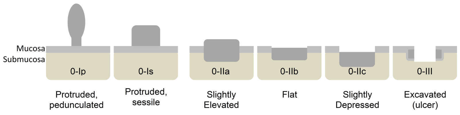
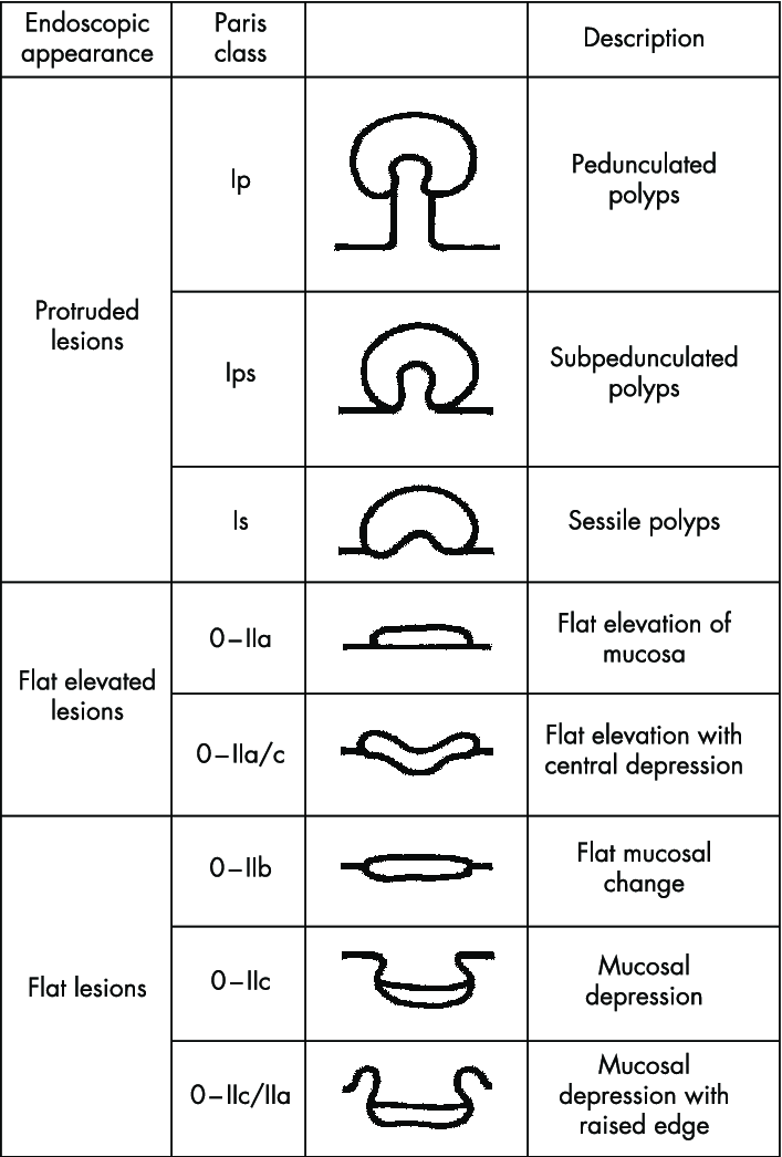
- Paris classification
- Vertical growth pattern, less likely to be invasive Ip and Is
- Flat are moderate increased risk, maybe high grade adenoma.
- Lateral spreading tumours LST. Paris II
- Homogenous type – low risk 0.5% SMI
- Nodular mixed and pseudo depressed type have high risk of deep SMI
- Depressed are high risk for carcinoma, Paris III
- Paris classification
- Signs of concerning lesions -should biopsy but not try endoscopic resection.
- Non-lifting sign – should not have to do in first place
- Fold convergence
- Demarcated depressed area
- Stalk or base swelling
- Spont bleeding
- Chicken skin appearance
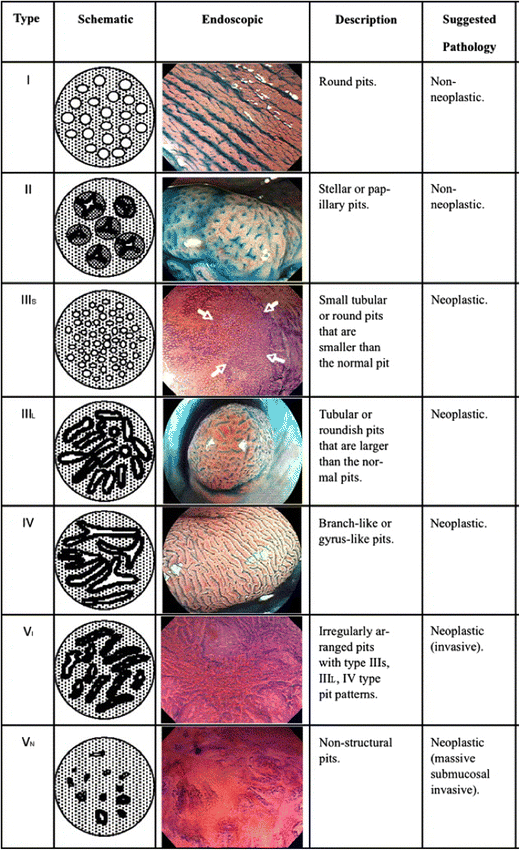
- Microscopic assessment
- Narrow band imaging – can use different wavelengths to highlight surface epithelial and underlying vessel features
- NBI International colorectal endoscopic (NICE) classification
- Type 1 to 3
- Colour, light to brown
- Vessels, none to disrupted
- Surface pattern
- Most likely pathology: hyperplasic, adenoma or deep submucosal invasive cancer.
- Narrow band imaging – can use different wavelengths to highlight surface epithelial and underlying vessel features
Histological assessment
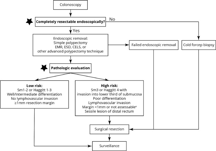
- Risk stratification based on pathology
- Level of invasion
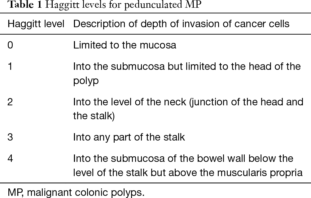
- Pedunculated: Haggitt classification level 1-4: head, neck, stalk or base
- Risk in 0-III <1%, risk in IV is 25%
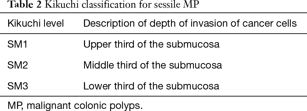
- Sessile: Kikuchi classification sm1-3: level of submucosa depth by thirds. Risk of nodal mets 2, 8 and 23% respectively.
- Sm1 local excision adequate
- Sm2/3 not recommended for local excision
- Note : 1mm submucosal invasion is often used instead of the above (given you need some muscularis propria in order to properly use Kikuchi)
- Pedunculated: Haggitt classification level 1-4: head, neck, stalk or base
- Margin status
- If < 1 mm, then treat as positive. Risk of residual disease 21-33%.
- Polyp architecture
- Risk of underlying malignancy by architectural variants
- Villous polyps 10%-18%
- Tubulovillous 6%-8%
- Tubular polyps 2%-3%
- Risk of underlying malignancy by architectural variants
- Adverse features:
- Poor differentiation
- Lymphovascular invasion
- Tumour budding
- Level of invasion
- Piecemeal resection
- may be unreliable margin.
| Histological feature | Grade of risk | Estimated risk of residual disease |
| Resection margin < 1 mm | Very high | 20% |
| Pedunculated Haggitt level 4 | Very high | 20% |
| Kikuchi sm3 | Very high | 20% |
| Poor differentiation | High | 8–15% |
| Kikuchi sm2 | Medium | 5–10% |
| Lymphovascular invasion | Medium | 5–10% |
| Resection margin 1–2 mm | Low | 5% |
| Tumour budding | Low | 5% |
Surveillance after adenoma
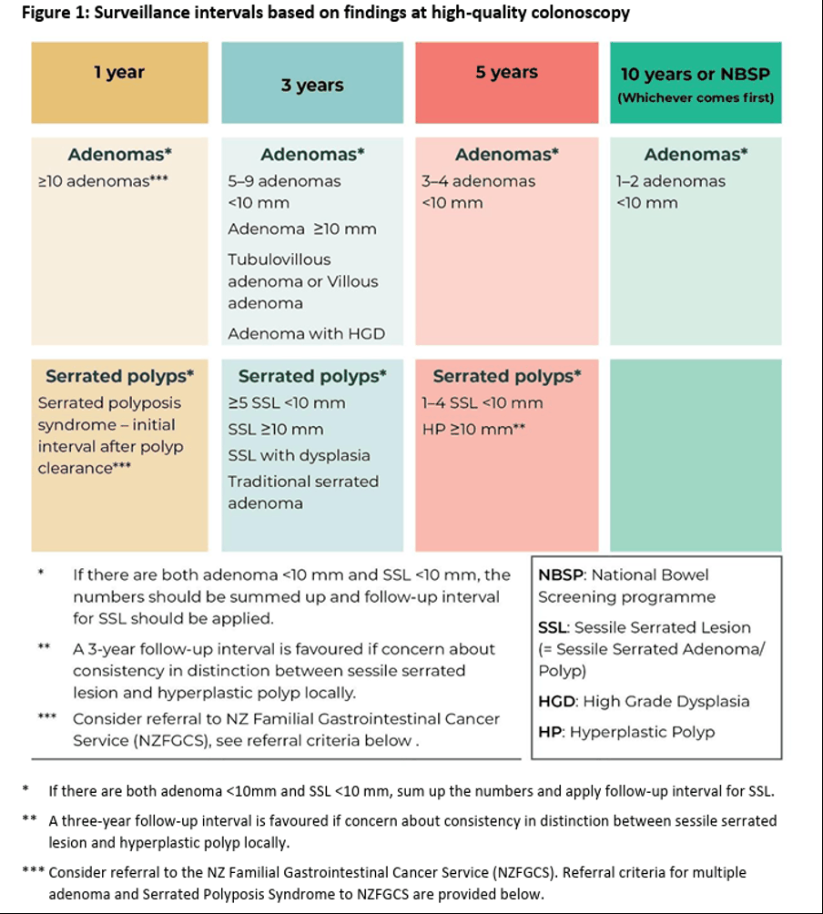
- Very low risk: 1-2 adenomas < 1 cm repeat in 10 years
- Low risk: 3-4 adenomas
- Intermediate: 5-9 adenomas or at least 1 > 1 cm, or TV or villous adenoma or HGD repeat in 3 years
- High risk: > 10 adenomas, or 3+ with 1 > 1 cm repeat in 1 year
Polyp classificaion
Paris classification
Kudo classification
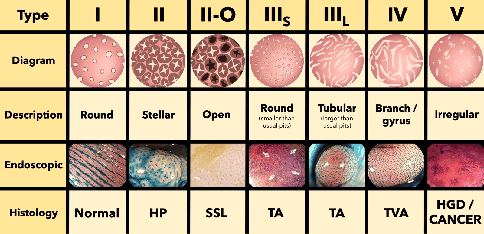
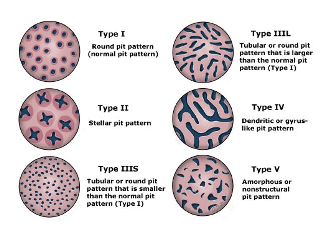
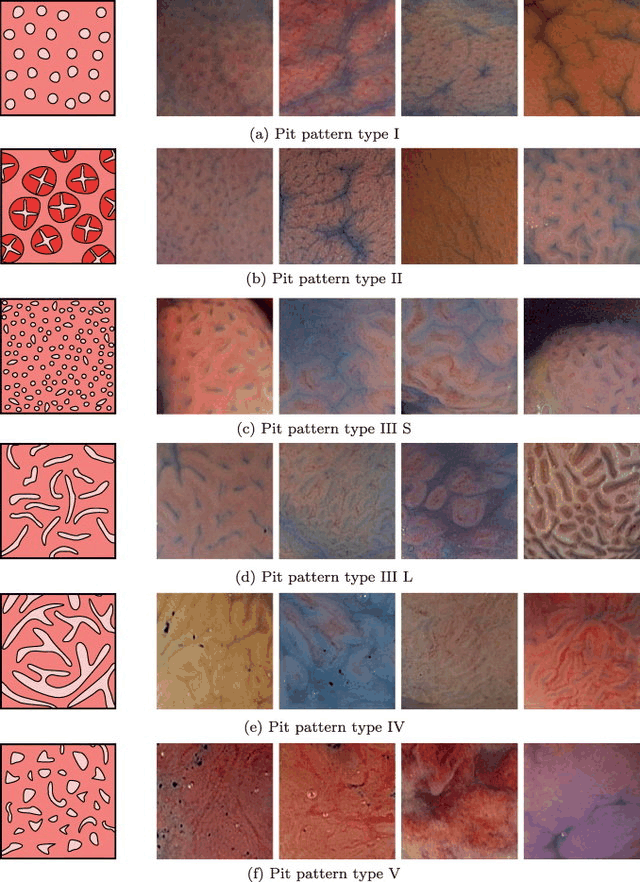
NICE NBI classification
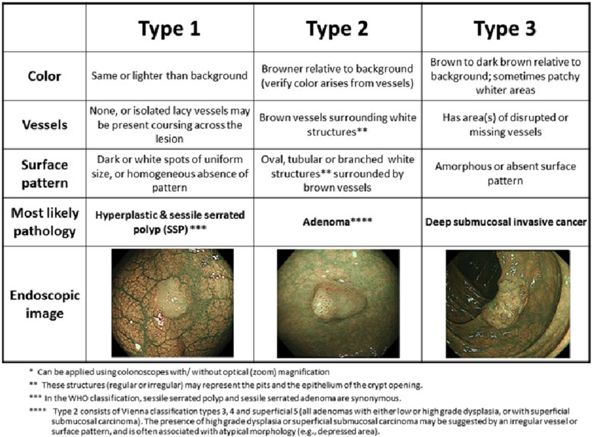
Source: Colorectal polypectomy and endoscopic mucosal resection- European Society of Gastrointestinal Endoscopy (ESGE) Guideline
Endoscopic mucosal resection
- Inject beneath the lesion to lift it and separate it from the muscularis propria.
- Use a needle to inject a solution, typically saline or a mixture with epinephrine (to reduce bleeding) and a dye like methylene blue or indigo carmine (to visualize the lift).
- Ensure an adequate and uniform submucosal cushion is created (“the lift”).
- Resection
- Snaring:
- Use a snare to encircle the lesion.
- Position the snare around the elevated lesion and close it gently to capture the base of the lesion.
- Electrocautery:
- Apply electrocautery energy to cut and cauterize simultaneously. Settings depend on lesion location and equipment.
- Resect in piecemeal fashion for larger lesions (piecemeal EMR) or en bloc for smaller lesions.
- Snaring:
Endoscopic submucosal dissection
- Define the borders:
- Use the ESD knife or argon plasma coagulation to mark the perimeter of the lesion at least 5mm beyond its visible margin.
- This ensures clear margins during dissection.
- Create a submucosal cushion:
- Inject a solution (e.g., saline, glycerol, hyaluronic acid, or a mixed solution with epinephrine and indigo carmine).
- This separates the lesion from the muscularis propria, facilitates dissection, and reduces the risk of perforation.
- Reinject as needed during the procedure.
- Circumferential mucosal incision:
- Use the ESD knife to incise the mucosa around the marked borders.
- Start with a small incision and extend it to encircle the lesion completely.