Section: Colorectal Sub-section: Other Curriculum: Curriculum, page 25
Stomal complications
- Early
- Retraction
- If full mucocutaneous seperation then revision is required immediately
- Necrosis
- Bleeding
- Retraction
- Late
- Peristomal skin issues
- Issues
- Erythema
- Wet
- Breakdown and ulceration
- Granulation tissue
- Bleed → apply silver nitrate
- Management
- Check fit and no leak
- Use barrier film to coat and protect the skin
- Stoma powder
- Issues
- Prolapse
- Hernia
- Bleeding
- Retraction
- Stenosis
- Peristomal pyoderma gangrenosum
- Peristomal skin issues
Difficult ileostomy
- Metabolic problems
- Sodium depletion
- Can then get drop in potassium in attempt to preserve sodium by producing aldosterone
- Sodium depletion if Urinary Na:K <1 (usually about 2)
- Volume
- Usually settles within 2 weeks of operation
- Related to body weight
- 70kg 500mL
- 100kg 900mL
- Same applies to pouches
- Sodium depletion
- Ileostomy/pouch dysfunction
- Definition
-
8 emptying’s/evacuations/ day
- Relative to weight
-
- Etiology
- Ileal resection
- Partial obstruction – active sodium secretion
- Intra-abdominal sepsis
- Crohn’s
- Foods – beans, leafy green vegetables, beer, coffee, chocolate, spicy food, too much fiber, alcohol
- Investigation
- Urinary Na:K
- Rule out infection
- Rule out major causes
- Rule out dietary factors
- Treatment
- Loperamide
- Sodium – IL ileostomy fluid = 1 teaspoon salt
- Pouches - isogel
- Definition
Parastomal hernia
- Incisional hernia related to an abdominal wall ostomy site
- Incidence
- 5-10% of colostomies
- 3-10% of ileostomies
- ≈10-20% require repair
Classification
- Devlin Classification
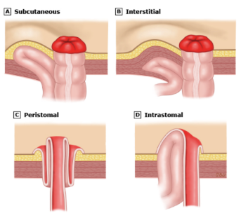
- Subcutaneous - hernia sac lies in subcutaneous plane
- Interstitial - hernia sac lies within the layers of the abdominal wall
- Intrastomal - sac penetrates the spout of an ileostomy
- Peristomal prolapse - where the sac is within a prolapsing stoma
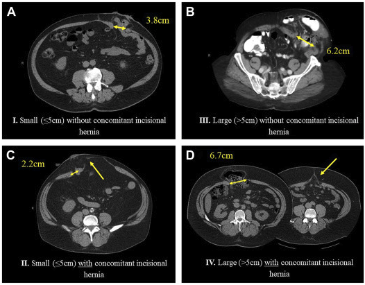
- European Hernia Society (EHS) classification
Etiology
- Only true risk factor is BMI > 30
- Risk factor most supported by evidence
- Patient specific
- Age
- Wound infection
- Chronic increased abdo pressure, COPD
- Obesity, weight gain (or malnutrition)
- Steroids/immunosuppression
- Malignancy
- IBD
- Technical Factors
- Emergency stoma
- Surgical technique
- Abdominal wall strength
Clinical
- Generally asymptomatic
- May present with difficulty placing stoma appliance
- Pain – usually mind
- +/- obstructive Sx
Management
- Indications for repair:
- Difficulty with stoma appliance – unable to see or fit
- Recurrent obstruction
- Skin breakdown
- Strangulation
- +/- cosmetic
- Options:
- Rejoin
- Resite
- Repair
Overview
- Primary vs. Mesh
- Primary
- Technically simple
- Avoids manipulation of intra-abdo contents
- Has low morbidity
- BUT has high recurrence rate
- Significantly higher than mesh repair (OR 8.9)
- Mesh
- Complications rare but difficult to manage, include
- Contamination
- Erosion
- Fistula formation
- Usually preferred technique, may be some instances where primary repair is better
- Complications rare but difficult to manage, include
- Primary
- Open vs Lap
- Not enough data to go other way
- May have presumed lower morbidity and possibly improved outcomes
- Best considered in patients without extensive intra-abdominal adhesions
- Also better for smaller (%3C 8-12cm) hernias
- Onlay vs Sublay
- Onlay
- Technically more straightforward
- Avoids intra-abdo dissection
- Higher risk wound infection
- Sublay
- Increased intra-abdo dissection
- Increases risk for adhesions and intestinal obstruction
- Associated with fewer recurrences
- Intra-abdo pressure cannot dissociate mesh from the repair
- Onlay
Techniques
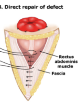
Primary Repair
- Reduce the size of the facial defect by re-approximating the fascial edges with permanent sutures
- Approach generally avoided because of the physics of parastomal hernia and nature of the defect
- Unavoidable to cause tension on the repair
- Leads to high recurrence rates
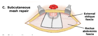
Onlay Mesh Repair
- Incision made well away from stoma (typically in the midline)
- Subcutaneous dissection along rectus and oblique fascia
- Circumferentially around stoma
- Hernia reduced
- Abdo wall closed using tension free mesh repair
- Note risk ischaemic injury to the skin
Sublay Mesh Repair
- Less infection risk
- Entails large dissection
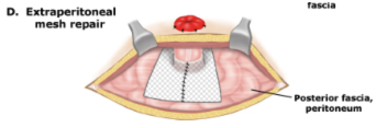
Intra-abdominal Mesh Repair
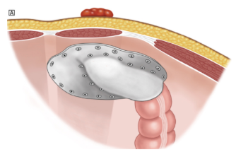
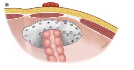
- Reduce hernia contents and close facial defect with mesh under the defect, and wide overlap
Sugarbaker
Keyhole
Parastomal Hernia-Contemporary Management, page 1 Parastomal Hernia Repair, page 1 Pauli, page 1
Prognosis / Natural Hx:
- Most hernias develop within first few years of stoma formation
- Mesh repair may lead to strictures (1/36 pts)
Stoma Siting Procedure
- Determination of optimal site; assessed in a lying, sitting and standing position
- Key Points
- Positioning issues: contractures, posture, mobility e.g. wheelchair confinement, use of walker etc.
- Physical considerations: large/protruding/pendulous abdomen, abdominal folds, wrinkles, scars/suture lines, other stomas, rectus muscle, waist line, iliac crest, braces, pendulous breasts, vision, dexterity, presence of hernia.
- Patient considerations: Diagnosis, history of radiation, age, occupation
- other: Surgeon and patient preference, type of ostomy or diversion, anticipated stool consistency.
- Multiple stoma sites: Mark fecal and urinary stomas on different horizontal planes/lines. Procedure:
- Marking pen, surgical marker, transparent film dressing, flat skin barrier
- Carefully examine abdominal surface. Begin with patient fully clothed in sitting position with feet on floor. Observe the presence of belts, braces and any other ostomy appliances.
- Examine patient’s exposed abdomen in various positions (standing, lying, sitting and bending forward) to observe for creases, valleys, scars, folds, skin turgor and contour.
- Draw an imaginary line where the surgical incision is going to be. Choose a point approximately 2 inches from the surgical incision where 2 – 3 inches of flat adhesive barrier can be placed.
- With patient lying on back identify the rectus muscle. (This can be done having the patient do a modified sit up (raise the head up off the bed). Placement within the rectus muscle can help to prevent peristomal hernia formation and/or prolapse.
- Choose an area that is visible to the patient, and if possible below the belt line to conceal the pouch.
- Large abdomen; choose the apex of the mound or in extremely obese, place in the upper abdominal quadrants.
- It may be desirable to mark sites on the right and left sides of the abdomen to prepare for a change in the surgical outcome.
- Clean the desired site with alcohol and allow to dry. Then proceed with marking the selected site with a surgical marker / pen. You may cover with transparent film dressing if desired to preserve the mark.
- Once marked have the patient assume sitting, bending and lying position to assess and confirm best choice. It is important to have the patient confirm they can see the site.