Section: Colorectal Sub-section: Proctology Curriculum: Curriculum, page 25
Definition
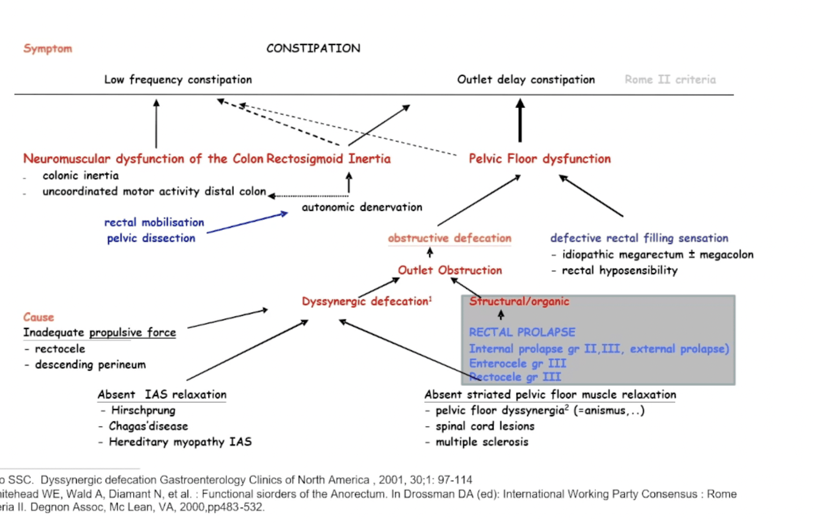
Obstructed defecation syndrome is a type of constipation characterized by fragmented stools, need for straining at defecation, sense of incomplete evacuation, tenesmus, urgency, pelvic heaviness and self-digitation
In a patient presenting with difficult defecating pelvic flood disorders i.e ODS can be a cause.
Constipation causes:
- Normal transit constipation i.e Functional Constipation
- Slow transit constipation - see Constipation
- Outlet delay constipation - see Obstructed defecation
- This can be divided into anatomical and functional
- Irritable bowel syndrome
- Secondary constipation
- Medications - opiates, anticholinergics, antihypertensives, iron supplements, antacids and non-steroidal anti-inflammatory drugs
- Neurological disease - Hirschsprung’s disease, multiple sclerosis, Parkinson’s disease and diabetic autonomic neuropathy
- Electrolytes - hypercalcaemia and hypokalaemia
- Hormonal - hypothyroidism
ROME IV
Functional defecation disorders
- The patient must satisfy diagnostic criteria for Constipation and/or Irritable bowel syndrome with constipation
- During repeated attempts to defecate, there must be features of impaired evacuation, as demonstrated by 2 of the following 3 tests:
- Abnormal balloon expulsion test
- Abnormal anorectal evacuation pattern with manometry or anal surface EMG
- Impaired rectal evacuation by imaging
Inadequate Defecatory Propulsion
- Inadequate propulsive forces as measured with manometry with or without inappropriate contraction of the anal sphincter and/or pelvic floor muscles
Dyssynergic Defecation
- Inappropriate contraction of the pelvic floor as measured with anal surface EMG or manometry with adequate propulsive forces during attempted defecation
Aetiology
Causes of ODS are:
- Structural
- Rectocele
- Intussusception
- Enterocele
- Perineal descent
- Rectal prolapse
- Functional
- Pelvic floor dyssynergia
- Poor propulsion
- Or combination of both
Clinical
History
What happens when they get to toilet?
- Slow passage of hard stool – likely SLOW TRANSIT
- Start to evacuate and then abrupt stop – likely RECTOCELE / INTUSSUSCEPTION
- Unable to evacuate despite straining – possible ANORECTAL DYSYNEGIA (Anismus)
Obstructive Defecation Syndrome Score (ODS-S)
| Symptoms/variables | 0 | 1 | 2 | 3 | 4 |
|---|---|---|---|---|---|
| Excessive straining | Never | Rarely | Sometimes | Usually | Always |
| Incomplete rectal evacuation | Never | Rarely | Sometimes | Usually | Always |
| Use of enema/laxative | Never | Rarely | Sometimes | Usually | Always |
| Vaginal/perineal digital pressure | Never | Rarely | Sometimes | Usually | Always |
| Abdominal discomfort/pain | Never | Rarely | Sometimes | Usually | Always |
| A score of ≥9 suggests a diagnosis of ODS |
(Renzi et al.) (https://link.springer.com/article/10.1007/s00192-022-05114-8#ref-CR10 “Renzi A, Brillantino A, di Sarno G, D’Aniello F. Five-item score for obstructed defecation syndrome: study of validation. Surg Innov. 2013;20(2):119–25.”)]
Investigations
| Investigation | Role | Advantages | Disadvantages |
|---|---|---|---|
| Colonic Transit Studies | To distinguish between slow transit constipation and evacuatory difficulties | Easily accessible, simple investigation | Crude investigation which may not appreciate patients with mixed pathology |
| Anorectal Physiology | |||
| Anorectal Manometry | To assess the function of the anal sphincters | Highlights concurrent anal sphincter weakness which should be addressed prior to surgical interventions | Requires specialist equipment and training Results may not correlate with symptoms |
| Rectal Balloon studies | To assess dyssynergy and rectal compliance and sensitivity | Highlights concurrent rectal hyposensitivity | Results may not influence treatments |
| Imaging | |||
| Endoanal Ultrasound | Assess the structure of the anal sphincter | Assess the integrity of the anal sphincter and examine for concurrent obstetric anal sphincter injury, sepsis and fistula | Requires specialist equipment and training |
| Integrated Total Pelvic Floor Ultrasound | Dynamic visualisation of entire pelvic floor as an alternative to defaecatory imaging | Dynamic visualisation of anatomical changes and changes to the anorectal angle Simple to perform, cheap, safe, portable, well tolerated by patients compared to proctography. Can be performed in a one stop clinic with simultaneous endoanal assessment of anal sphincter integrity | Does not observe defaecatory dynamics. User dependent |
| Defaecation Proctography | Dynamic assessment of anatomical and functional aspects | Dynamic visualisation of anatomical changes and rectal emptying. Performed in the upright physiological position | Multicompartmental visualisation is invasive. May overestimate pathology There is debate regarding normal parameters. Radiation exposure |
| MRI | Defaecation MRI - dynamic assessment of anatomical and functional aspects Dynamic MRI – Dynamic assessment of anatomical aspects | Multicompartmental assessment No radiation | Posterior pathology underestimated without rectal evacuation Anterior pathology underestimated in the supine position Limited access to open configuration systems |
Pelvic dyssynergy
The measurement of rectal and anal pressures during balloon expulsion can be used to evaluate dyssynergy.
- Type I – adequate increased rectal pressure with paradoxical rise in anal pressure
- Type II – inadequate increase in rectal pressure with paradoxical rise in anal pressure
- Type III – adequate increased rectal pressure with failure of reduction in anal pressure
- Type IV – inadequate increase in rectal pressure with failure of reduction in anal pressure
Proctography
- Dynamic investigation of rectal emptying
- Allows the visualisation of:
- Anatomical abnormalities (namely rectocele, intussusception, rectal prolapse, enterocoele, sigmoidocele and perineal descent)
- Functional problems (changes in the anorectal angle, the extent and duration of rectal emptying and ‘trapping’ of stool within a rectocele)
Causes and treatment
Rectocele
- Hernia of anterior rectal wall bulging into the rectovaginal septum.
- Posterior rare (due to trauma or surgical intervention breaching anococcygeal ligament) Causes:
- Muscular/nerve damage during vaginal delivery
- Hormonal changes during menopause
- Paradoxical contraction of puborectalis.
- Previous vaginal repair Treatment:
- Dietary modification, the use of laxatives, suppositories, adjuncts such as rectal irrigation and biofeedback training
- Surgical: transanal, perineal, transvaginal, abdominal – incl. lap ventral rectopexy
Enterocele
- Peritoneum of pouch of Douglas may herniate – marker of global pelvic floor weakness
- Symptoms are vague and non-specific
- The relevance of an enterocoele is controversial as an enterocoele, which descends onto the rectum during defaecation proctography does not necessarily impede evacuation.
Rectal intussusception:
- Invagination of rectal wall during defaecation.
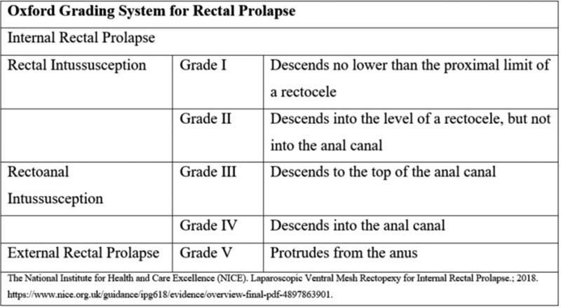
- Classification according to Oxford Radiological Grading System:
- Grade I-II: recto-rectal intussusception
- Grade III-IV: recto-anal intussusception
- Grade V: high take-off external rectal prolapse
- Half of patients with intussusception suffer with incontinence.
- The reasons behind this are not clear but it may be due to the infolding rectal folds leading to rectal distention, the associated chronic straining causing pudendal neuropathy, inflammation from a solitary rectal ulcer causing urgency or prolapsing rectal mucosa opening the anal canal.
Treatment:
- Dietary modification, the use of laxatives, suppositories, adjuncts such as rectal irrigation and biofeedback training
- Surgical: transanal, perineal, transvaginal, abdominal – incl. lap ventral rectopexy
Solitary rectal ulcer syndrome
- Caused by paradoxical contraction of anal sphincter muscle during defaecation.
- Associated with anal digitation, results in anterior mucosal trauma and ulceration.
- Treatment: dietary changes, bulking agents and biofeedback.
- Surgery rarely indicated, only for concomitant prolapse or refractory symptoms.