Section: Colorectal Sub-section: Proctology Curriculum: Curriculum, page 54
Colorectal Surgery A Companion to Specialist Surgical Practice 1, page 250
Definition
- Pilonidal disease is an acquired disease driven by the embedding of loose hair within hair follicles usually located in the natal cleft. This triggers an inflammatory response leading to the formation of midline pits, sinuses, or abscesses.
Epidemiology
- Young adults
- Male > Female
Pathophysiology
- Congenital hypothesis
- Born with pits
- Not really supported by evidence
- Acquired hypothesis
- Karydakis described three factors associated with sinus development: Hair, forces present in the natal cleft, skin vulnerability
- Hair:
- Chisel-like roots inserts into the natal cleft, leading to a foreign body tissue reaction and subsequent infection.
- Forces present in the natal cleft:
- Friction caused by movement (marching or truck driving)
- Friction of the movement of the buttocks
- Skin vulnerability:
- Poor healing in the area
- Hair:
- Karydakis described three factors associated with sinus development: Hair, forces present in the natal cleft, skin vulnerability
Risk factors
- Obesity
- Family history
- Hormonal changes at puberty
- Hair
- Poor hygiene
- Occupational - sitting
- Excessive sweating
- x4 increased risk with hydratanitis and crohns disease
Classification
International pilonidal Society classification - Berlin 2017
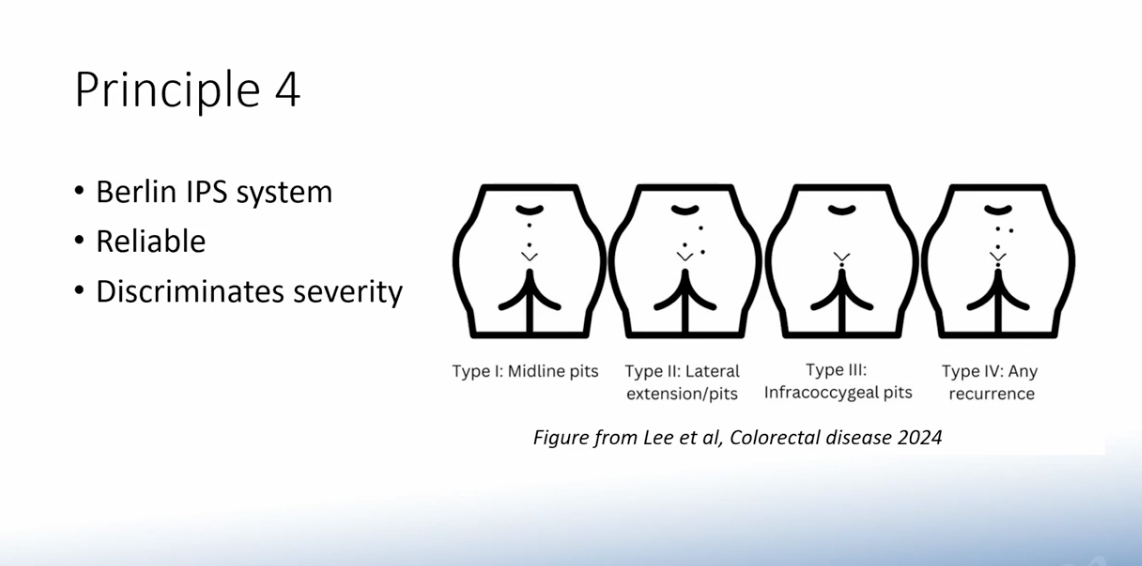
Clinical
- Typically presents an acute abscess in the midline of the sacrococcygeal region.
- The primary tract may become epithelialised and form a small pit in the midline of the natal cleft.
- Some patients develop a secondary tract that may run ;atera; and cephalad
Differential Diagnosis
If no primary pits are seen or if the sinus drains either lateral to the sacrum or appears caudal to the primary pits, other diagnoses should be considered. It is very unusual to find multiple sinuses that open into both buttocks simultaneously. In such a case, the differential diagnosis would include:
- hydradenitis suppurativa
- complex anal fistulas
- osteomyelitis with draining sinuses to the skin
- infective conditions, such as tuberculosis or actinomycosis.
Treatment
Goals of treatment
- Acute setting - managage sepsis
- Chronic setting - treat if symptomatic/impact on life
- Balance between recurrence and time off work/complexity of procedure/complication rate as demonstrated by the PITSTOP study
Non-operative management
- Appropriate if minimal symptoms
- Antibiotics - limited and conflicting evidence
- Hair removal
- Dressings - minimal evidence that specialised dressings treat disease
Operative management
Abscess: Incision and drainage - up to 60% will have complete healing.
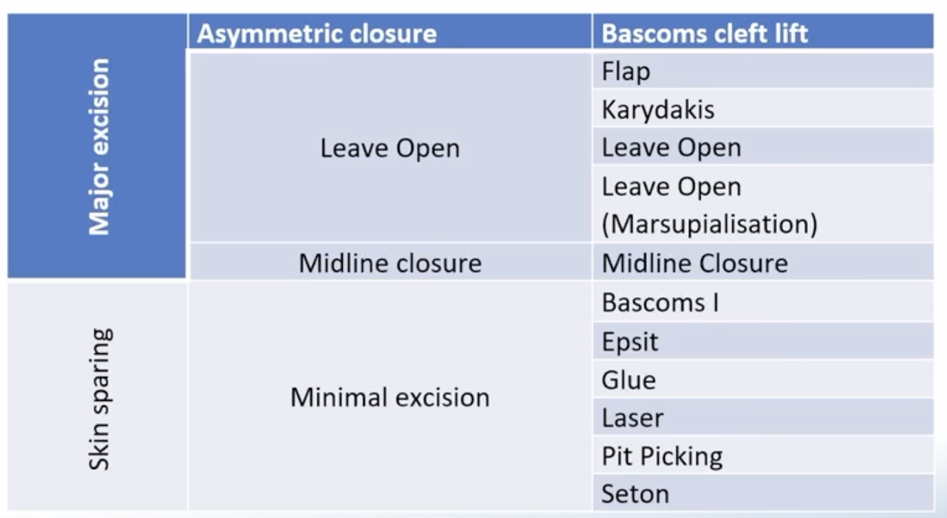
Skin sparing
Phenol for pilonidal disease
- Phenol 80% solution can be injected into the sinus tract following removal of sinus hairs and debris, achieving similar results to various other surgical procedures with the advantages of a shorter length of stay.
- Fibrin glue has been used either as monotherapy or as an adjunct to surgery. Efficacy uncertain.
Fibrin glue for pilonidal disease
Bascom I
- Excision of midline pits (can be done with a punch biopsy)
- Laterally-placed parallel incision
- Explore the underlying cavity and remove and debride, curettage of the track
- Each midline wound is then closed with a single suture
- 5 year recurrence rate of 15%
Others
- Endoscopic Pilonidal Sinus Treatment (EPSiT
- Glue
- Laser
- Pit picking
- Seton
Major excision
Pilonidal excision midline closure is generally not recommended as it takes long to heal and has a higher rate of recurrence.
Pilonidal excision and secondary intension
Modified Karydakis flap
Line drawn 2cm off midline
- The ellipse is based on a axis 2cm lateral to the midline with the caudal point modified to sit 3cm lateral to the midline. This means the scar does to angle to the anal verge. Incision
- On the medial aspect the incision is vertical down to pre-sacral fascia making sure to completely remove the disease
- On the lateral aspect the incision is 45 degrees
- A flap is raised which is 1cm thick and 2 cm deep
Bascom cleft lift procedure
- An off-centre, elliptical incision mark is drawn vertically to make the cleft shallower and flatter.
- The first incision incorporates all of the damaged skin to be removed.
- An advancement skin flap, approximately 1 cm thickness, is elevated from the medial side of the incision.
- Once the flap has been raised, the tapes on the buttocks are removed, and the flap is checked for sufficient stretching.
- Subsequently, the island of the skin on the opposite side, is removed and all sinus tracts, debris and hair are removed.
- Closed in layers
Limberg flap
- Rhomboid marked out. All sides equal. Includes all disease
- Flap marked out - lateral line the same length as rhomboid edges and a perpendicular line caudally.
- Caudal point off midline
- Blue dye injected into sinus to confirm all sinus is excised
- Dissection down to pre-sacral fascia
- Flap is then raised and includes fasica of glut maximus
- Drain in cavity
- Corners sutured in place first
- Mattress sutures