Fluids
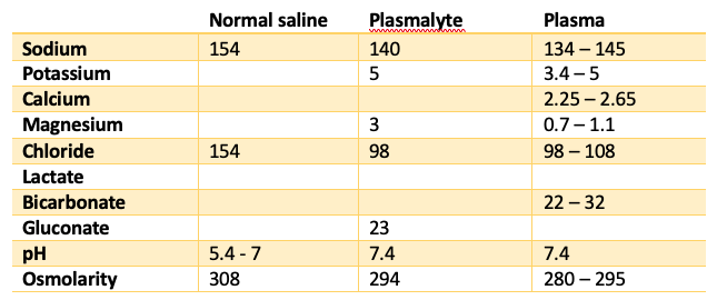
Composition of fluids compared to plasma:
Body water:
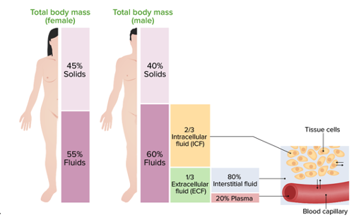
- Total ~50-70% body weight
- Intravascular approx. 10% of total body water. 5-7% of body weight
Maintenance fluids
- Rate:
- 4ml/kg/hr for first 10 kg
- 2ml/kg/hr for next 10 kg
- 1ml/kg/hr for every kg over 20 kg
- For any adult > 40 kg, fluid is weight + 40 ml/hr. e.g. 70kg is 110 ml/hr
- However needs to vary depending on hydration status of patient.
- Requirement:
- Total water = 2000 ml
- Sodium = 80 – 100 mmol (1-1.2 mmol/kg/day)
- Potassium = 70 mmol (1 mmol/kg/day)
- Type:
- No ideal fluid
- Traditionally 4% dextrose + 0.18% saline + 40 mmol KCl per 1L.
- Reasonable to use plasmalyte in short term, but need to ensure replacement/monitoring of electrolytes
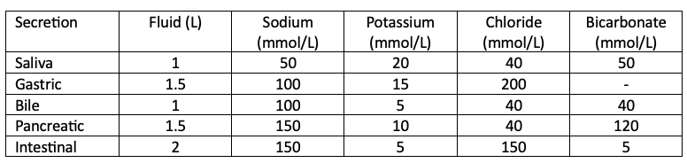
GI secretions:
Electrolytes
Sodium
- Vital for homeostasis and action potential. Predominant molecule controlling water movement.
- Hyponatraemia
- Severe (< 120 mmol/L):
- Symptoms: headaches, lethargy, comatose, seizures
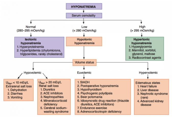
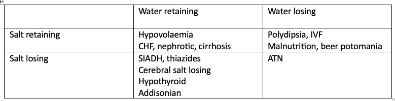
- Causes – see below chart
- Hypovolemic
- Euvolemic
- Hypervolemic
- Fluid status is vital. High ADH in hypovolaemia is a normal response. SIADH is only if euvolemic.
- Diagnosis of SIADH
- Urine osmolarity > 150 mmol/kg
- Urine sodium > 20 mmol/L
- Serum osmolarity < 270 mmol/L
Check if real or pseudo: glucose, triglycerides, uraemia proteins
- Hypernatraemia
- Severe (> 160 mmol/L)
- Symptoms: muscle weakness, restlessness, lethargy, insomnia, coma
- Causes: endocrine syndromes (ADH synthesis or release fails), failure of renal tubules to respond to ADH, DI, increased salt intake/infusion, loss of water.
- Treatment of DI is desmopressin (DDAVP) – synthetic analogue of ADH which does not have vasoconstrictive properties of ADH.
Potassium
- Main intracellular ion. R-A-A axis key regulator of potassium clearance.
- Hypokalaemia
- Symptoms:
- Generalised fatigue, weakness, ileus, atrial arrhythmia, acute renal insufficiency, rhabdomyolysis (K < 2.5), flaccid paralysis, respiratory compromise (K < 2)
- ECG changes:
- Increased P wave amplitude
- ST depression
- T wave flattening/inversion
- U waves
- Causes:
- Renal losses
- Extrarenal losses – vomiting, NG tubes, diarrhoea, fistula, alkalosis, catecholamine secretion
- Medications – CHF medications, diuretics, insulin
- Hyperthyroidism
- Hyperaldosteronism, renin-secreting tumours
- Treatment:
- IV or oral replacement. Rate of >10 mmol/hr IV needs cardiac monitoring.
- Magnesium level should be monitored → hypomagnesemia can produce refractory hypokalaemia due to failure of ATP Na/K pump.
- Symptoms:
- Hyperkalaemia
- Symptoms
- Cardiac arrhythmia
- ECG changes:
- Peaked T waves
- Flattening P wave
- Bradyarrhythmias, conduction blocks
- Widened QRS
- Causes:
- Acute renal failure (most common)
- Cellular injury (sepsis or ischaemia-reperfusion), rhabdomyolysis
- Treatment:
- Calcium gluconate
- Sodium bicarbonate
- Glucose-insulin infusion, salbutamol, calcium resonium.
- Dialysis
- Symptoms
Calcium
- Most abundant electrolyte in the body. 99% in bones, 1% in blood.
- Hypocalcaemia
- Symptoms: paraesthesia, muscle spasms (tetany), seizures
- Causes: hypoparathyroidism postop, pancreatitis (sequestration in the abdomen), rhabdomyolysis, tumour lysis syndrome, post resuscitation from shock, post RBC transfusion
- Treatment:
- IV calcium infusion
- Oral calcium gluconate or chloride + vitamin D
- Treat coexisting hypomagnesaemia
- Hypercalcaemia
- Symptoms: see Parathyroid Gland
- Causes:
- Hyperparathyroidism
- FHH
- Malignant causes (SCC, renal, bladder, ovarian, bone tumours)
- Medications: thiazide diuretics, lithium, vitamin A & D overdose, antacid overdose.
- Thyrotoxicosis
- Treatment:
- IVF
- Loop diuretic
- Haemodialysis
- Bisphosphonates
Magnesium
- 2nd most prevalent cation. Cofactor for ATP reactions. Primarily intracellular, < 1 % in ECF.
- Hypomagnesaemia
- Symptoms: neuromuscular irritability, convulsions, severe can lead to ventricular arrhythmias such as torsades de pointes
- Causes:
- Renal (most common diuretics), GI, skin losses, hungry bone syndrome
- Treatment:
- Oral or IV replacement
- Hypermagnesaemia
- Symptoms: ileus, urinary retention, loss of reflexes, flaccid paralysis, apnoea, lethargy, confusion, coma
- Causes: iatrogenic – oral or IV overdose, ingestion of antacids
- Treatment:
- Calcium to stabilise heart – calcium gluconate 10% (10-20 ml for 10 min)
- Normal saline
- Diuretics
- Dialysis if renal impairment
Gastric outlet obstruction
- Underlying pathophysiology
- Pyloric stenosis causing gastric outlet obstruction, leading to inability to tolerate oral intake and vomiting of gastric secretions.
- Dehydration
- Leads to activation of ADH from posterior pituitary with conservation of water in the renal tubules and collecting system. Also activation of RAA pathway as below.
- Hypokalaemia
- Dehydration causes activation of renin-angiotensin-aldosterone pathway. Aldosterone conserves sodium in the kidney tubules, while wasting potassium in the urine, with the Na-K ATP transporter pump.
- Hypochloraemia
- Vomiting causes loss of hydrochloric acid from the stomach, therefore depleting chloride from the body.
- Metabolic alkalosis with paradoxical aciduria
- Vomiting causes loss of hydrochloric acid, depleting H+ leading to alkalosis. Conservation of sodium due to dehydration leads to loss of H+ in the kidneys with further alkalosis and production of acid urine.
- Catabolic state
- Hypoglycaemia and reduction in nutrition leads to activation of catabolic pathways. Insulin is reduced and glucagon is secreted from the pancreas. Glucose is mobilised from liver by glycogenolysis and mobilised from liver and muscles from protein by gluconeogenesis and proteolysis.
- Fats are broken down by lipolysis leading to ketones and free fatty acids which are utilised as energy source.
- Catabolism of adipose tissue and muscle leads to weight loss.
- Basal metabolic rate is lower with reduction of ATP production.
- This pathophysiology is understood, considering that emesis results in hydrogen and chloride ion losses. The pancreas will secrete bicarbonate when stimulated by hydrogen reaching the duodenum but without this stimulus, there results an increase in serum bicarbonate and subsequent alkalosis.
- To buffer this pH change, 1) hydrogen exits the cells in exchange for potassium, which causes subsequent hypokalemia and 2) the kidneys excrete excess bicarbonate.
- However, emesis results in a decrease in extracellular volume, causing contraction alkalosis which stimulates the renin angiotensin aldosterone pathway to reabsorb sodium in exchange for potassium, thereby worsening the hypokalemia and causing an overall increase in bicarbonate reabsorption maintaining the alkalosis.
- With prolonged emesis, the kidneys attempt to maintain volume via sodium reabsorption in exchange for hydrogen ions, which can then produce the phenomenon of paradoxical aciduria.
- https://www.sciencedirect.com/science/article/pii/S2213576618303567