Section: Critical care Curriculum: Curriculum, page 30, Curriculum, page 46
Definitions
- Sepsis
- Clinical syndrome of organ dysfunction due to a dysregulated host immune response
- Sepsis 3 guideline state diagnosed in the setting of suspected or confirmed infection with a SOFA of 2 or more or a qSOFA score of 2 or more
- Clinical syndrome of organ dysfunction due to a dysregulated host immune response
- Septic Shock
- Definition
- Sepsis with cellular, circulatory and metabolic abnormalities
- High risk of mortality
- Clinical
- Satisfy criteria of sepsis
- Requirement of vasopressor to maintain MAP of >/= 65 despite adequate fluid resuscitation
- Lactate >2mmol/L
- Satisfy criteria of sepsis
- Definition
- SIRS
- Nonspecific Inflammatory response - may represent a normal or dysregulated response to infection or injury
- 2/4 of:
- HR >90
- Temp <36 or >38
- RR >20 or PaCO2>32mmHg
- WBC <4, >12 or >10% bands
- 2/4 of:
- Nonspecific Inflammatory response - may represent a normal or dysregulated response to infection or injury
- SOFA
- Assessment of organ dysfunction by scoring derangement of clinical and biochemical parameters as representative features of organ function
- Brain - GCS
- Heart - BP and pressor requirement
- Lungs - PaO2:FiO2
- Kidney - creatinine
- Haematology - platelets
- Liver - bilirubin
- Assessment of organ dysfunction by scoring derangement of clinical and biochemical parameters as representative features of organ function
- qSOFA
- Rapid bedside assessment to identify early sepsis or those at high risk of developing sepsis
- ⅔ indicates high risk and prompts screening of SOFA score
- GCS 14 or less
- SBP <100
- RR >22
- ⅔ indicates high risk and prompts screening of SOFA score
- Rapid bedside assessment to identify early sepsis or those at high risk of developing sepsis
- Sepsis 6
- 3 in - IVF, IV abx, oxygen
- 3 out - urine output, blood culture and lactate
- MODS
- Progressive organ dysfunction involving 2 or more organs
- Homeostasis unable to be maintained without intervention
Inflammatory cascade
1. Initiation phase
- Stimulus:
- Infection (bacteria, viruses, fungi).
- Tissue injury (trauma, ischemia, toxins).
- Immune reactions (allergies, autoimmune conditions)
- Activation of Innate immune cells
- Mainly macrophages - recognise and bind microbial parts or injured tissue
- Host immune cells have pattern recognition receptors - PRR
- Pathogen associated molecular patterns (PAMPs) on pathogenic toxins, pathogenic DNA etc
- Danger associated molecular patterns (DAMPs) on injured tissues E.g heat shock protein
- Host immune cells have pattern recognition receptors - PRR
- Mainly macrophages - recognise and bind microbial parts or injured tissue
- Release of proinflammatory mediators
- Cytokines - TNF alpha, Interferon- gama, IL-1, IL- 6, IL-8
- Prostaglandins and leukotrienes - Amplify inflammation.
- Histamine (from mast cells): Increases vascular permeability and vasodilation.
2. Amplification phase
- Vascular Phase
- Vasodilation: Increased blood flow (hyperemia) leads to redness and warmth.
- Increased Vascular Permeability: Leakage of protein-rich fluid (exudate) into the interstitial space causes swelling (edema).
- Stasis: Slowing of blood flow in capillaries facilitates leukocyte recruitment.
- Cellular Phase
- Leukocyte Recruitment: Margination, adhesion, transmigration and chemotaxis
- Leukocyte Activation: Neutrophils and macrophages become activated, enhancing their ability to phagocytose pathogens and secrete inflammatory mediators (e.g., IL-1, IL-6, TNF-α)
- Systemic effects
- Fever - IL-1, TNF-alpha
- Stress hormones - cytokines
- RAAS system
- Activation of Complement cascade
- Bone marrow response - increased WBC
- Acute phase reactants - CRP, fibrinogen amyloid A
- Metabolic - hyperglycaemia and insulin resistance
3. Resolution
- CARS - compensatory anti-inflammatory response
- Inflammation subsides after the threat is neutralized:
- Anti-inflammatory mediators (e.g., IL-4, IL-10, TGF-β) dampen the immune response.
- Repair processes (angiogenesis, fibroblast activity) restore tissue integrity
4. Transition to Sepsis/MODS
- Proinflammatory mediators exceed the boundaries of the local environment
- Concepts → malignant intravascular inflammation
- Malignant - uncontrolled, unregulated and self sustaining
- Intravascular - haematogenous mediator spread
- Inflammatory - exaggerated normal inflammatory response
- Concepts → malignant intravascular inflammation
- This is due to:
- Endothelial damage and dysfunction
- Increased vascular permeability, edema, and impaired blood flow.
- Leads to fluid loss and decreased oxygen delivery
- Microcirculatory Dysfunction
- Inflammatory mediators disrupt the balance between oxygen delivery and consumption.
- Leads to tissue ischemia, hypoxia, and cellular injury.
- Cellular Energy Failure
- Mitochondrial dysfunction impairs ATP production, leading to cellular apoptosis or necrosis.
- Inflammation and proinflammatory factors
- Cause direct damage to tissue
- Endothelial damage and dysfunction
- This leads to
- Progressive Organ Failure
- Initial damage to one organ increases the risk of dysfunction in others through systemic effects
- Progressive Organ Failure
Organ Effects of Sepsis
Cardiovascular
- Hypotension
- Vasodilatation e.g. NO
- Vasoactive mediators cause widespread dilatation
- Attempt to improve metabolic autoregulation
- Makes tissue perfusion easier
- Impaired ADH
- Redistribution of intravascular fluid
- Increased endothelial permeability
- Reduced arterial vascular tone
- Vasodilatation e.g. NO
Lung
- Pulmonary oedema → ARDS
- Interstitial and alveolar oedema
- Endothelial injury (pulmonary vessels)
- Increased vascular permeability
- Neutrophils trapped in lung microcirculation
- Amplifies inflammation
- Causes VQ mismatch → hypoxaemia and hypoxia
- Interstitial and alveolar oedema
GIT
- Loss of barrier function → bacterial translocation (PV and lymphatic)
- Amplifies septic response
- Gastroparesis to ileus to bowel ischaemia and GI bleeding
Liver
- Failure of the reticulendothelial system
- Reduce ability to clear endotoxins and pathogenic products
- Spills into systemic circulation
- Shock liver/Hepatic ischaemia
- Acute and LFT deterioration with elevated INR.
- Transaminases, ALP and bilirubin are markers of hepatic injury
- Synthetic function - measured by INR, albumin and bilirubin.
- Non-shock liver
- Milder form which usually resolves as MODS resolves
- Treatment for both is to avoid hepatotoxins
Kidney
- AKI
- RIFLE
- Risk - 1.5x baseline creatinine rise. Urine output < 0.5mls/kg/6 hours.
- Injury - 2x baseline creatinine rise. Urine output < 0.5mls/kg/12 hours.
- Failure - 3x baseline creatinine rise. Urine output < 0.5mls/kg/24 hours.
- Loss of renal function - persistent failure for 4 weeks.
- ESRD - Renal failure at 3 months.
- RIFLE
- ATN
- Hypoperfusion and hypoxia
CNS
- GCS should be measured
- Failure to treat results in brain death - occurs due increased intracranial pressure resulting in decreased cerebral perfusion pressure.
- Encephalopathy
Endocrine
- Relative adrenal insufficiency occurs commonly
- Glucocorticoids should be given to patients who demonstrate refractory shock (> 1 hour)
Hematological
- Anaemia is caused by anaemia of inflammation or anaemia of chronic disease
- Likely occurs due to decreased iron availability - which may be a defense mechanism
- Restricted transfusion practices should be used (Hb < 70 or <80 for patients with CAD)
- Thrombocytopenia is common
- Can be mild (100-150), moderate (50-100), or severe (<50)
- Should give platelets if < 10
- If needing surgery - give platelets if < 50
CRISP algorithm
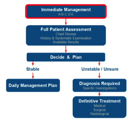
Goal directed therapy
- Supportive (advanced organ and system support)
CV support
- Indications
- MAP <60mmHg
- Reduced SBP from baseline resulting in end organ dysfunction from hypoperfusion
- Principles/Considerations
- Need to replaced IV volume prior to commencement
- 2-3L of IVF prior
- Given centrally - rapid delivery, less chance of extravasation
- Except Metaraminol can be given peripherally
- Monitor in ICU
- Need to replaced IV volume prior to commencement
- Drugs
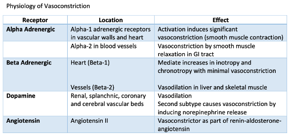
- Metaraminol
- Alpha agonist
- Vascoconstriction
- NA
- Alpha 1 and 2
- Potent vasoconstriction
- Adrenaline
- Adrenergic receptor stimulation
- Vasoconstriction
- Inotropic
- Chronotropic
- Dopamine
- Dopaminergic, alpha 1 and beta 1 adrenergic R
- Renal vasodilation
- Vasopressin = only vasoconstriction
- Metaraminol
- Complications
- Hypoperfusion
- Excess vasoconstriction (high SVR)
- Poor perfusion to non preferential areas
- Mesenteric hypoperfusion - poor healing of anastomosis
- Arrhythmia
- Tachy, AF
- Myocardial ischaemia
- Increased myocardial oxygen consumption
- Local effects
- Extravasation - local VC and skin necrosis
- Inject local phentolamine to reduce risks if it occurs
- Extravasation - local VC and skin necrosis
- Hypoperfusion
Respiratory support
- Ventilator
- ETT
- Tracheostomy
Renal support
- Dialysis (continuous renal replacement therapy or CRRT)
- Indications
- A: Acid-base issues
- Metabolic acidosis refractory to medical therapy
- E: Electrolyte disturbances
- Hyperkalaemia refractory to medical therapy
- I: Intoxication (Toxins)
- Intoxication amenable to dialysis - e.g lithium
- O: Overload (Fluid overload)
- Diuretic resistant pulmonary oedema
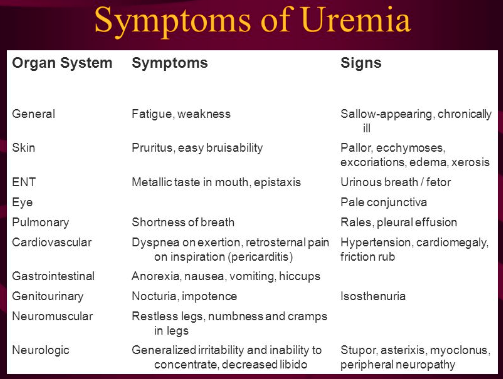
- U: Uremia
- Uraemia and complications
- A: Acid-base issues
- Complications
- Vascular access related
- Bleeding
- Vessel thrombosis/fistula probs
- Infection
- Air embolism
- Non vascular
- Hypotension
- Haemorrhage
- Hyperkalaemia
- Seizures
- Vascular access related