Sub-section: Adrenal Section: Endocrine
Incidence and presentation
- Rare – 1-2 per million per year
- Almost all present between 40-50yrs
- Minor peak in children < 5
- M = F
- Presentation = Late
- Tend to be large (mean 9-13cm) and spread beyond Adrenal Gland
- Most are functioning (57%)
- 48% Cushing’s syndrome
- 18% Multiple (Cushing’s and Virilisation)
- 6% Pure virilisation
- 3% Feminisation
- Rarely hyperaldosteronism
- Non-functioning (43%)
- 34% Abdominal discomfort
- 9% Incidentaloma
Aetiology
- Most sporadic
- Can be associated with some syndromes
- MEN 1 syndrome
- Beckwith-Wiedemann Syndrome in childhood
- Exophthalmos, microglassia, nephromegaly
- Li-Fraumeni syndrome
Biochemical Assessment
- Screen for steroid horome secretory profile
- As per individual disease workup
- Characteristic profile of adrenocortical carcinoma
- Excess secretion of multiple steroid hormones
- Particularly Glucocorticoids and Androgens
- Elevated DHEAS and Oestradiol in men and post-menopausal women
- Accumulation of steroid precursors
- 17α- Hydroxyprogesterone, Androstenedione
- Because enzyme function often defective
- Need to exclude Phaeochromocytoma
- Can’t do this on imaging alone
- Excess secretion of multiple steroid hormones
Imaging
- Unenhanced CT scan
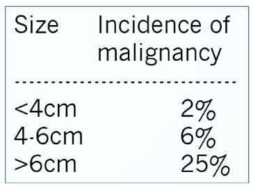
- Usually large (> 90% are > 4cm)
- 4cm cut-off for malignancy
- 93% sensitivity
- 76% specificity
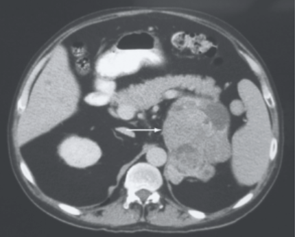
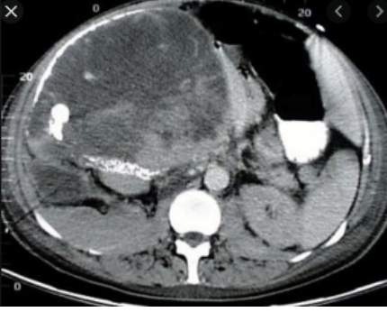
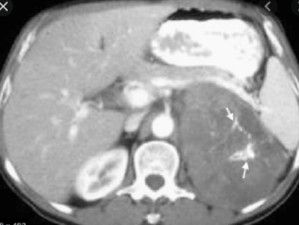
- Heterogenous +/- central necrosis
- Irregular margins
- Denser than adenomas (Hounsfield units > 10)
- If contrast added – delayed washout
- MRI
- High intensity in T2 weighted images
- With gadolinium – heterogenous enhancement, delayed washout
- FDG-PET
- If can’t characterise by CT/MRI
- 100% sensitivity, 88% specificity in differentiating malignant from benign lesions
Staging
- Percutaneous biopsy not recommended
- Difficulty differentiating benign and malignant disease
- Tumour seeding
- Only absolute criteria for malignancy:
- Invasion into local structures
- Distant metastatic disease
| T category | T criteria |
|---|---|
| TX | Primary tumor cannot be assessed |
| T0 | No evidence of primary tumor |
| T1 | Tumor ≤5 cm in greatest dimension, no extra-adrenal invasion |
| T2 | Tumor >5 cm, no extra-adrenal invasion |
| T3 | Tumor of any size with local invasion but not invading adjacent organs |
| T4 | Tumor of any size that invades adjacent organs (kidney, diaphragm, pancreas, spleen, or liver) or large blood vessels (renal vein or vena cava) |
| N1 and M1 - present |
WEISS score
- Differentiate adenomas from ACC
- The Weiss score is currently the most widely used.
- It is based on 9 criteria rated 0 or 1 each depending on their absence or presence
- (1) diffuse architecture >1/3 of the tumor surface
- (2) Tumor necrosis
- (3) ≤ 25% of clear cells of spongiocyte aspect
- (4) Führman nuclear grade III or IV
- (5) mitotic count > 5 mitoses / 50 fields x400
- (6) presence of atypical mitosis
- (7) capsular invasion
- (8) venous invasion
- (9) sinusoidal invasion
- Tumors with a score ≥ 3 are considered to have metastatic potential and are diagnosed as ACC
Management
Surgical Management
- Surgery offers only potential cure
- Open Adrenalectomy +/- En-block resection of adjacent organs and regional lymphadenectomy
- Laparoscopy may increase risk of recurrence or carcinomatosis
- Complete resection in 70%
- Incomplete resection has very poor prognosis (mean < 1yr)
- Take care with right sided tumours > 9cm
- Potential IVC invasion (+/- right heart invasion)
- Should do these cases on cardiopulmonary bypass
- Prevents risk of potentially fatal tumour embolism
- Palliative surgery (esp. if functional)
Adjuvant treatment
- Chemo with Mitotane
- Derivative of insecticide DDT
- Direct adrenocortical toxin
- Overall response usually poor
- Indications
- High risk disease
- Ki-67 >10
- R1 resection
- Tumour spillage
- Vascular invasion
- High risk disease
- Dose dependent GI and neurotoxic side effects
- Narrow therapeutic window
- Adrenal insufficiency
- Need to start glucocorticoid replacement
- Also need to monitor for aldosterone deficiency
- Hepatic toxicity
Unresectable disease
- Mitotane alone + addition of chemotherapy
- Can consider ablation for small tumours (RFA) <5cm to provide short term local control
Prognosis
- 5yr survival 16-38% depending on stage at presentation
- Increasing with use of mitotane
- Local recurrence and metastases common
- Typically occurs within 2yrs from surgery
- Mean survival following non-curative surgery or metastatic disease
- < 12 months