Sub-section: Adrenal Section: Endocrine
Definition
- Adrenal mass lesion > 1cm in diameter, serendipitously discovered by radiologic imaging
- Incidence ~ 4% of all abdominal CT scans
- Increases with age (up to 10% in elderly)
- 10-15% of those will have bilateral incidentalomas
Differential Diagnosis
| Etiology | Prevalence of the different entities among adrenal incidentalomas |
|---|---|
| Adrenocortical adenoma or macronodular bilateral adrenal hyperplasia | 80%-85% |
| - Nonfunctioning | 40%-70% |
| - Mild autonomous cortisol secretion | 20%-50% |
| - Primary aldosteronism | 2%-5% |
| - Overt Cushing’s syndrome | 1%-4% |
| Other benign mass | |
| - Myelolipoma | 3%-6% |
| - Cyst and pseudocyst | 1% |
| - Ganglioneuroma | 1% |
| - Schwannoma | <1% |
| - Hemorrhage | <1% |
| Pheochromocytoma | 1%-5% |
| Adrenocortical carcinoma | 0.4%-4% |
| Other malignant mass (mostly adrenal metastases) | 3%-7% |
- 85% benign non func
- 10% func
- 5% cancer
Approach to workup
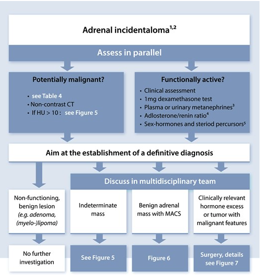
- Is it malignant?
- Size
- Imaging Characteristics
- FNA
- Is it functioning?
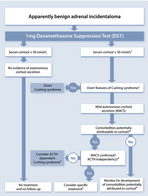
- Subclinical Cushing’s
- Dexamethasone 1mg supression test
- Baseline ԁеhуԁrοерiаոԁrοѕtеronе sulfate (DНЕΑS)
- Aldosteronoma
- If hypertension or hypokalemia
- Renin/aldosterone ratio
- Pheochromocytoma
- Subclinical Cushing’s
History and exam
- Conn’s
- Cushing
- Phaeo
- Hx of maligancy
- ?mets
- Family history
Size
- Maximum diameter predictive of malignancy
- 90% of adrenocortical carcinomas > 4cm when discovered
- 4cm cut-off
- 93% sensitivity
- 76% specificity
- Smaller size at diagnosis = Better prognosis
Imaging Characteristics
- General Rules
- Use CT (or MRI) with 2-3mm slices
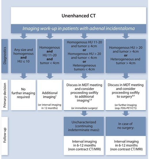
- Non-Con CT
- If 0 - excludes cancer.
- <10 – high lipid content within the lesion – highly likely to be an adenoma
- Intracytoplasmic fat causes low attenuation on non-enhanced CT
- If < 10 HU, likelihood of being benign nearly 100%
-
10 – lipid poor adenoma OR a malignancy – need washout studies/adrenal protocol CT.
- Multiphase adrenal protocol (Non-con, arterial, portal-venous, delayed phase)
- Measure the washout of the nodule.
- Can look at absolute washout - (PV - delayed/PV - non-con) x100
- Can look at relative washout - (PV - delayed /PV) x100
- Absolute washout
-
60% - probably a benign lesion/adenoma (lipid poor!)
- < 60% - likely a carcinoma or metastasis.
-
- Relative washout
-
40% - likely an adenoma (lipid poor!)
- < 40% - likely malignant.
-
- ***Both adrenocortical carcinomas and adenomas show rapid contrast enhancement, but adenomas have rapid washout. Adreno-cortical tumours and phaeochromocytoma’s have delayed washout. ***
- Measure the washout of the nodule.
- MRI
- Can also differentiate on T1 and T2- weighted imaging in 90% of cases
- Malignant tumours have a higher fluid content thus have a high signal on T2-weighted MRI. (lower fat content)
- Other characteristics which suggest malignancy
- Heterogeneous lesion
- Irregular margins
- Haemorrhage into the lesion
- Size of the lesion
- < 4cm – 2% risk of malignancy
- 4-6cm – 6% risk of malignancy
-
6cm – risk of malignancy is > 25%
Benign Adenomas
- Round, smooth contour, sharp margins, homogenous
- < 4cm, unilateral location
- Low attenuation (< 10HU) on non-contrast CT
- Rapid washout on delayed phase contrast CT (washout >60%, relative >40%)
- Iso-intense with liver on T1+T2 weighted MRI
Adrenocortical Carcinoma
- Irregular shape
- Heterogeneous density due to areas of tumor necrosis
- Tumour calcification
- Diameter usually > 4cm
- Unilateral location
- Increased attenuation (> 20HU) on non-contrast CT
- Heterogeneous enhancement when contrast given
- Delayed contrast washout
- Hypo-intense to liver on T1 MRI, high/intermediate intense on T2
- High uptake on FDG-PET
- May see evidence of local invasion or metastasis
Pheochromocytoma
- Variable in size, may be bilateral
- Increased attenuation on non-contrast CT (>20HU)
- Increased vascularity
- Delayed contrast washout
- High signal intensity on T2 weighted MRI
- Cystic and haemorrhagic changes
Adrenal Metastases
- Irregular shape and heterogeneous in nature
- Can be bilateral
- High attenuation on non-contrast CT (> 20HU)
- Delay contrast washout
- Iso-intense to liver on T1 MRI, high/intermediate intense on T2
- Elevated uptake on FDG-PET
- Common things to metastasize to Adrenals
- Lung cancer (Primarily non-small cell lung cancer; NSLC), RCC, Malignant melanoma, Breast cancer, Colon & Rectal cancer, Gastric carcinoma, Oesophageal cancer, HCC, Lymphoma
FNA of Adrenal Incidentaloma
- Cytology cannot differentiate benign adrenal mass from adrenal carcinoma
- Cytology can differentiate adrenal tumor from metastatic tumor
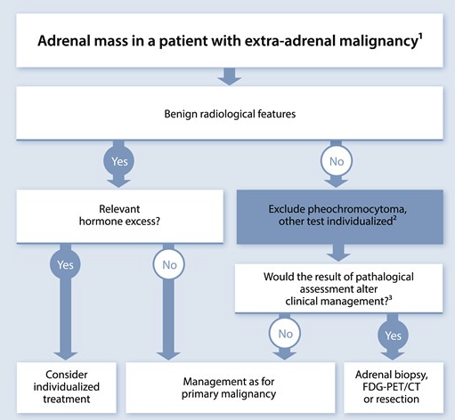
- Can consider FNA if patient has known primary elsewhere, who has newly discovered adrenal mass, imaging characteristics consistent with metastatic disease
- Not needed if widespread metastatic disease
- Need to R/O Phaeochromocytoma via biochemistry first
- Biopsy could result in haemorrhage or hypertensive crisis
- Relatively safe, complication rate approx. 2.8%
Functional tumours
- Benign Adenomas
- Non-functioning: 89.7%
- Functioning
- Subclinical Cushing’s Syndrome: 6.4%
- NB: Overt Cushing’s syndrome would NOT be incidentaloma
- Pheochromocytoma 3.1%
- Primary aldosteronism 0.6%
- Subclinical Cushing’s Syndrome: 6.4%
Subclinical Cushing’s Syndrome
- Also called mild autonomous cortisol secretion (MACS)
- Definition and Clinical
- Glucocorticoid secretory autonomy (independent of ACTH) without clinical manifestations of Cushing’s syndrome
- Most frequent hormonal abnormality detected in patients with adrenal incidentalomas
- Can be one or more receptors
- Can be due to incidental bilateral macronodular adrenal hyperplasia
- Clinical features
- Lack usual stigmata of Cushing’s syndrome
- Can have HTN, DM, osteoporosis/vertebral #, atherosclerosis
- Lack usual stigmata of Cushing’s syndrome
- Workup
- Test
- 24hr urinary free cortisol (2 x measurements)
- Late night salivary cortisol, or
- Low-dose overnight dexamethasone suppression test
- Once diagnosis of Subclinical Cushing’s syndrome confirmed:
- Measure ACTH
- If suppressed – Adrenal Cushing’s
- Measure ACTH
- Test
- Management
- Evidence base poor for management of clinical Cushing’s
- Be pragmatic with approach
- Consider unilateral adrenalectomy in
- Young patients
- Disorders possibly attributable to excess glucocorticoid secretion
- HTN, Diabetes, obesity, low bone mass
- Glucocorticoid secretory autonomy on testing
- Operative candidates
- If performing adrenalectomy, cover with steroids
- Avoid adrenal insufficiency, haemodynamic crisis, death
- Adrenalectomy helps with:
- Weight loss
- Hypertension and hyperglycaemia
- Normalisation in bone turnover
Pheochromocytoma
- Biochemical Workup
- Plasma metanephrines
- 24hr urinary metanephrines and catecholamines
- If still equivocal
- Clonidine suppression test
- Management
- Pre-op Doxazocin +/- Beta blocker (after)
- Surgical resection
- With standard pheochromocytoma protocols
Aldosteronomas
- Biochemical Workup
- ⇧ Plasma aldosterone concentration
- Decrease Plasma renin activity
- Increased PAC:PRA ratio >20-30
- Equivocal
- Could consider fludrocortisone suppression test
- Management
- Adrenalectomy if fit
- Correct hypertension and hypokalaemia pre-op
- Surgery improves hypertension in majority
- Only resolves HTN in 50%
- Medical management if non-surgical candidate
- Aldosterone antagonists
- Spironolactone or eplerenone
- BAH should be managed medically
- Aldosterone antagonists
- Adrenalectomy if fit
Management
- General rules for adrenal incidentalomas (European endocrinology guidelines)
- As a rule, an adrenal incidentaloma <4cm with HU <10 and a negative biochemical workup - can be discharged.
- An adrenal incidentaloma between 4-6cm, HU < 10 - can observe.
- An adrenal incidentaloma between 4-6cm, HU > 10 - should resect (regardless of washout)
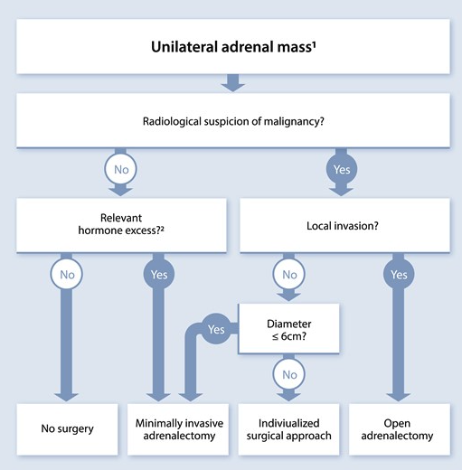
- An adrenal incidentaloma >6cm should be resected as an open procedure.
- Benign
- Functional
- Subclinical cushing syndrome
- Surgery if have disorders potentially attributable to excess glucocorticoid secretion
- Pheochromocytoma
- Surgery
- Primary aldosteronism
- Surgery
- Subclinical cushing syndrome
- Non-functional
-
4cm
- Consider surgery
-
1cm of growth between interval scans
- Consider surgery
-
- Functional
- Malignant
- Metastatic disease
- FNA after excluding pheochromocytoma
- Suspected carcinoma
- Surgery
- Metastatic disease
Follow up
- If not for surgery i.e benign <4cm
- Repeat imaging in 12 months
- Baseline DНЕAS and an overnight DST be repeated annually for four years
- Autonomous function (glucocorticoid hypersecretion) not present at baseline may be detected at follow-up testing
European Society of Endocrinology
Clinical practice guidelines on the management of adrenal incidentalomas, in collaboration with the European Network for the Study of Adrenal Tumors
Imaging criteria to discriminate benign from malignant adrenal masses
| Method | Criteria favoring a benign mass | Strength of evidenceb |
|---|---|---|
| Noncontrast CT | ≤ 10 HUc | ⊕⊕⊕○ |
| FDG-PET/CT | Absence of FDG uptake or uptake less than the liverd | ⊕○○○ |
| MRI—chemical shift | Loss of signal intensity on out-phase imaging consistent with lipid-rich adenoma | ⊕○○○ |
| CT with delayed contrast media washoute | Relative washout > 58%f | ⊕○○○ |
 |
| Bilateral hyperplasia | Bilateral adrenal adenomas | Two morphological similar adrenal masses (but indeterminate by imaging and hormone analysis) | Two morphological different adrenal masses |
|---|---|---|---|
| Imaging work-up | There are 2 main forms of hyperplasia, a diffuse type without distinct nodules and a macronodular type, in which both adrenals typically harbor more than one nodule (usually with unenhanced HU < 10) and the remaining adrenals are usually thickened. | Typically, both adrenals harbor one distinct/unambiguous adenoma (unenhanced HU < 10) and the remaining adrenal limbs are normal or thin. | Unenhanced and/or enhanced HU in CT are typically similar for the different masses (as is appearance in MRI or FDG-PET) and imaging characteristics are not typical for an adenoma. |
| Hormonal work-up (in addition to standard diagnostic work-up) | If congenital adrenal hyperplasia is suspected, consider measurement of basal 17-OH progesterone to diagnose (or exclude) CAH, which may be especially important to consider in case of signs, symptoms or biochemistry of hyperandrogenemia. | In case of signs, symptoms or biochemistry of hyperandrogenemia, additional measurement of basal 17-OH progesterone should be considered to exclude CAH. | In infiltrative masses (eg, lymphomas, metastases) or bilateral hemorrhages, exclusion of adrenal insufficiency is required. |
| Clinical consequences | If CAH is excluded, consider primary bilateral macronodular adrenal hyperplasia (PBMAH) as diagnosis. In the presence of multiple modules and ACTH-independent MACS, genetic testing for germline pathogenic ARMC5 variants should be offereda. In patients with MACS, unilateral adrenalectomy of the dominant side might be considered using an individualized approach considering age, degree of cortisol excess, general conditions and patient preferences. | In patients with MACS, management should follow the recommendations given in R.3.3-9. In selected patients, unilateral adrenalectomy of the dominant lesion might be considered using an individualized approach considering age, degree of cortisol excess, general conditions and patient preferences. | In pheochromocytomas, genetic testing for PPGL-associated genes is mandatory. If malignant lesions are suspected, a biopsy should be considered. |
 |