Section: Endocrine Sub-section: Parathyroid
Overview
- Primary hyperparathyroidism
- One of more glands are making to much PTH
- About 85% of cases there are a single parathyroid adenoma
- Secondary hyperparathyroidism
- Physiological secretion of PTH due to chronic hypocalcaemia
- Seen in CKD and Vitamin D deficiency – in chronic renal failure reduced production of calcitriol which leads to reduced intestinal absorption which increases PTH release.
- Tertiary hyperparathyroidism
- Persistent autonomous secretion of PTH hormone in CKD.
Primary Hyperparathyroidism
Definition
- Inappropriate secretion of PTH from Parathyroid glands relative to serum Ca2+
- Classic - elevated calcium + PTH
- Normocalcaemic - normal calcium + elevated PTH
- Due to intrinsic abnormality of one or more parathyroid glands
- Enlarges and secretes excessive PTH
- New set point in affected glands
- Higher circulating Ca2+ maintained
- Most commonly presents as asymptomatic hypercalcaemia
- Presentations vary on the spectrum of signs/symptoms of hypercalcaemia
Incidence
- 3rd most common endocrine disorder
- Behind diabetes mellitus and thyroid disease
- Incidence increases with age (> 45)
- F > M
- 3-5 per 1000
- Incidence increasing with time due to increased availability of serum Ca2+ measurement
Aetiology
- Single adenoma 85%
- Multi-gland adenomas 5%
- Four gland hyperplasia 10%
- Parathyroid carcinoma 1 % (very rare)
Risk factors
- Most patients have no known risk factors
- Could consider hx of irradiation, or familial syndromes
- MEN 1 syndrome associated with multi-gland hyperparathyroidism
- MEN 2A syndrome associated with single gland adenoma
Genetic testing
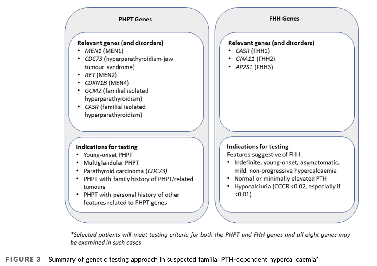
- Genetic testing is recommended in patients with
- PHPT with young‐onset multi‐gland disease
- Parathyroid carcinoma
- Family history of PHPT and/or related tumours, and/or a personal history of other features related to PHPT predisposition syndromes.
- Genetic testing should also be considered in hypercalcaemic patients with features suggestive of FHH.
Pathology
Single Adenoma
- Gross appearance
- Large, tan or beefy red (like “little kidney”)
- Other glands appear normal or atrophic
- Microscopic
- Normal has predominantly chief cells with scattered oxyphil cells only
- Adenoma had solid sheets of chief cells, oxyphil cells, or combination of both (in a fibrous capsule)
- Classically has rim of compressed normal parathyroid surrounding adenoma (seen in 20-30%)
- Features:
- Hypercellularity
- Loss of fat
- Loss of lobulation
- Oxyphilic change
- Can get pleomorphism and multinucleation, but mitosis are rare
Double Adenoma
- Uncommon
- May lead to recurrent or persistent hyperparathyroidism if have diseased gland missed because only do unilateral exploration
- If find two abnormal glands on one side, must do bilateral exploration
Hyperplasia
- Polyclonal expansion of parathyroid cells
- More typical of familial hyperparathyroidism
- Can be in sporadic cases
- Gross appearance - typically non-uniform
- Can get one gland larger than the rest, gives false impression of single adenoma
- Microscopy
- Chief cells mostly affected
- Absence of fat ++
- Hypercellularity
- Retained lobulation
- If surgery needed:
- 4 Gland Exploration and Subtotal Parathyroidectomy
Carcinoma
- Rare, difficult pre-op diagnosis
- Often made in retrospect after metastatic disease
- Tends to be younger patients (50s) than benign (60s)
- M=F
- Palpable mass 30-75% patients
- Far more than benign disease
- RLN involvement suggestive of malignancy
- Classically find adherence to surrounding structures or invasion intra-op, with dense scarring
- Histology
- Bizarre nuclear atypia, mitotic figures, capsular invasion
- Only definitive criteria for malignancy
- Local invasion
- Metastatic disease (lung, liver, LNs)
- Recurrence rate 66%
- 5yr survival 69%
- Death caused by metabolic sequelae of hypercalcaemia
Diagnosis
- Primarily Biochemical
- Elevated serum Ca2+
- Albumin adjusted should be test
- Gold standard for measuring calcium in the blood is ionised calcium corrected for pH
- Ionised Ca2+ more accurate (e.g. in hypoalbuminaemia)
- If normal calcium and increase PTH then test ionised (may be false normal)
- Elevated PTH
- High normal, need to do urine Ca2+ to confirm
- Low Phosphate (except in renal failure)
- Chloride: Phosphate ratio ≥ 33
- Specific indicator of Hyperparathyroidism
- Hyperchloraemic state due to exchange with phosphate
- Elevated serum Ca2+
- Familial hypocalciuric hypercalcemia
- 24hr Urinary Ca2+
- Hypercalciuria
- Rules out FHH, Ca2+ spills over
- Hypercalciuria
- Spot Urine Ca2+/Cr ratio
- Rule out FHH
- 24hr Urinary Ca2+
- Also needs
- Normal Vitamin D
- Check to rule out secondary hyperparathyroidism
- Off medications that interfere with calcium excretion such as thiazides
- Normal Vitamin D
Axillary studies
- In patients with a diagnosis of primary hyperparathyroidism
- DXA scan of the hip, lumbar spine, and distal radius
- Thoracolumbar imaging at baseline
- Renal tract imaging should be considered for patients with PHPT without other indications for parathyroidectomy. Available modalities include renal tract ultrasound and X‐ray KUB. Non‐contrast CT scan (CT‐KUB), has relatively little radiation exposure and excellent sensitivity
- Even in the absence of a history of urinary tract calculi or detection on imaging, urinary calcium excretion may be assessed. Parathyroidectomy may be considered in patients with confirmed hyperparathyroidism and hypercalciuria
Imaging Studies and Localisation
- Rational for use for pre-operative planning
- Localise single adenoma to be able to perform minimally invasive surgical approach
- Should not be used for diagnosis (this is biochemical)
- May diagnosis concurrent thyroid disease
- And perform dual surgery
- Helps to localise pathology in re-operative situations
- Non-invasive localisation
- USS
- CT
- MRI – rarely indicated (could use in pregnancy if USS non-diagnostic)
- T99 – Sestamibi scans
- Invasive localisation
- Angiography and venous sampling
- For the exam say - you would do a neck USS and sestamibi, followed by a 4D-CT if necessary. If still not finding it – do a 4 gland exploration.
USS
- Benefits
- Fast
- Inexpensive
- Non-radiating
- Non-invasive
- Can visualise adjacent structures
- Thyroid, carotids etc
- Issues
- Operator dependent
- Difficulty locating if:
- Retro-oesophageal, Retrosternal, Retrotracheal and deep cervical lesions
- False +ve 15-20%
- Sensitivity 71-80%
- Falls to 40% for re-operative cases
- Best used combined with Sestamibi
- Findings:
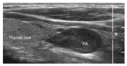
- Hypoechoic lesion posterior to thyroid gland, with solitary feeding vessel
CT
- Use fine slice (2.5mm)
- Best if dedicated radiologist
- 3D CT has long been used
- Results have been extremely variable (sensitivity 40% to 89%).
- 4D CT (4th dimension = Time)
- Accounts for differences in perfusion characteristics between the hyperfunctioning parathyroid gland and surrounding structures, such as the thyroid gland
- Improved sensitivity of 88% for four-dimensional CT compared with the sensitivities of Sestamibi and neck ultrasound (65% and 57%, respectively) was demonstrated
- Recently, a combination of 4D-CT and USS demonstrated a sensitivity of 94% and specificity of 96% for lateralizing hyperfunctioning parathyroid glands
- Sensitivity was 82% for localization to the correct quadrant of the neck
T99 Sestamibi Scintigraphy
-
Sestamibi
- The drug is a coordination complex consisting of the radioisotope technetium-99m bound to six (sesta=6) methoxyisobutylisonitrile (MIBI) ligands.
- Passively diffuses across cell membranes
- Accumulates in mitochondria
-
Selective affinity for abnormal parathyroids due to abundance of mitochondria
-
Two primary techniques to differentiate sestamibi uptake by abnormal parathyroid and thyroid tissue.
- Dual isotope approach with sestamibi and either iodine-123 or 99mTc pertechnetate (subtraction imaging)
- Sestamibi alone with dual (early and delayed) phase imaging
- Also taken up by thyroid tissue - washes out thyroid quickly therefore initial scan and delayed scan performed.
-
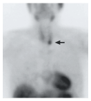
Sestamibi single photon emission computed tomography (SPECT)
- Allows 3D images, better anatomical localisation
- Sestamibi tracer used in SPECT
- Fuse a CT scan with Sestamibi scan
Selective Venous Sampling
- SVS is the most sensitive localization procedure, use in re-operative patients
- Procedure
- Catheterize common femoral vein for baseline PTH
- Then SVS of small venous branches from the neck and mediastinum
- 2 x increase in PTH value compared with baseline defines a positive localization study
- With rapid PTH assay in the interventional radiology suite, near real-time assay results are available
- Disadvantages
- SVS is expensive and time-consuming
- Requires an experienced interventional radiologist
- Serious complications are rare
| Sensitivity | Specificity | Advantages | Disadvantages | |
|---|---|---|---|---|
| USS | Mod-high | Mod-high | Fast and cheap. Can combine with FNA Allows thyroid to be assessed | Operator dependent Difficulty with posterior areas. Cannot detect lesion < 5mm. |
| Sestamibi-Tc scan | Very high | Very high | Minimal radiation. Widely available. Combining with SPECT improves ectopic localisation | May miss multiple adenomas. SPECT not widely available |
| 4D-CT | Moderate | High | Good at looking at retro-oesophageal/retro-tracheal areas. Can combine with FNA. | Difficult with lower neck/should area Very high dose of radiation. Previous neck surgery can yield artifact. |
| MRI | Mod-high | Very high | Very good at localising ectopic glands Does not require IV contrast Good modality for re-operative cases (not subject to artefact) | Expensive Cannot be combined with FNA Cannot detect lesions <5mm |
| Selective venous sampling with angiography | Very high | Very high | Good for patients requiring re-do surgery. | Is an invasive modality - risk of groin complications. Experienced interventional radiologists not widely available. Has largely been superseded by the above imaging modalities. |
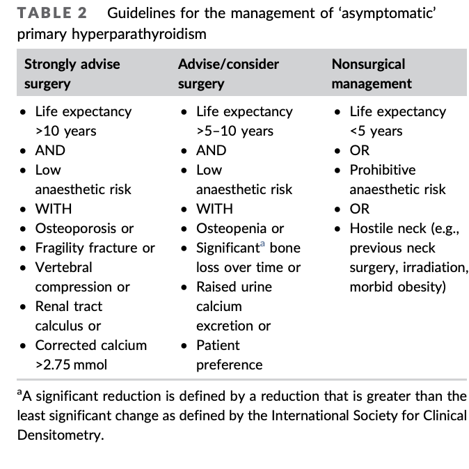
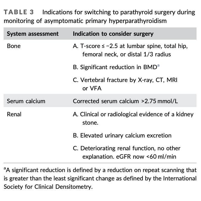
Indications for Surgery
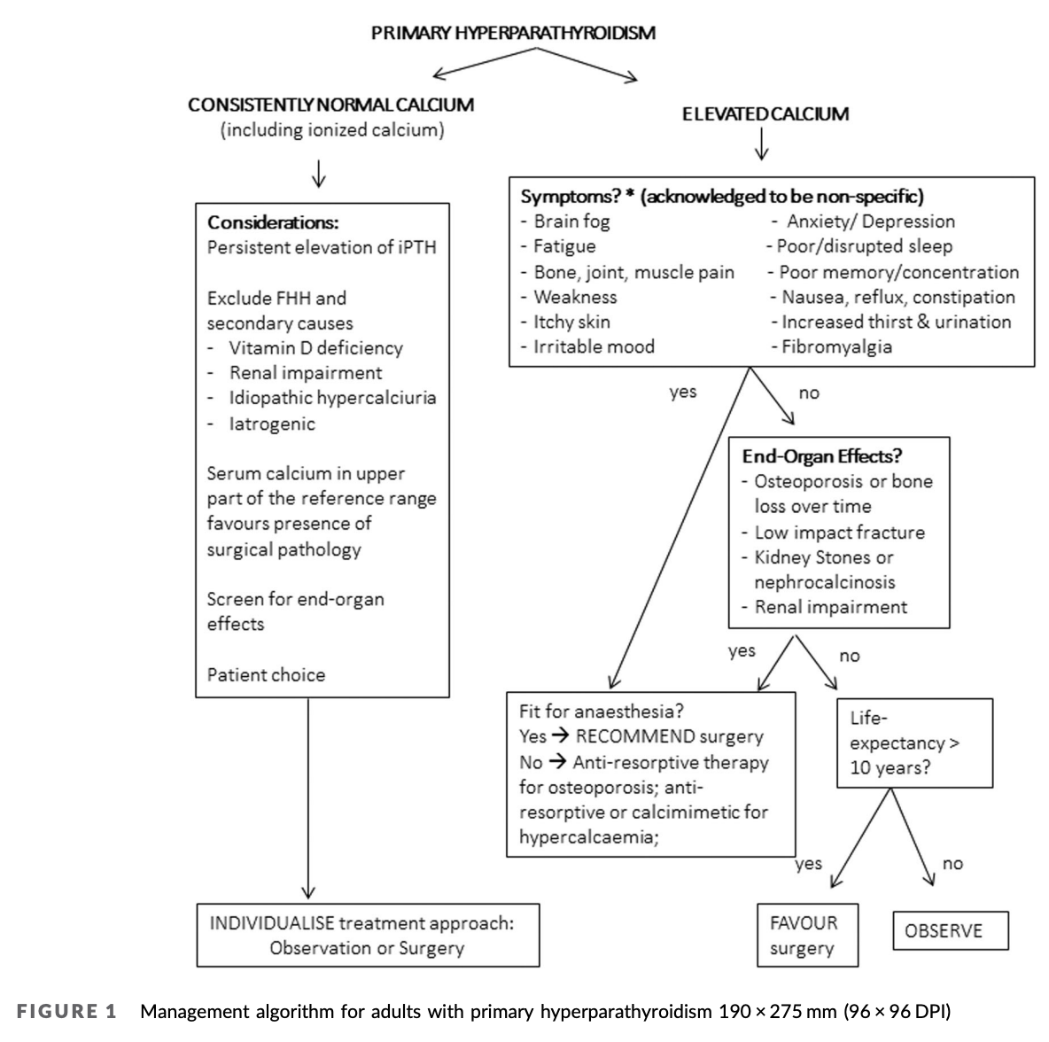
- All symptomatic patients
- Only cure for 1° Hyperparathyroidism
- Surgery unless unfit
- Asymptomatic patients who have raised calcium
- End organ effects
- Renal stones on imaging
- Renal impairment
- Crcl < 60mL/min
- Osteoporosis
- T-score < -2.5 at any site or
- Previous osteoporotic fractures
-
10 year life expectancy
- Previously guidlines stated Age < 50
- Patient choice or those not suitable for surveillance
- End organ effects
- What is meant by “increased calcium”
-
0.25mmol/L above upper limit normal
- Patients with mildly elevated levels of calcium should be assessed and managed in a similar manner to those with more severe hypercalcaemia
- There is evidence that BMD decline in PHPT occurs independent of degree of hypercalcaemia
- Although no strong correlation between severity of hypercalcaemia and PHPT symptoms and end‐organ effects, patients with higher calcium levels may be more likely to achieve symptomatic benefit after surgery.
- Therefore calcium >0.25 mmol/L above the local reference range swings the risk/benefit ratio further towards surgery
- Patients with mildly elevated levels of calcium should be assessed and managed in a similar manner to those with more severe hypercalcaemia
-
Guidlines from: Primary hyperparathyroidism in adults—(Part I) assessment andmedicalmanagement: Position statement of the endocrine society of Australia, the Australian & New Zealand endocrine surgeons, and the Australian & New Zealand bone and mineral society. Published 2021
Normocalcaemic Primary Hyperparathyroidism
- Variant of 1°HPT (? early presentation)
- Typically identified in the setting of an evaluation for low bone mineral density or renal calculi
- PTH elevated but Ca2+ normal
- In order to make a diagnosis
- Ionized calcium levels should be normal
- All secondary causes for hyperparathyroidism must be ruled out
- E.g. Vit D deficiency and CKD
Hyperparathyroidism in pregnancy
- Diagnosis
- Same as above
- Ionised calcium more important due to low albumin
- Complications
- Maternal
- Hyperemesis
- Nephrolithiasis
- Hypercalcaemic crisis
- Hypertension
- Pre‐eclampsia and eclampsia
- Foetal complications
- Intrauterine growth restriction
- Premature delivery
- Stillbirth
- Neonatal tetany
- Maternal
- Management
- Ideal surgery prior to pregnancy
- In pregnancy
- Mild <2.7
- Can monitor
- Mod-severe >2.7
- Operate - ideally second trimester
- USS preferred localisation technique
- Operate - ideally second trimester
- Mild <2.7
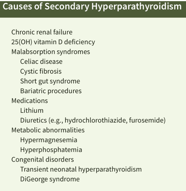
Secondary Hyperparathyroidism
Overview
- Hyperparathyroidism caused by factors other than primary parathyroid disease
- Usually physiologic secretion of PTH by the parathyroid glands in response to hypocalcaemia
Cause
- Vitamin D Deficiency
- A low serum 25(OH)D leads to a decrease in 1,25(OH)2D
- PTH secretion increases in an effort to normalize 1,25(OH)2D
- Leads to a higher rate of bone resorption, and cortical bone loss
- Get increased markers of bone turnover (urine hydroxyproline)
- Eventually get resistance, leading to decreased Ca2+ levels
- Chronic Renal Failure (Uraemic Hyperparathyroidism)
- Initial cause
- Leads to hyperphosphataemia
- ⇒ Hypocalcaemia (inverse relationship)
- Also decreased renal conversion of 25(OH)D to 1,25(OH)2D
- Thus, reduced intestinal Ca2+ absorption
- Subsequent hypocalcaemia and stimulation of PTH
- Mechanism multifactorial
- Mutations in calcium-sensing receptors
- Alterations in calcium set-points
- Decreased metabolic clearance of PTH
- Increased phosphate retention
- Leads to hyperphosphataemia
- Progression
- Over time get hyperplasia of all glands and PTH elevation in response
- This autonomous function is termed “refractory hyperparathyroidism” or tertiary hyperparathyroidism by some authors - although tertiary is often received for patients who have had a renal transplant and have autonomous function
- Initial cause
Management
- Usually managed medically
- For Vit D defincey
- Vit D supplementation
- For CKD
- Start and titrate active Vitamin D (Calcitriol)
- Often causes hypercalcaemia
- If this happen requires starting Calcimimetic to decrease calcium
- Often causes hypercalcaemia
- Restrict dietary phosphorus and treat hyperphosphateamia
- Start and titrate active Vitamin D (Calcitriol)
- For Vit D defincey
- If fail, offer Subtotal Parathyroidectomy
- Definitive rx for 2° Hyperparathyroidism = Renal transplant
Autonomous function
- Patient who have autonomous function of their parathyroid glands will develop hypercalceamia have complication of this
- High PTH alone (secondary) may causes symptoms associated with high bone turn over but the hypercalceamic complications will be absent
Presentation and Management
- Many pts asymptomatic
- May get
- Osseous lesions (bone pain due to increased remodelling)
- Osteitis fibrosis cystica (up to 30% dialysis patients)
- Chaotic matrix deposition and woven bone appearance
- Osteitis fibrosis cystica (up to 30% dialysis patients)
- Pruritus (up to 85% on dialysis), may be disabling
- Symptomatic relief after subtotal parathyroidectomy
- Metastatic calcification
- Can affect any organ, most commonly vasculature
- Also heart valves, kidneys, GIT, penis
- Calciphylaxis
- Less than 5% of patients require surgical intervention
- Osseous lesions (bone pain due to increased remodelling)
Indications for surgery
- Subtotal or total with implant
- Indications
- Failure of medical mgmt
- Intractable bone pain
- Intractable pruritis
- Fractures
- Symptomatic Ectopic Calcifications
- On transplant list
- PTH cut off of >100 is often used
- If <100 then often secondary and will resolved with transplant
- PTH cut off of >100 is often used
Tertiary hyperparathyroidism
Definition
- Long-standing chronic renal failure who have resolution of renal disease
- Usually patients who have persistent autonomous secretion of PTH after renal transplantation
- Loss of response to serum Ca2+ levels leads to four-gland hyperplasia with autonomous activity
- Can see marked asymmetry in parathyroid gland size
- Renal transplant recipients often have additional risk factors for the development of tertiary HPT
- Alterations in GFR
- Transplant-associated drugs (corticosteroids, cyclosporine and thiazide diuretics)
Implications
- Results in elevated serum Ca2+ and intact PTH levels
- Can be life-threatening
Management
- Surgical Subtotal Parathyroidectomy indicated if persistent hypercalcaemia after 12 months of observation
- In addition, patients with secondary HPT who are being considered for renal transplantation should have serum calcium and PTH levels screened pre-op
Calciphylaxis
- Calcific uremic arteriolopathy - Vascular calcification, thrombosis, and skin necrosis
- Rare but serious, seen mainly in ESRF pts
- Commonly who are on HD or who have recently received a kidney transplant
- Not exclusive to patients with ESRD (Non-uraemic calciphylaxis)
- Seen in 1°HPT, breast cancer (treated with chemo), EtOH liver cirrhosis, cholangioca, Crohn’s, RA, and SLE
- Chronic non-healing wounds and can be fatal
- Clinical diagnosis
- Characteristic ischaemic skin lesions with areas of skin necrosis associated with severe pain
- Appears as violaceous, leathery lesions and can be extensive
- Confirm with skin biopsy
- Small arterial calcification and occlusion in the absence of vasculitis
- Bone scintigraphy may show increased tracer accumulation in the soft tissues
- Treatment
- Results for calciphylaxis are variable, prevention is crucial
- Rigorous and continuous control of phosphate and calcium balance
- Necrotic skin areas may become infected, leading to sepsis
- Overall, clinical prognosis poor
- Urgent Subtotal Parathyroidectomy is usually recommended