Section: Hepatobiliary Sub-section: Biliary Curriculum: Curriculum, page 89
Aetiology
- Mostly lap chole
- Primary cause – visual perceptual illusion (97%), technical (3%)
- Occasionally abdominal trauma, injection of scolicidal agent for hydatid cyst, ablation of hepatic tumours or radiotherapy.
Techniques to avoid injury
- Cholecystectomy
- Correct identification of biliary anatomy
- Dissection at correct level – junction of gallbladder and cystic duct, staying close to gallbladder.
- Structures traced into gallbladder
- Critical view of safety
- Avoiding diathermy in Calot’s triangle. Avoid blind cauterising or clipping of arterial bleeding.
- Connor et al. five-point checklist
- Confirm gallbladder lies in the hepatic principal plane and is retracted to 10 o’clock position
- Confirm Hartmann’s pouch is lifted up and toward the segment IV pedicle
- Identify Rouviere’s sulcus
- Confirm the release of the posterior left of the peritoneum covering the hepatobiliary triangle.
- Confirm the critical view
- IOC
- Reduction in biliary injuries when used routinely
- Correct IOC interpretation involves:
- Duodenal flow
- Proximal filling of the common bile duct
- Three proximal hepatic ducts (right anterior, posterior sectoral and left main ducts)
- Absence of filling defects within the common bile duct
- The presence of spiral valves within the cystic duct
- Subtotal or opening gallbladder.
Strasburg classification
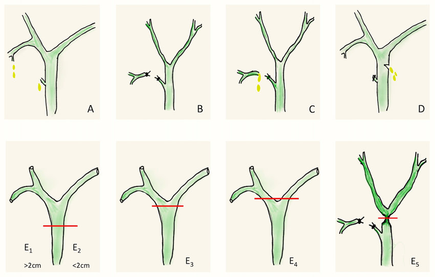
- A– leakage from cystic or subvesical ducts
- B – occlusion of part of the biliary tree e.g. aberrant right hepatic duct
- C – transection without ligation of part of the biliary tree
- D – lateral injury to biliary tree
- E – subdivided into E1-5 by Bismuth
- E1 – Low CHD transection or stricture – hepatic duct stump > 2 cm
- E2 – proximal CHD transection or stricture – hepatic duct stump < 2 cm
- E3 – hilar transection or stricture with right and left ducts in communication.
- E4 – destruction of hepatic confluence – R & L ducts separated
- E5 – combined injury to confluence and right sectoral duct.
Presentation
- Best recognised at time of surgery (<1/3)
- Early post-op
- Biliary fistula
- Biliary peritonitis
- Jaundice
- Symptoms of peritoneal or diaphragmatic irrigation or associated LFT derangement.
- Late post-op
- Presentation may be stricture due to partial injury, localised inflammation or ischaemic insult
- Ligation of sectoral ducts may cause late atrophy of the drained liver segments which may become infected secondarily.
- Occasionally liver resection or transplant may be required. May have late liver failure due to secondary biliary cirrhosis.
Management
Intraop recognition
- Should be referred to experienced surgeon immediately.
- Success of repair by initial surgeon is unlikely.
- If help unavailable, cholangiogram if possible to understand anatomy, T-tube or similar drain placed and drains left in subhepatic space.
- Complete transection should not be attempted to repair
- Partial injury can be closed directly with placement of T-tube through separate choledochotomy.
Postop recognition: biliary fistula
- If not fit for discharge at 24 hours due to abdo pain, vomiting, fever or bile in drain should be considered to have a biliary leak.
- CT better than USS – more objective information and allows assessment of liver vasculature.
- Laparoscopy – visualise source of bile leak. If cystic duct clips dislodged, can apply further clips or suture. However, any other source should be specialist referral.
- ERCP allows anatomical definition, sphincterotomy or stent placement.
- CT IV cholangiogram or MRCP can determine continuity of biliary tree prior to endoscopy (CBD transection precludes ERCP).
- PTC
- May be required if ERCP unsuccessful or bile duct ligated.
- Can decompress biliary system
Postop recognition: biliary obstruction
- Ligation or clipping of biliary tree presents early with jaundice.
- If stricture formation, may present later.
- If diagnosis of ductal obstruction made within first week postop and only moderately elevated bilirubin with no coexisting coagulopathy or sepsis, immediate repair is best chance of success.
- USS may indicate level of obstruction or exclude presence of retained stone
- CT IVC not indicated as contrast agent not excreted.
- MRCP will identify stricture or transection.
- ERCP will also identify stricture or transection.
- Stent placed only after consultation with specialist unit – undrained biliary tree may allow proximal biliary dilatation facilitating later reconstruction.
- Endoscopic stent or balloon dilation may be beneficial but usually short duration
- Indications – sepsis, severe itch or significant hepatic dysfunction
Timing of repair
- Ideally at the time or soon within 48h.
- Early repair
- If recognised early with minimal peritoneal contamination, definitive repair by experienced surgeon can be successful.
- Delayed repair
- 1st priority is drainage of biliary sepsis
- CT required to ensure resolution of intraabdominal collections and exclude liver atrophy. Atrophy may be due to prolonged obstruction or presence of vascular injury.
- Once recovered and nutritionally optimised, can consider repair in 2-3 months.
Operative techniques
- Right subcostal incision.
- Retraction with Omni-Tract®.
- Injuries involving separation of confluence, can anastomose right and left together before formation of hepatojejunostomy.
- For isolated right sectoral duct, best repaired or drained into Roux limb of jejunum.
- 70 cm Roux limb, with fine absorbable interrupted sutures 4/0 or 5/0 PDS, end to side.
- Hepatoduodenostomy prone to enteric reflux.
- If no recognisable bile ducts visible in porta hepatis, variation of portoenterostomy (Kasai procedure) can be considered with Roux limb sutured to fibrous structure of the hilar plate.
- Partial injury can be repaired with fine interrupted sutures although if diathermy injury, formal hepato-jejunostomy may be necessary as thermal injury may cause later stricture formation.
Revisional surgery
- If primary repair fails, usually from stricture, may need Roux-en-Y hepaticojejunostomy
- Major vascular injury or unrecognised segmental biliary obstruction may lead to atrophy of liver, chronic intrahepatic infection, abscess formation or secondary biliary cirrhosis.
- CT should be done to identify associated areas of liver atrophy and exclude portal vein thrombosis.
- If ongoing sepsis or drainage of extrahepatic biliary tree not possible due to sectoral duct damage or fibrosis, may need liver resection.
- Failed reconstruction and persistent cholangitis may lead to end-stage liver failure.