Using a standardized method for laparoscopic cholecystectomy to create a concept operation-specific checklist, page 1
Definitions
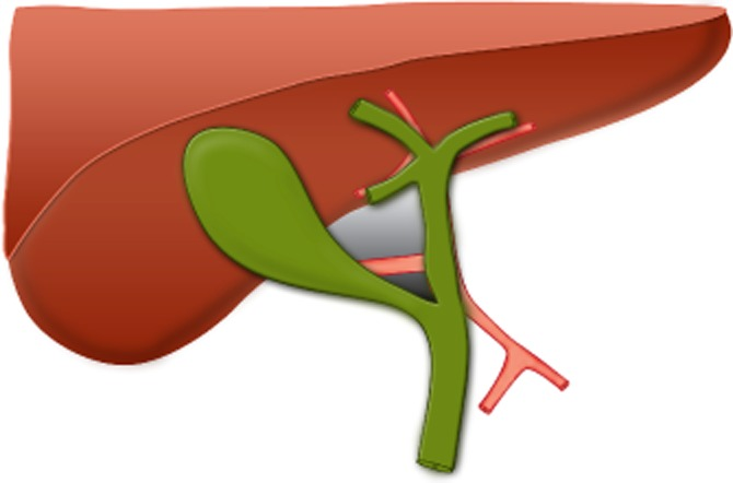
Hepatobiliary triangle
- Three borders
- Common hepatic duct
- Cystic duct
- Inferior border of the liver
- Contents include the cystic artery and branches thereof and the cystic node
- This space is covered by anterior and posterior layers of peritoneum
Calot’s triangle
- Three borders
- Cystic duct
- Cystic artery
- Common hepatic duct
Laparoscopic
Step 1
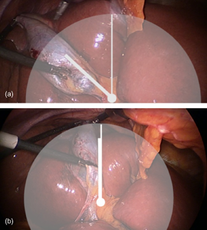
Confirm the gallbladder lies in the hepatic principal plane and is retracted cephalad to the 10 o’clock position
- This will expose the maximum surface area of the posterior peritoneum
- Avoid operating ‘front on’ to the gallbladder–cystic duct–common hepatic duct junction
- Important to place lateral port as lateral as possible
- Liver stiffness can be an issue - consider low calorie diet in overweight patients
Step 2
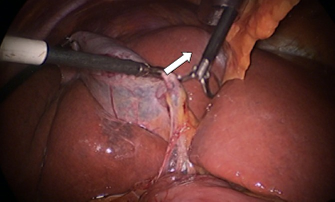
Confirm the lifting and positioning of Hartmann’s pouch
- Hartmann’s pouch should be grasped and lifted up and across toward the origin of the segment IV pedicle
- If this step is prevented by a large impacted stone, it may be possible to ‘milk’ the stone back into the gallbladder or alternatively to open the gallbladder and remove the stone.
- This maximizes the exposure of the posterior peritoneum of the hepatobiliary triangle.
Step 3
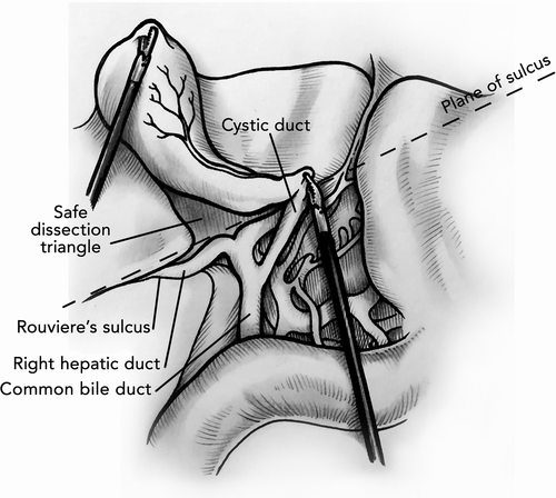
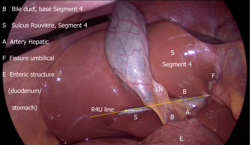
Identify Rouvière’s sulcus
- Marks the level of the right posterior portal pedicle, is such a landmark and is identifiable in at least 80% of patients
- When viewed laparoscopically from the umbilicus with normal upward retraction of the gallbladder, an imaginary line drawn along the sulcus and carried across to the base of segment IV shows the level ventral to which dissection is ‘safe’ and dorsal to which it is not
- R4U line - Rouvieres, base of segment 4, Umbilical fissure
Step 4
Confirm the release of the posterior leaf of the peritoneum covering the hepatobiliary triangle
- Allows the hepatobiliary triangle to increase in size as the gallbladder is lifted away
Step 5
Confirm the critical view and consider intraoperative cholangiography
- Anterior leaf of the peritoneum covering the surface of the hepatobiliary triangle can now be dissected from an anterior aspect
Intraoperative cholangiogram
- Five features
- Duodenal flow
- Proximal filling of the common bile duct
- Three proximal hepatic ducts (right anterior, posterior sectoral and left main ducts)
- Absence of filling defects within the common bile duct
- The presence of spiral valves within the cystic duct
- Head down for poor liver filling
- 1 mg glucagon or 10-20mg Buscupan for poor duodenal filling or to help pass a stone
Outline the pros and cons of performing this investigation on a routine basis.
- Pros
- Detects previously unexpected CBD stones
- Helps identification of abnormal duct anatomy
- Has been shown to reduce CBD injury rate
- Without = 0.4-0.6%
- With = 0.2-0.4%
- Has been shown to improve identification of CBD injuries and picked up early
- Improves operative Skill
- Improves interpretation
- Cons
- Cannot be performed in up to 10%
- Doesn’t eliminate CBD injury
- Just part of process and no replacement for meticulous dissection
- Small unexpected stones are likely clinically insignificant anyway Increased operating time and cost?
- False positives and negatives
Bail out options
- When anatomy unclear:
- Subtotal is safe technique
- If area is ‘frozen’ with dense adhesions, cholecystostomy is option
Open Cholecystectomy
- Positioning
- Patient supine
- Stand on right
- Incision
- Transverse subcostal incision through fat down to fascia, ~6-10cm
- Incise fascia 3-4cm below costal margin to allow for closure and avoid chronic pain
- Pass swab beneath rectus to assist division of rectus, ligating vessels along the way i.e. superior epigastric vessels
- Grasp posterior layer of fascia with two clips and incise between before extending length of wound
- Operative details
- Inspect abdominal contents
- Swab above right liver to bring gallbladder and hilum into operative field
- Divide omental/visceral adhesions retracting colon and small bowel caudally
- Use Kelly retractor as required
- Rampleys to fundus retracted towards right shoulder
- Two broad dissection techniques:
- Fundus first
- Calot’s dissection first (this is what i do)
- Transfix cystic duct and artery
- Remove from cystic plate
- Check bile leaks from cystic plate
- Mass closure
Intra-operative issues
•Obese patient o Consider optifast in elective o Setup Appropriate table Body strap Foot plate Arms strapped o Close liaison with anaesthetist about pressures o Consider transumbilical entry or Palmers point visiport o Extra port for liver retraction or caudal omental/LB/SB retraction with fan retractor o Fundus grasper lower than normal • Unable to dissect Calot’s triangle / obtain critical view of safety o Options are to convert to open operation to continue dissection or to perform a subtotal cholecystectomy. Subtotal cholecystectomy involves opening the gallbladder, removing the stones and removing the majority of the gallbladder at the level of Hartmann’s pouch. Ideally try to control the cystic duct with a purse-string suture or place a drain into the GB fossa. If posterior wall of gallbladder left behind ablate mucosa with diathermy o If bile leak post subtotal wait 5 days, if ongoing perform ERCP and stent • Bleeding o Apply pressure with swab or gallbladder and wait. Do not blindly clip o Inform anaesthetist o If significant consider conversion to open procedure and enlisting help from colleague. o If injury to hepatic artery or portal vein obtain proximal and distal control and close with 5-0 prolene o If bleeding from middle hepatic vein (which can lie superficially in GB fossa) inform anaesthetist as risk of air embolism and apply pressure. Figure of 8 suture either laparoscopically or open o If ooze from GB fossa consider surgicel or haemostatic agents • Suspected Bile duct injury o If suspected intraoperatively pause and obtain advice from HPB colleague, do not attempt primary repair yourself o Obtain cholangiogram to clarify anatomy/injury o Typically repair by HPB surgeon, if none available, place large drains and transport patient. Do not continue cholecystectomy
Complication
- Bile duct injuries
- Benign biliary strictures
- 5% get loose bowel habits or urgency
- Adaption over 3-6 months
- If continues then Loperamide
- Spilled stone 7%
- Uncommon to have complications
- post cholecystectomy syndrome