Section: Hepatobiliary Sub-section: Biliary Curriculum: Curriculum, page 88
Definition
Epidemiology
Risk factors
5 Fs
-
Female
-
Fair (caucasian)
-
Fat (high cholesterol excreters, rapid weight loss, obesity)
-
Fertile (post pregnancy and gallbladder stasis)
-
Forties
-
Genetic (25% of gallstones) / demographic
- Strong family history.
- 3x more frequent in 1st degree relative than spouse or unrelated control.
- Caucasian. Intermediate in Asian. Least common in African
- Female to male 10:1.
- Oestrogen increases cholesterol secretion, diminishes bile salt secretion.
- Pregnancy
- Strong family history.
-
Environmental (cholecysterol stones)
- Rapid weight loss in obese
- Obesity (BMI >45 have 7x risk)
- TPN
- Medications promoting cholesterol secretion in bile e.g. fibrates
- Dyslipidaemia
- Sedentary lifestyle
- T2DM
-
Disruption of enterohepatic circulation
- Excessive bile salt excretion
- Higher concentration of cholecterol
- Cholesterol stones
- Higher conc and decreased solubilisation of cholesterol and precipitation as crystals
- Crohn’s disease or intestinal resection
- Excessive bile salt excretion
-
increased enterohepatic circulation (pigmented stones)
- Chronic haemolytic anaemias (e.g. hereditary spherocytosis, sickle cell disease, B thalassaemia)
- Ineffective erythropoiesis (e.g. pernicious anaemia) and cirrhosis
- Cystic fibrosis
Aetiology
- 3 types
- Cholesterol
- Pigmented 10%
- Mixed
- Brown
- Bile is composed of bile salts, cholesterol, phospholipids (Lecithin) and various minerals; if any in excess, solution becomes supersaturated and cholesterol crystals form.
- Loss of gallbladder motility and excessive sphincteric contraction associated with gallstone formation
- Hypomotility leads to bile stasis and decreased reservoir function
Cholesterol
- Cholesterol supersaturation can be caused by excessive hepatic secretion of cholesterol or decreased hepatic secretion of bile salts or phospholipids with relatively normal cholesterol secretion.
- Mucus is nidus for stone formation.
- Decrease bile salt concentration (and therefore incrase cholecysterol)
- Interrupted bile circulation
Black pigmented
- Excess of unconjugated bile due to increased enterohepatic circulation of bilirubin (excessive haemolysis)
- Precipitation of calcium bilirubinate
- Commonly seen in chronic haemolytic anaemias (e.g. hereditary spherocytosis, sickle cell disease, B thalassaemia), ineffective erythropoiesis (e.g. pernicious anaemia) and cirrhosis
Brown Pigment stones
- Brown pigment stones – usually other areas of biliary tract, mostly SE Asia, associated with parasite and E.coli
- Thought to secondary to infections
- Bacterial glucuronidase deconjugates bilirubin to insoluble form
- Calcium binds to the unconjugated bilirubin → Calcium Bilirubinate
- Cholesterol incorporated in these (= brown colour)
- Stones tend to be found in the bile ducts
The formation of stones falls into three phases; supersaturation, nucleation, and stone growth; the imbalanced mixture generates a precipitant which forms a nidus for further stone formation and crystallisation, once formed the stones persist and enlarge or consolidate over time.
Gallstone complications
-
Biliary pain
- Viscerally innervated pain poorly localised, referred T9
- Further episode 38–50% per annum
- 1-2% per risk of complications in symptomatic gallstones
-
Cholecystitis
- When pain persists beyond a few hours
- Prolonged cystic duct obstruction causes release of prostaglandins within gallbladder mucosa resulting in fluid secretion producing cycle of increased distension and further mucosal damage and inflammation
- Complication
- Emphysematous cholecystitis
- Secondary infection in the gallbladder wall occurs with gas-forming bacteria, such as Clostridium welchii , E. coli or anaerobic streptococci. Imaging may reveal the presence of gas within the gallbladder wall.
- It is most commonly seen in elderly men with diabetes.
- Emphysematous cholecystitis
- Cholecystoenteric fistula
- Gallstone ileus
- gallbladder may become adherent to an adjacent loop of bowel (duodenum, jejunum or colon) and eventually rupture into it, discharging its contents
- Bouveret’s syndrome
- Gastric outlet obstruction due to impaction of a gallstone in the duodenum
- Cholecysto-duodenal fistula.
- Gallstone ileus
- Perforation into the peritoneal caivty
- Pericholecystic abscess
- Gallbladder Mucocele
- Obstruction at Hartmanns pouch causes a tense distended gallbaldder
- Gallbladder empyema
-
Obstructed and fluid becomes infected
-
-
Mirizzi syndrome
- Stone impacted in cystic duct
- Stone compressing the common hepatic duct
-
Choledocholithiasis
- Stone impacted in the bile duct
- Causing pain and jaundice
- Risk of cholangitis
- Can cause gallstone pancreatitis
- Stone impacted in the bile duct
-
- Chronic inflammation
Cholecystitis
TG18 Cholecystitis Severity Grading
- Mild
- Mild inflammatory changes in the Gallbladder
- Moderate
- Elevated WBC >18
- Palpable tender mass
- Duration >72hours
- Marked local inflammation (gangrenous cholecystitis, pericholecystic abscess, hepatic abscess, biliary peritonitis, emphysematous cholecystitis)
- Severe
- Organ dysfunction
Choledocholithiasis
Presentation
- Many are asymptomatic
- Obstructed jaundice (conjugated Bilirubin)
- Raised ALP/GGT
- Pale stool, dark urine
- Can cause gallstone pancreatitis
Cholangitis
- Charcot’s triad
- Jaundice
- Fevers
- RUQ pain
- Secondary infection by enteric bacteria
- Early IV abx + biliary decompression
Criteria for acute cholangitis (TG18)
- Systemic inflamation
- Fever
- Labaratory tests
- Cholestasis
- Icterus
- Raised LFTs
- Imaging
- Bile duct >7mm
- Evidence of stones or obstruction
Bile duct exploration
- Need a dilated duct, ideally > 1 cm to ensure closure without a stricture.
- Open
- Longitudinal incision mid part of anterior wall
- Stones removed with Desjardin forceps
- Fushing and use of flexible choledochoscope
- Laparoscopic
- Transcystic – limited to stones < 5 mm
- 3-mm choledochoscope
- Caught in a basket under fluoroscopy
- Transductal – larger stones.
- Longitudinal incision on anterior surface.
- Can manipulate duct,
- Basket and balloon extraction with 5 mm flexible choledochoscope.
- Can be closed longitudinally (absorbable suture)
- Transcystic – limited to stones < 5 mm
CBD stones in post-operative patients
- No access to the pylorus
- Roux-en-Y gastric bypass
- Billroth II gastrectomy
- Roux-en-Y HJ
- The afferent limb of the bypass is relatively short, then ERCP may still be possible
- If the pylorus is still patent (as in gastric bypass surgery for obesity), laparoscopic-assisted ERCP
- inserting a port directly into the stomach at laparoscopy
- Via a mini-laparotomy locating the stomach and inserting a purse string suture.
- Percutaneous approach
- If the bile ducts are dilated and in patients with a previous HJ
- Radiological basket
- SpyGlass or choledochoscope over the previously inserted percutaneous wire
Mirizzi syndrome
-
Mirizzi syndrome – stone impacted in Hartmann’s causes adherence to CBD, loss of Calot’s triangle.
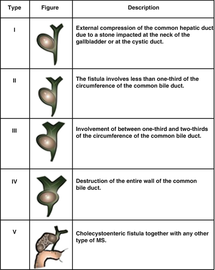
-
Type 1 - External compression
-
Type II - <1/3 bile duct
-
Type III – 1/3 – 2/3 bile duct
-
Type IV >2/3 bile duct
-
Type I
- Subtotal cholecystectomy
- Leave posterior wall where it is adherent to bile duct
- Ligate cystic duct if identified and still patent
- If not found, drain left in gallbladder fossa.
-
Type II-IV
- Presence of fistula due to erosion of impacted gallstone in Hartmann’s pouch.
- CBD reconstruction with Roux-en-Y hepaticojejunostomy
- If you find this intra-op and no HPB around → cholangiogram to define anatomy, leave drains, refer to HPB
Intrahepatic stone disease
- Usually Asian
- 10% risk of cholangiocarcinoma
- Brown stones
- Soft and friable.
- Result from anaerobic-bacteria-secreting enzymes that hydrolyse ester and amide linkages in biliary lipids as insoluble anions or calcium salts.
- Presentation are often related to CBD stones
- Therefore CBD decompresion with stent.
- Definitive management
- Clear the biliary tract of stones, provide adequate biliary drainage and, where necessary, provide adequate access to the biliary duct
- Percutaneous, endoscopic and surgical options
- Biliary strictures, which are found in 35% to 96% of patients with hepatolithiasis, are a major factor in the recurrence
- When extrahepatic or hilar duct strictures
- Hepaticojejunostomy
- When extrahepatic or hilar duct strictures
- Clear the biliary tract of stones, provide adequate biliary drainage and, where necessary, provide adequate access to the biliary duct
Management
- Symptomatic - surgery
- Asymptomatic - no surgery
Cholecystectomy
- Open via Kocher’s incision
- IOC
- Routine
- Increase the rate of intra-operative recognition of BDI
- Conflicting evidence suggest it may decrease rate of BDI
- Increased cost
- Will find stones that are not clinical apparent
- Selective
- Decreased cost
- Decrease time of operation
- Won’t find clinical insignificant stones
- My exam answer
- This is a controversial topic without clear evidence either way. I perform a routine IOC. Although there is conflicting evidence there is some suggestion that IOC may decrease the rate of BDI and increase the rate of immediate recognition of a BDI. Immediate recognition is important as this improves patients outcomes. The negative of this is that it increase the operative time, the operative cost and also you may identify clinical occult gallstones which will then need to be managed or followed up.
- Routine
Prophylactic cholecystectomy:
- Consider in sickle cell anaemia or hereditary spherocytosis combined with splenectomy.
- Organ transplant recipient or neuroendocrine tumours on somatostatin analogues
- Controversial but can consider in: bariatric surgery, concurrent polyps
Acalculous cholecystitis
- Life threatening condition that occurs in critically ill patients.
- Due to poor perfusion.
- Cystic artery is an end-organ artery with no collaterals.
- Thought to be due to microvascular occlusion of end arteries within wall resulting in ischaemia.
- Risk factors:
- Severe trauma
- Burns
- Major surgery
- Prolonged fasting
- TPN, sepsis
- Diabetes
- Atherosclerotic disease
- Systemic vasculitis
- Acute renal failure
- AIDS.
- Diagnosis:
- Often hindered by obtundation, presence of pre-existing diseases or recent surgery.
- USS confirms diagnosis.
- Management:
- Percutaneous cholecystostomy
- Reduces tension on the wall.
- Preferred alternative to cholecystectomy in the treatment of critically unwell patients.
- Early cholecystectomy
- May be appropriate depending on patient condition and if failure to improve with cholecystostomy
- Risk of gangrene and perforation of gallbladder.
- Percutaneous cholecystostomy
Chronic acalculous cholecystitis
- Poorly understood.
- Biliary pain without cholelithiasis.
- Occasionally biliary sludge may be responsible or tiny stone in spiral valve of cystic duct.
- Investigations: HIDA or DISIDA (cholescintigraphy) have been used to improve outcomes.
- CCK or fatty meal following uptake of isotope given to stimulate emptying of gallbladder and calculate EF.
- Low EF (< 40%) may get long term symptom relief in 65-80%
- Treatment with cholecystectomy may be justifiable, however informed consent that 50% best likelihood of pain resolution.
Primary infections of gallbladder
- Infective cholecystitis is rare.
- Seen in immunocompromised patients.
- Causative organisms – Salmonella typhi, Campylobacter jejuni and Vibrio cholera.
- Presentation: similar to acalculous cholecystitis but often history of gastroenteritis like illness.
- AIDS: susceptible with low CD4 count.
- Opportunistic infection: CMV, cryptosporidium, Candida and Mycobacteria tuberculosis.
- 30-day mortality in AIDS with acute cholecystitis is 20%
- Treatment: IV antibiotics followed by laparoscopic cholecystectomy
Adenomyomatosis of the gallbladder
- Adenomyomatosis of the gallbladder is a hyperplastic cholecystosis of the gallbladder wall.
- It is a relatively common and benign cause of diffuse or focal gallbladder wall thickening, most easily seen on ultrasound and MRI.
- Hyperplasia of the wall with the formation of Rokitansky-Aschoff sinuses (intramural diverticula lined by mucosal epithelium) penetrating into the muscular wall of the gallbladder, with or without gallbladder wall thickening.
Management
- Patient symptomatic with right upper quadrant pain (often due to gallstones)
- Appearances (especially when focal) may be difficult to distinguish from malignancy
- Simple adenomyomatosis is not a premalignant condition.
- In absence of symptoms, no treatment.