Section: Hepatobiliary Sub-section: Biliary Curriculum: Curriculum, page 88
Definition
Malignant epithelial tumours arising from the biliary tree
- Intrahepatic - 10%
- Extrahepatic
- Hilar - 60%
- Mid-duct and distal duct - 20-30%
Mid-ductal - upper border of the duodenum and the cystic duct Distal bile duct - duodenum to the ampulla of Vater.
Epidemiology
- Incidence 0.3 – 3 per 100,000.
- Equal M & F.
- Age 55 – 75.
Risk factors
- Majority are sporadic
- Chronic biliary inflammation (5%):
- Primary sclerosing cholangitis
- Chronic choledocholithiasis
- Hepatolithiasis
- Parasitic biliary infestation
- Caroli’s disease
- Choledochal cyst
- Enteric reflux due to anastomosis
- NAFLD
- Hep B/C
- T2DM & metabolic syndrome
Rare association with UC, and PSC is the greatest risk factor for its development.
- 12–15% of patients transplanted for PSC have cholangiocarcinoma
Clinical presentation
- Symptoms at advanced stage:
- Abdominal pain
- Malaise
- Night sweats
- Asthenia
- Nausea
- Weight loss
- Jaundice
- Non-specific LFTs
- Serum markers CEA and Ca 19-9 lack sensitivity and specificity
Pathology
- Precursor:
- Flat or micropapillary growth of atypical biliary epithelium (biliary dysplasia or biliary intraepithelial neoplasia)
- Intraductal papillary neoplasm
- Mucinous cystic neoplasm
- Intraductal tubular papillary neoplasm
- Majority are adenocarcinoma 95% with 5% other variants
- Adenocarcinomas are divided into three subtypes
- Sclerosing
- Nodular
- Papillary
Intrahepatic cholangiocarcinoma
Arises from peripheral intrahepatic biliary radicles (different from hilar and CBD cholangio)
3 morphological subtypes :
- Mass forming (commonest)
- Periductal-infiltrating which spreads along bile ducts
- Intraductal-growth type with intraluminal spread
- Can have mixed components
Staging
TNM
| T1a: < 5 cm without vascular invasion |
|---|
| T1b: > 5 cm without vascular invasion |
| T2: solitary tumour with vascular invasion or multiple tumours |
| T3: perforating visceral peritoneum |
| T4: involving local extrahepatic structures by direct invasion |
| N0 or 1 |
| M0 or 1 |
Investigations
Imaging
-
CT
- Arterial phase: variable rim-like enhancement
- Delay: gradual centripetal enhancement
-
MRI
- Hypointense on T1,
- hyperintense on T2.
- Large, non-encapsulated, heterogeneous, associated with narrowing of adjacent portal veins and retraction of liver capsule. May have satellite nodules.
- MRCP useful - biliary dilation
-
FDG-PET
- Distant mets
-
Diagnostic laparoscopy - consider
Diagnosis
- Main differential is colorectal metastases.
- Biopsy shows adenocarcinoma of biliary phenotype (CK7+ CK20- ) in contrast to colorectal (CK7- CK20+)
Management
Resectable disease
- Surgical resection only curative treatment.
- PVE can be performed with FLR inadequate
- Portal lymphadenectomy indicated given prognostic information gained
- 5y survival 25-40% in R0
- Major hepatectomy in 75-80%.
- Postop mortality 6%.
- R1 or R2 resection 25%.
Prognostic factors:
- Lymph node invasion
- R1 resection
- Infiltrating worse than mass-forming due to spread along Glisson’s capsule and high incidence of LN involvement. Intraductal growth are better.
Adjuvant treatment
Unresectable disease
- Gemcitabine and Cisplatin
- TACE, TACE-DEB, SIRT with yttrium-90 microspheres, radiation therapy
- Hepatic arterial infusion (HAI) chemotherapy with floxuridine
- Possible to down stage
- Liver transplant not typically done
Perihilar
Investigations
- CEA and Ca 19-9 may be elevated (but nonspecific)
Imaging
- USS
- Doppler US – good for delineating level of tumour, extension within bile duct and venous invasion.
- CT
- Arterial phase: variable rim-like enhancement
- Delay: gradual centripetal enhancement
- MRI
- Hypointense on T1,
- hyperintense on T2.
- Large, non-encapsulated, heterogeneous, associated with narrowing of adjacent portal veins and retraction of liver capsule. May have satellite nodules.
- MRCP useful
- FDG-PET
- Useful for identifying nodal or distant mets. Can differentiate between benign disease and cholangiocarcinoma
Invasive modalities
- Percutaneous transhepatic cholangiography (PTC)
- Can also get brushings + stent placement
- useful for obstructive perihilar lesions
- ERCP
- Brushings
- Skyglass
- Targeted biopsies
- Staging laparoscopy should be performed
Stage
Bismuth-Corlette classification:
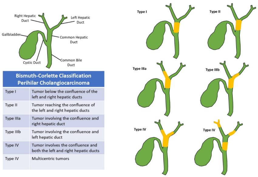
Blumgart/Memorial Sloan-Kettering Cancer Center (MSKCC) (determines resectability for hilar cholangiocarcinoma)
| T1 | Confluence +/- unilateral extension to 2nd order biliary radicles |
|---|---|
| T2 | Confluence +/- unilateral extension to 2nd order biliary radicles |
| AND ipsilateral portal vein involvement +/- ipsilateral hepatic lobar atrophy | |
| T3 | Confluence +/- unilateral extension to 2nd order biliary radicles AND |
| contralateral portal vein involvement | |
| OR contralateral hepatic lobar atrophy | |
| OR main or bilateral portal venous involvement | |
| Only T1 and T2 can be considered for resection - but the outcomes from resecting a patient with T2 disease is poor (high chance of finding metastatic disease at exploration and poor median survival) |
The median survival is 20, 13, and 8 months for T1, T2, T3 respectively.
TNM stage (AJCC 8th adition)
- T1: tumour confined to the bile duct, with extension up to the muscle layer or fibrous tissue
- T2:
- T2a: tumour invades beyond the wall of the bile duct to surrounding adipose tissue
- T2b: tumour invades adjacent hepatic parenchyma
- T3: tumour invades unilateral branches of the portal vein or hepatic artery
- T4:
- Tumour invades the main portal vein or its branches bilaterally, or
- unilateral second-order biliary radicals with contralateral portal vein or hepatic artery involvement
- N1 - 1-3
- N2 - 4 or more
- M1 - mets
Irresectability
- Patient factors
- Anatomical
- Local tumor factors
- Bilateral spread to secondary biliary radical
- Encasement or occlusion of the main portal vein proximal to its bifurcation
- Atrophy of one lobe with - contralateral portal vein branch encasement or occlusion - contralateral tumour extension to secondary biliary radicles
- Unilateral tumour extension to secondary biliary radicles with contralateral portal vein branch encasement or occlusion
- Local tumor factors
- Biology
- Metastatic disease
Management
Preoperative considerations:
- Preop tissue dx
- Not mandatory if no prior biliary tract operation, PSC, hepatolithiasis
- Preop biliary stenting:
- Controversial.
- Pros
- Possibly improved periop outcomes in patients with predicted FLR < 30%
- May improve hepatic function and patient nutrition
- Cons
- Contamination by enteric bacteria
- Tumour seeding
- Cholangitis
- Bile leak
- Pros
- Controversial.
- Neoadjuvant therapy
- Limited data on efficacy
- May be a role for chemoradiotherapy, particular for locally advanced, to help achieve R0 and improve survival.
- PVE
- For patients with anticipated FLR < 30%
Operative considerations:
- Negative margin
- R0 most predictive for survival
- Usually Bismuth IIIb need left hepatectomy and I, II, IIIa require extended right
- Frozen section proximal and distal margins
- Portal lymphadenectomy
- Survival benefit not proven. Diagnostic information gained may guide decisions about adjuvant therapy
- LN status is independent predictor for long-term survival
- Median number of nodes is 3, despite target of 7
- Caudate lobectomy
- Standard for hilar cholangiocarcinoma (particularly on the left)
- Vascular resection
- Portal vein resection and even IVC resection are not contraindications
- Transplantation
- Only select cases, but not done in most centres
Postoperative oncological outcomes
- 5-year survival
- 20-40% for intrahepatic;
- 30-40% for perihilar cholangio;
- 50% for distal/extrahepatic.
Palliative therapy
- Intrahepatic biliary-enteric bypass:
- Most often to segment III duct – usually sufficient to relieve jaundice.
- Systemic therapy:
- Gemcitabine + cisplatin standard of care.
- Median overall survival of 18 months.
- Targeted therapy: no difference in survival
- Regional chemotherapy/hepatic artery infusion: may prolong survival
- Yttrium Y-90 radioembolisation: show survival benefit
Distal-duct
Staging
T1: tumour invades the bile duct wall with depth <5 mm T2: tumour invades the bile duct wall with depth 5–12 mm T3: tumour invades the bile duct wall with depth >12 mm T4: tumour involves the coeliac axis, superior mesenteric artery, and/or common hepatic artery N1 - 1-3 N2 - 4 or more M1 - Mets
Managment
Operative
- Pancreatoduodenectomy
- Compared with pancreatic cancer
- Often amenable to resection
- Less often have microscopic disease at the resection margin,
- Less frequently have spread of tumor to adjacent lymph nodes
- Compared with pancreatic cancer
Adjuvant treatment
- Adjuvant capecitabine survival benefit compared to observation.
- Radiotherapy after R1 resection survival benefit (compared to no radiotherapy)
BILCAP Trial — Comprehensive Summary
- Full name: Capecitabine Compared with Observation in Resected Biliary Tract Cancer
- Published:** Lancet Oncology, 2019 (Primrose et al.) | Long-term update: JCO, 2022
- Only ~20% of biliary tract cancer (BTC) patients are eligible for curative-intent resection; 5-year OS for all BTC patients is <10%
- Prior to BILCAP, no RCT had demonstrated a survival benefit for adjuvant therapy in resected BTC
- BILCAP was the first phase III RCT with positive results for adjuvant chemotherapy in BTC
- The trial established adjuvant capecitabine 1250 mg/m² bd, days 1–14, 21-day cycle × 8 cycles as the standard of care
- Despite a technically negative ITT primary endpoint, the totality of evidence strongly supports benefit — a clinically meaningful 14.7-month OS improvement in the ITT analysis
- ASCO, ESMO, NCCN, and eviQ (Australia/NZ) all recommend adjuvant capecitabine for 6 months post-resection