Section: Colorectal Sub-section: Colitis Curriculum: Curriculum, page 20
Definition
Ulcerative colitis (UC) is an idiopathic relapsing inflammatory bowel disease (IBD) involving the mucosa and lamina propria of the rectum and variable segments of the proximal colon.
- Remissions and exacerbations
- Medical management is generally effective in controlling ulcerative colitis, but ultimately 30–40% of patients will require surgical intervention.
- Removal of the colon and rectum is essentially curative
Epidemiology
- Uncommon disease with varying incidence rates (0.5–24.5/100,000)
- Western countries
- Jewish and caucasians - highest incidence
- Age distribution is biphasic - 20-40 and 50-70
- Male incidence same as female
- Urban populations, indoor living, smaller families, and individuals of middle and upper socioeconomic status, who primarily reside in more sanitary surroundings
Pathophysiology
The pathogenesis remains unclear
Environmental factors
- decreased risk with smoking and alcohol
- Increased risk in urban populations, indoor living, smaller families, and individuals of middle and upper socioeconomic status, who primarily reside in more sanitary surroundings Immune system
- Hygiene hypothesis - cleaner living environments reduce the amount of organ- isms one is exposed to early in life, thus reducing the ability of the immune system to become toler- ant, and subsequently causing an aberrant response when thus exposed
- Antibiotic usage - resulting gut flora dysbiosis Family history
- UC has been reported to be as high as 29% in those with a positive family history, and between 10% and 20% of affected individuals have a first-degree relative with IBD
- 50% identical twins
Histopathology
- Inflammation in UC is confined to the rectum and colon.
- The mucosal columnar glandular epithelium extends into the anal canal to the level of the anal transitional zone.
- Segmental or skip areas do not occur, rather the inflammation in the colon and rectum is diffuse without intervening normal mucosa.
- The rectum is always involved, although the appearance of relative rectal sparing can occur in patients receiving transanally applied anti-inflammatory agents.
- A spared rectum not associated with local treatment should raise the suspicion of Crohn’s disease.
- Backwash ileitis occurs only in cases with colonic extension to the ileocaecal junction. Mild
- mucosal inflammation, goblet cell depletion, crypt of Lieberkuhn distortion, and vascular congestion.
- Mucin within goblet cells is expectorated, making them appear less evident or absent (goblet cell depletion).
Moderate
- As severity progresses the lamina propria will exhibit infiltration by neutrophils, plasma cells, lymphocytes, eosinophils and mast cells.
- Neutrophils present within the epithelium of crypts (cryptitis) can aggregate in the crypt lumen, forming abscesses.
- Mucosal destruction, ulceration and subsequent atrophy are partly the result of rupturing of crypt abscesses.
Severe
- Crypt destruction and loss occur as a result of damage to the crypt basal epithelium.
- Deeper submucosal or transmural inflammation with ulceration can also be observed, leaving large areas of exposed muscularis propria covered with granulation tissue giving the appearance of pseudo- polyps.
- In the more chronic and quiescent phase a distorted architectural pattern with crypt distortion, branching and foreshortening can be identified.
Summary
- Mucosal inflammation, ulcers, atrophy
- Goblet cell depletion (doesn’t occur in Crohn’s)
- Distorted and atrophic glandular architecture
- Vascular congestion.
- Crypt abscesses
- Neutrophilic and lymphocytic infiltration of the lamina propria.
- Pseudopolyps – granulation tissue over exposed muscularis propria.
Diagnosis
- pANCA - pANCA is present in 50% to 65% of patients with UC and 10% to 25% of patients with CD
- WBC, CRP, ESR
- FBC - anaemia if on going bleeding
- LFT - screening for PCS
- Faecal calprotectin - found in neutrophils and therfoer shows neutrophils have migrated into the lumen of the bowel
- Stool spec + c.dif + parasites
- Endoscopy
Differential diagnosis for Colitis
- Infectious - viral, bacterial, protozoal
- Inflammatory bowel disease - Crohn’s disease, Indeterminate colitis
- ischaemic colitis
- radiation colitis
- diversion colitis
- pharmacotherapy- induced colitis
- Microscopic colitis - collagenous colitis, lymphocytic colitis
Stool specimen should be taken to exclude infectious causes
Endoscopy should be performed for direct mucosal visualisation as well as providing an avenue for obtaining tissue biopsies
- Quiescent phase the mucosa will appear relatively normal
- with the exception of neovascular changes.
- Mild inflammation
- Oedema, erythema and an abnormal mucosal vascular pattern
- Oedema can also cause fine granularity in which there is a delicate regular stippled
- Moderate
- Superficial erosions, ulcerations and contact bleeding secondary to scope trauma are observed.
- Severe
- Development of pseudopolyps
- Chronic
- ‘featureless microcolon’ with mucosal atrophy, muscular hypertrophy, a decreased luminal diameter and loss of haustral folds
- The acronym to remember the endoscopic findings is FUCE(K)ER
- F - friability
- Ulceration (superfical)
- Contact/spontaneous bleeding
- Erosions
- Erythema
- Reduced vascular pattern
Classification
Ulcerative Colitis Endoscopic Index of Severity” (UCEIS)
| Category (the most serious change is evaluated) | Definition (Likert Scale) |
|---|---|
| Vessels | 1 = normal 2 = partial loss 3 = total loss |
| Bleeding | 1 = none 2 = mucosal 3 = luminal bleeding (mild) 4 = luminal bleeding (severe) |
| Erosions/ulcerations | 1 = none 2 = erosion 3 = superficial ulcerations 4 = deep ulcerations |
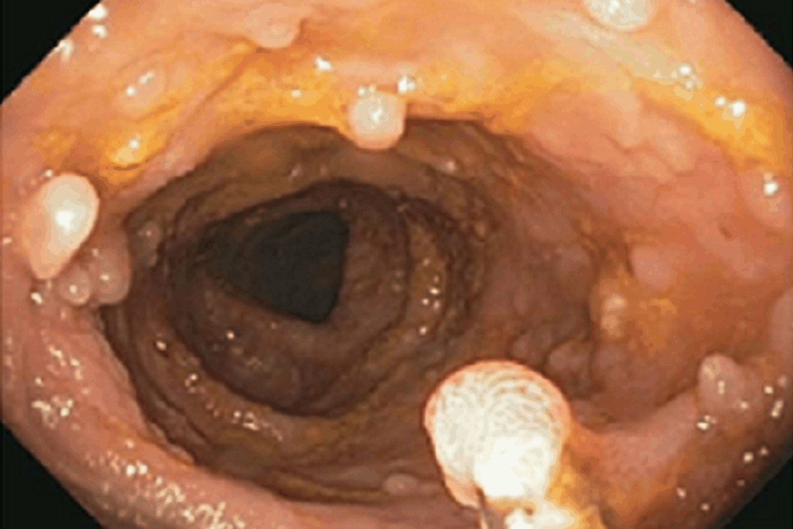
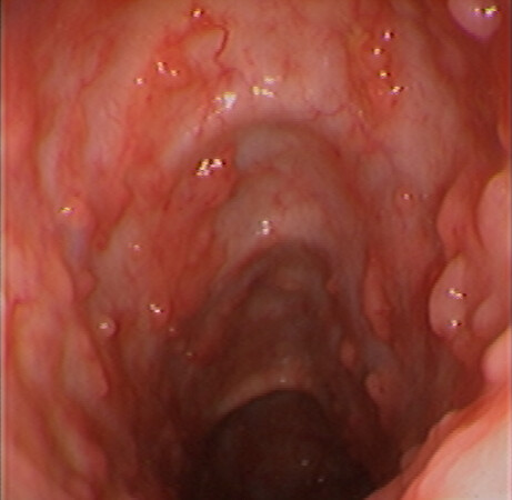
Examples are shown in Figures 3a–e.
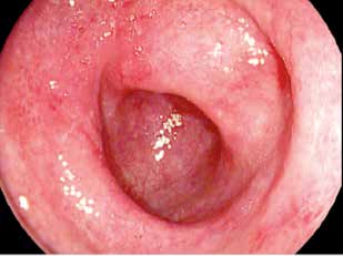
partial loss of vessels
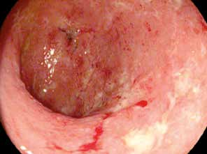
luminal bleeding, mild
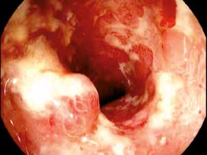
luminal bleeding, severe
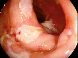
superficial ulcerations
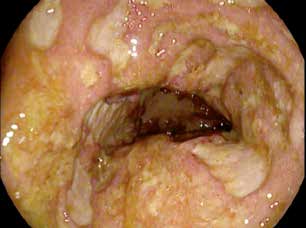
deep ulcerations
pseudopolyps
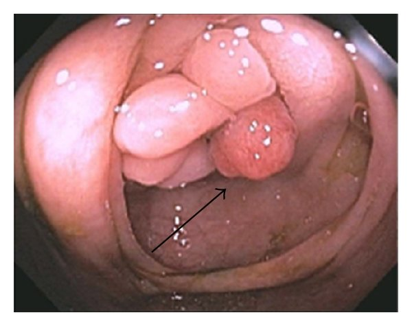 An adenoma-like DALM (black arrow)
Mayo score
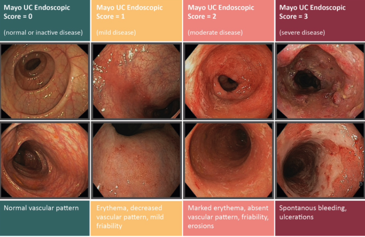
Truelove and Witts
Disease severity is classified as mild, moderate or severe, and is based on the original descriptions provided by Truelove and Witts.
| Mild | Moderate | Severe | |
| Bowel movements (no. per day) | Fewer than 4 | 4-6 | 6 or more plus at least one of the features of systemic upset (marked with * below) |
| Blood in stools | No more than small amounts of blood | Between mild and severe | Visible blood |
| Pyrexia (temperature greater than 37.8°C) | no | no | yes |
| Pulse rate greater than 90 bpm | no | no | yes |
| Anaemia (< 10g/100mL) | no | no | yes |
| Erythrocyte sedimentation rate (mm/hour) | 30 or below | 30 or below | above 30 |
Montreal Classification
The Montreal Classification for UC incudes Severity and Extent
| Extent | E1 | Ulcerative proctitis | Confined to the rectum |
| E2 | Left sided UC | Distal to splenic flexure | |
| E3 | Extensive UC | Proximal to splenic flexure | |
| Severity | S0 | Clinical remission | |
| S1 | Mild UC | Similar to Truelove Witts | |
| S2 | Moderate UC | ||
| S3 | Severe UC |
Clinical
- 50% present with proctosigmoiditis, 25% present with pancolitis and 25% with left-sided colitis
- Common symptoms associated with UC include urgency, diarrhoea, tenesmus and haematochezia
- Protein losing enteropathy may lead to loss of lean body mass and anaemia, and growth retardation in children.
- Haemorrhage
- Perforation - rare
- Fulminant colitis
Extraintestinal manifestations
Upwards of 20% of patients with UC will develop extra-alimentary manifestation
Musculoskeletal
Peripheral arthropathy
- Affecting up to 20% of patients
- involves numerous small and large joints (knees being the most common)
- Rheumatoid factor negative (seronegative)
- Typically fleeting
- Non-deforming
- Dependent on activity of colitis
Axial arthropathy (ankylosing spondylitis)
- 5% of patients
- Involving the sacroiliac joints and one or more vertebrae
- majority of cases are HLA-B27 positive
- Independent on activity of colitis
Asymptomatic sacroileitis
- limited to the sacroiliac joint
- Independent on activity of colitis
- Radiographically detected in 24%
Hepatopancreatobiliary
Primary sclerosing cholangitis
- Idiopathic chronic and progressive disorder manifesting as stricturing, inflammation, and fibrosis of intra- and extrahepatic bile ducts.
- 5% of patients with UC develop it
- 75% of patients with PSC are found to have concurrent UC
- Patients with coexisting PSC and UC are at a markedly increased risk for colonic neoplasia (five times)
- Independent on activity of colitis
- An increased risk of development has been demonstrated in patients with human leucocyte antigen (HLA) B8, DR2, DR3 or DR6 haplotype positivity.
- Treatment of PSC with steroids, colectomy or antibiotics is ineffectual.
- Patients undergoing restorative proctocolectomy have a higher subsequent in- cidence of pouchitis and dysplasia in the ileal pouch mucosa
- Ultimately, the disease progresses to liver cirrhosis and eventual failure, which may prompt consideration for liver transplantation
- Rare association with UC, and PSC is the greatest risk factor for its development.
- 12–15% of patients transplanted for PSC have cholangiocarcinoma
Dermatological
- 10–20% of patients with UC
- tender, inflamed, red nodules mainly on the anterior surfaces of the lower extremities
- Dependent on activity of colitis
- 1–3% of patients with UC
- plaques or pustules that break down and form painful ulcerations with undermined borders and necrotic centres
- Legs are the most commonly affected area, though it can oc- cur anywhere, including peristomally.
- Can be dependent or Independent on activity of colitis
Thromboembolic
- 3x higher rate of deep venous thrombosis and pulmonary embolism
- Though unproven, a hypercoagulable state in UC is hypothetically related to corticosteroid usage, activation of the coagulation cascade during a systemic inflammatory state, or up-regulation of acute phase reactants with flares
Ophthalmological
Ophthalmological manifestations can occur in up to 5% of patients
Episcleritis
- most common ophthalmopathy
- Presents with pain, burning and scleral injection.
- Dependent on activity of colitis
Uveitis
- presents with pain, blurred vision, photophobia and headaches.
- Classically, the redness is most prominent centrally and dissipates radially.
- Independent on activity of colitis
- Prompt treatment is necessary to decrease the risk of visual impairment.
Scleritis
- Scleritis presents similarly to episcleritis, though is more severe
- Necessitates aggressive treatment in order to minimise retinal detachment and optic nerve impairment.
Colorectal cancer
- Prolonged duration, continuously active disease, severity of inflammation, PSC and diffuse involvement (pancolitis) are cumulative risk factors for the development of colorectal cancer in the setting of UC.
- Cumulative probabilities of 2% by 10 years, 8% by 20 years and 18% by 30 years
- 10 years after the diagnosis of UC, the incidence of colorectal cancer increases by approximately 1% per year
- x8 increase risk
- Dysplasia-associated lesion or mass (DALM)
- Visibly raised dysplastic lesion within an area of inflammation
- confers a high risk of malignant potential
- Needs to be excised, if not able to be achieved endoscopically then colectomy
- Progression from colitis without dysplasia to colorectal cancer does not necessarily follow a sequence of low-grade dysplasia, high-grade dysplasia and ultimately carcinoma. Rather, LGD can progress directly to colorectal cancer.
- Low Grade
- LGD has been shown to be a strong predictor of progression to advanced neoplasia (53% at 5 years) in surveillance colonoscopy, and in patients who underwent a colectomy an unexpected advanced neoplasia (high-grade dysplasia or cancer) was found in nearly 24%
- Other studies have shown the presence of LGD is as likely as high-grade dysplasia (54% vs. 67%) to be associated with an already established cancer
- A large meta-analysis of 20 surveillance studies showed the risk of developing cancer in patients with LGD is high. When LGD is detected on surveillance there is a ninefold risk of developing cancer and 12-fold risk of developing any advanced lesion
- Management is controversial but options include close surveillance in unifocal disease.
- If multifocal – should proceed to a colectomy.
- High grade
- 45% incidence of CRC at time of colectomy. Thus if flat high grade dysplasia is seen – colectomy is mandatory.
- Colonoscopic surveillance should be every 1–2 years starting 8–10 years after a diagnosis of pancolitis, or 15 years after left- sided colitis.
- Recommendations are also advocated for random non-targeted biopsies performed every 10 cm in all four quadrants, equating to 20–40 biopsies per colon.
New Zealand screen guidelines
Low risk - every 5 years
- extensive but quiescent ulcerative colitis or
- extensive but quiescent Crohn’s colitis or
- left-sided ulcerative colitis (but not proctitis alone) or Crohn’s colitis or similar extent.
Intermediate risk - every 3 years
- extensive ulcerative or Crohn’s colitis with mild active inflammation that has been confirmed histologically or
- post-inflammatory polyps or
- family history of colorectal cancer in a first-degree relative aged 50 years or over.
High risk - every 1 year
- extensive ulcerative or Crohn’s colitis with moderate or severe active inflammation that has been confirmed histologically or
- primary sclerosing cholangitis (including after liver transplant) or
- colonic stricture in the past 5 years or
- any grade of dysplasia in the past 5 years or
- family history of colorectal cancer in a first-degree relative aged under 50 years.
Baseline colonoscopy should be offered 8-10 years after diagnosis
Management
Medical management
Options
- Aminosalicylates/5ASA
- Brand names
- Both Metabolised by bacteria to sulfapyridine and 5-ASA
- MOA: Decreases synthesis of prostaglandin and leukotriene, modulating the inflammatory response derived from the cyclooxygenase and lipooxygenase pathways.
- These preparations are more useful in UC than Crohns – can be given orally or rectally.
- First line in mild/moderate UC.
- Corticosteroids
- Inhibit the production of inflammatory mediators including phospholipase A2.
- Are used for induction of treatment in severe disease.
- Can also be used in acute flares.
- Budesonide – good for ileal Crohns disease.
- Complications include weight gain, osteoporosis, increased risk of infection, acne, hypertension, sleep and mood disturbance, cataracts.
- Immunomodulators
- Thiopurines
- 6-mercaptopurine
- Azathioprine
- Notes
- Take 2-3 months to start working thus not good for induction.
- Both purine analogues – inhibit cell proliferation and suppress the production of cytotoxic T cells and NK cells.
- Patients need to be tested for the enzyme thiopurine S-methyltransferase (TPMT) prior to starting these drugs. If they do not have TPMT, they will not break down the drugs and they can get bone marrow suppression.
- Main complications are bone marrow suppression, pancreatitis, hepatitis, fevers and rash.
- Methotrexate
- Not commonly used.
- Folic acid antagonist
- Inhibits T cell activation and down regulates B cells.
- Potential side effects include hepatotoxicity, ulcerative stomatitis, bone marrow suppression, fatigue, pneumonitis, and rarely pulmonary fibrosis and kidney failure
- Is teratogenic.
- Thiopurines
- Biologic agents
- MOA
- Monoclonal antibiotics which target mediators of the inflammatory response.
- Anti-tumor necrosis factor agents
- Infliximab (Remicade)
- Adalimumab (Humira)
- Golimumab (Simponi)
- Notes
- You can measure serum anti-TNFa drugs levels and anti-drug antibodies while on treatment
- Anti-integrin antibody
- Vedolizumab
- Notes
- prevents lymphocyte migration into intestinal tissue. Slower onset than other biologic treatments.
- Interleukin inhibitor
- Ustekinumab (Stelara)
- Notes
- inhibits inflammatory mediators IL12 and IL23
- Complications of biologic treatments.
- Infection – increased risk of opportunistic infections.
- Development of lymphomas
- Immunogenicity – can get paradoxical development of immune mediated disorders
- MOA
- Calcineurin inhibitor
- Tacrolimus
- Ciclosporin
- Notes
- Rarely used
- Can be used as rescue therapy in hospital
- Used in transplant
Medical treatment
Goal to induce and maintain clinical remission
Mild to moderate distal colitis
Clinical remission:
- Step 1
- Mesalazine suppositories 1g/day
- Maximum response in 4-6 weeks
- Mesalazine suppositories 1g/day
- Step 2
- Add topical Corticosteroids daily (Hydrocortisone suppository or enema)
- Step 3
- Oral Mesalazine 2g/day or Oral Sulfasalazine 2–6 g/day
- Step 4
- Oral Prednisone 40-60mg/day or Budesonide 9mg daily (preferred as colonic release)
Mild to moderate extensive colitis
Extensive colitis involves the colon beyond the reach of topical therapy and necessitates oral pharmacotherapy.
Clinical remission:
- Step 1
- Oral Mesalazine 2g/day - better tolerated
- or Oral Sulfasalazine 2–6 g/day
-
- rectal Mesalazine 1g/day
- Step 2
- Oral Prednisone 40-60mg/day or Budesonide 9mg daily
- Topical steriods
Maintenance
- Step 1
- Oral Mesalazine 2g/day - better tolerated
- or Oral Sulfasalazine 2–6 g/day
-
- rectal Mesalazine 1g/day
Moderate to severe colitis
Clinical remission:
Option 1:
- Anti-tumor necrosis factor agents
- +/- Immunomodulators
- Thiopurines
- 6-mercaptopurine
- Azathioprine
- clinical response ranges from a few days to up to eight weeks
- Methotrexate + 1mg folic acid
- If Thiopurines are not tolerated
- Thiopurines
Option 2:
- Biologic agents medication alone
Oral prednisone at a dose of 40 mg daily is also used for inducing remission in patients with moderate to severe UC or may be given to provide more immediate symptom relief
Non-responders to initial management
- Switch to a different biologic regime
- Intravenous glucocorticoids
Severe colitis requiring hospitalisation
- Glucocorticoids or an anti-tumor necrosis factor (TNF) agent (infliximab) is an alternative initial treatment for some patient
- methylprednisolone (16 to 20 mg intravenously IV every eight hours) or hydrocortisone (100 mg IV every eight hours
- Also start broad spectrum antibiotics
Patients with acute severe UC who fail to improve within three to five days of intravenous glucocorticoids are given either infliximab or Ciclosporin as second-line medical therapy, or they undergo colectomy.
Maintenance
- Typically a patient will remain on the biologic agent that achieved remission long term.
- If steroids are used to induce remission then these should be tapered as a Biologic agents or Thiopurines medication is introduced.
Surgical management
Indications
- medical intolerability or unresponsiveness, intractability
- life-threatening complications (perforation, bleeding, toxicity)
- dysplasia or malignancy
- growth impediment in children
- Attempted improvement of some extraintestinal manifestations refractory to medical treatment
Emergent
- Toxic fulminant colitis, Toxic Megacolon, haemorrhage and perforation
- Turnbull ‘blow-hole’ procedure
- Loop ileostomy, a transverse colostomy +/- sigmoid colostomy
- Does not eliminate the colitis nor the physiological impact of the inflammation
- Total abdominal colectomy + ileostomy with preservation of the rectum
- Irrigate remaining rectum +/- mucus fistula
- Panproctocolectomy
- Restorative procedures more difficult with a retained short stump + tissue planes already opened
Elective
- Medical unresponsiveness intolerability or intractability
- Dysplasia or malignancy
- Need to do an oncological resection
- Growth retardation in children,
- Attempted improvement of some extraintestinal manifestations.
Depending on patient preference, continence, age, concerns of fertility and dysplastic changes, operative inverventions include:
- Proctocolectomy
- with Brooke ileostomy
- with continent ileostomy (Kocks pouch)
- ileo-anal reservoir/ileal pouch
- J (2), S (3) or W (4 limb) - J most common
- ileo-anal anastamosis
- Total abdominal colectomy with ileorectal anastamosis (IRA)
- Need for surveillance
- Good options for dysplasia if minimal disease, can have rectal 5-ASA
- Decrease the risk of sexual dysfunction
Ileo-anal pouch
- 3 stage or two stage
- Important to cut ileum flush of caecum and preserve ileal branches of ileocolic pedicle to maintain adequate blood supply to the pouch anastomosis
- Ways to decrease pouch anastomosis tension (allow it to reach)
- SB mesentery mobilisation to D3
- Proximal ileocolic vessel ligation (origin off SMA)
- Excise peritoneal tissue to right of SM vessels
- Score peritoneum over SM vessels ant and post
- Pouch complications
- Pouchitis (50%), most common
- Increased frequency, cramping, bleeding urgency, tenesmus, incontinence and fevers
- Clinical, endoscopic and histological diagnosis
- Cause unclear, likely related to bacterial overgrowth/local factors/ischaemia
- Extra-intestinal manifestations (PSC) and high pANCA linked with increased risk of pouchitis and chronic pouchitis
- If chronic or recurrent ?is dx actually Crohn’s
- Smoking and probiotics reduce risk
- Treatment - abx (Cipro and Met) +/- topical Mesalazine or steroid
- Surgery seldom helps and pouch excision is rarely needed
- Diversion doesnt always help
- Excision and recon can result in recurrence
- SBO (30%)
- Adhesions = most common cause
- 10% of patients require surgery for this
- Anastomotic stricture
- Up to 40%
- Pouch outlet obstruction and incomplete pouch emptying
- Dilation with DRE or sequential dilatation
- Stricture excision and pouch advancement OR removal is last line
- Pouch-vaginal fistula (rare)
- Importance of a pre-ileostomy reversal rectal contrast CT/pouchogram + EUA rectum/vagina prior
- Management dependent on low or high
- Seton, diversion, meds and surgery
- Pelvic sepsis (20%)
- Early postoperative pelvic sepsis = 5x increased risk of pouch failure
- Aggressive mx percutaneous or surgical drainage to preserve the pouch
- If the pouch is saved in this context it is often linked to subsequent pelvic fibrosis and compromised pouch function
- Pouch failure (10%)
- Pouch excision or permanent diversion
- Fertility
- Females who undergo pouch surgery → 40-50% infertility rate
- Likely secondary to adehsional tubal occlusion
- No impact on ability to carry a fetus to term or birth
- Men - increased sexual quality and function Independent predictors of pouch survival
- Pouchitis (50%), most common
- Patient diagnosis
- Prior anal pathology
- Abnormal anal manometry
- Patient comorbidities
- Pouch-perineal or pouch-vaginal fistula
- Pelvic sepsis
- Anastomotic stricture or separation Functional pouch outcomes
- 6 BM per 24 hrs
- 10% day and 20% night incontinence
- 50% have nocturnal leakage and minor spotting for 6/12 → improves
PUCCINI trial showed that Anti-TNF agents did not increased the risk of infectious complications or return to theatre
Prospective Cohort Study to Investigate the Safety of Preoperative Tumor Necrosis Factor Inhibitor Exposure in Patients With Inflammatory Bowel Disease Undergoing Intra-abdominal Surgery, page 1