Section: Colorectal Sub-section: IBD Curriculum: Curriculum, page 22
Definition
Chronic relapsing and transmural inflammatory bowel disease affecting any part of the gastrointestinal tract in a discontinuous pattern
Epidemiology
- Increased incidence in the West, urban environment, and higher standards of domestic hygiene
- Higher incidence in females
- Family history. This is highlighted by 50% concordance for IBD in monozygotic twins.
- Genome wide scanning has led to the identification of loci implicated (e.g. NOD2)
- Smoking increases the risk by two times
Pathophysiology
The pathophysiology of Crohn’s disease is poorly understood. The most widely accepted hypothesis suggests that CD is an immune mediated condition in genetically susceptible individuals. Disease onset is triggered by environmental factors that disrupt the mucosal barrier, alter the normal balance of gut microbiota and abnormally stimulate gut immune response.
In Crohn’s disease:
- Excessive activation of effector T cells (Th1)
- → Proinflammatory cytokines released by effector T cells stimulate macrophages to release tumour necrosis factor (TNF)-α, interleukin (IL)-1 and IL-6. I
- Due to immune dysregulation the inflammatory response in the intestinal mucosa proceeds unchecked, producing a chronic inflammatory state
- There are a number of genes which have been identified including NOD2 gene – mostly associated with ileal disease
Histopathology
Distribution of Crohn’s disease
- 30% - Ileal
- 30% - Colonic
- 30% - Ileocolonic
- 5% - oral/gastroduodenal
- 50% perianal and can overlap usually with ileocolonic
Macroscopic appearance of Crohn’s disease
-
Stiff and thick walled
-
Fat creeping
-
Cobblestone appearance
- Narrow linear ulcers with intervening islands of oedematous mucosa
-
Deep linear ulcers that can lead to fistulation
-
Aphthous ulcers - develop on surface of submucosal lymphoid nodules and are usually the 1st macroscopic appearance of Crohn’s
-
Pseudopolyps (more colon than SB)
-
Lymphadenopathy
-
Strictures - stiff with turgid oedema (hose-pipe) or fibrotic
-
Microscopic appearance Crohn’s disease
-
Transmural inflammation with lymphoid aggregates
-
Focal crypt abscesses
-
Aphthous ulcers - mucosal lymphoid aggregates with overlying ulceration
-
Goblet cell - preservation of mucin (unlike UC)
-
Thickened muscularis mucosa and muscularis propria
-
Patchy with lymphoid aggregates
Microscopic diagnostic hallmarks of Crohn’s disease:
- Deep non-caseating granulomas
- present in 60–70% of patients
- may be in the mesentery, regional lymph nodes, peritoneum, liver or contiguously involved tissue
- Intralymphatic granulomas
- Granulomatous vasculitis
Pitfalls diagnosis Crohn’s vs ulcerative colitis on histology
- Crohn’s usually patchy - UC as well if partially treated
- Perianael disease usually Crohn’s - exception is if a patient has UC and perianal disease of cryptoglandular origin
- Goblet cell mucin depletion usually in UC excpet in fulminant colitis where it can be preserved
Features help distinguish between Crohn’s and UC
- Normal number of goblet cells in CD
- Lack of crypt abscesses in CD
- Preserved glandular architecture in CD
- Patchy lymphocytic infiltrate in CD as opposed to uniformly heavy in UC.
- Muscularis mucosa is thickened in UC but normal in CD.
- Submucosal inflammation – heavy in CD and minimal in UC
Clinical manifestations
Gastrointestinal
- Diarrhoea
- Diarrhoea can be from Short gut (resection), malabsorption (disease), bile salt malabsorption (TI disease), fistula (bypass), overgrowth (obstruction), mucosal inflammation
- Tenesmus and frequent BM from reduced rectal compliance, proctitis and colitis
- Abdominal pain
- Colicky from obstruction
- Constant from ongoing active inflammation with peritoneal irritation
- Rectal bleeding
- Uncommon from ileal disease
- 50% of colonic involvement
- Perianal disease
- Faeculent leakage
- Discharge and pain from undrained sepsis
- Fissure which are usually large and painless - can be at sites other than 6 and 12
- Bladder and vaginal fistulae produce expected symptoms
- Apthous ulcers of the mouth
Systemic symptoms
- Weight loss (food fear, anorexia, diarrhoea, malabsorption)
- Malabsorption - active inflammation, bacterial overgrowth (fistula), blind loops and stasis (chronic obstruction)
- Fat soluble vitamin malabsorption
- Osteomalacia - vitamin D
- Bleeding disorder - vitamin K
- Anaemia
- Fe deficiency from blood loss
- B12/folate deficiency from malnutrition
- Anaemia of chronic disease
- Ileal resection
- 50cm = B12 malabsorption
- 100cm = bile salt and fat malabsorption → diarrhoea
- Low grade fever
- If spiking/high/rigors ?intra-abdominal collection
Diagnosis
Investigations
Bloods
- ASCAs (Anti-Saccharomyces cervisiae antibodies)
- Positive in 35% of patients with Crohn’s compared to less than 1% in ulcerative colitis.
- p-ANCA (Perinuclear antineutrophil cytoplasmic anti- body)
- Raised in ulcerative colitis, with a sensitivity of 55% and specificity of about 90%. If p-ANCA is elevated in Crohn’s, it is only with colitis.
- FBC - anaemia, infection/abscess
- U+E - renal stones
- Nutrients/electrolytes - Vit D, Ca, magnesium, zinc and selenium
- Coags - Vit K
- Quantiferon Gold (TB), LDH (lymphoma)
Stool
- Faecal calprotectin
- Spec to rule out Yersinia etc Imaging
- MR enterography
- Wall thickening >3mm and bowel wall oedema
- DISCRIMINATE BETWEEN FIBROSIS AND INFLAMMATION
- Mesenteric and bowel adjacent fat
- LN involvement
- Vascularity
- Fibrotic strictures
- Sinuses and fistulae
- Colonoscopy +/- gastroscopy/push endoscopy
- Pill cam - be careful ?obstruction
Endoscopy Appearances:
- Early features: small aphthous ulcers (in contrast to loss of vascularity and erythema in UC)
- Severe disease: deep fissuring ulceration to give cobblestone appearance
- Cancer surveillance similar to UC
- Gastroscopy if symptoms, or abnormalities on barium meal
- Findings: rugal hypertrophy, deep longitudinal ulcers, cobblestone mucosa (differentiate from peptic ulcer disease)
Differential diagnosis Small bowel Crohn’s
- Inflammatory/infective: yersinia, eosinophilic gastroenteritis, appendicitis, appendix abscess, caecal diverticulitis, pelvic inflammatory disease, TB
- Neoplastic: ovarian cyst/tumour, caecal carcinoma, ileal carcinoid, lymphoma
- Vasculitic: Behçet’s disease, systemic vasculitis affecting small bowel
- Metabolic: amyloidosis
- Iatrogenic: radiation enteritis Large bowel Crohn’s
- UC
- Diverticulitis
- Malignancy
Classification
Crohn’s disease activity index (CDAI)
Liquid stool, abdominal pain, general wellbeing, extraintestinal manifestations, anti-diarrhoea agents, abdominal mass, haematocrit, weight.
| Variable | Description | Scoring | Multiplier |
|---|---|---|---|
| Number of liquid stools | Sum of 7 days | ×2 | |
| Abdominal pain | Sum of 7 days’ ratings | 0 = none 1 = mild 2 = moderate 3 = severe | ×5 |
| General well-being | Sum of 7 days’ ratings | 0 = generally well 1 = slightly under par 2 = poor 3 = very poor 4 = terrible | ×7 |
| Extraintestinal complications | Number of complications listed | Arthritis/arthralgia, iritis/uveitis, erythema nodosum, pyoderma gangrenosum, aphtous stomatitis, anal fissure/fistula/abscess, fever > 37.8 °C | ×20 |
| Antidiarrhoeal drugs | Use in the previous 7 days | 0 = no 1 = yes | ×30 |
| Abdominal mass | 0 = no 2 = questionable 5 = definite | ×10 | |
| Haematocrit | Expected–observed haematocrit | Men: 47 observed Women: 42 observed | ×6 |
| Body weight | Ideal/observed ratio | [1 − (ideal/observed)] × 100 | ×1 (not < −10) |
CDAI <150 is a marker of remission of Crohn’s disease. CDAI >450 is a marker of severe Crohn’s disease.
Harvey-Bradshaw index:
general wellbeing, abdominal pain, liquid stool, abdominal mass, complications. Used a lot by gastros.
Health-related quality of life:
useful for subjective perception of health state including emotional and social aspects.
Phenotyping
- Different disease behaviours occur with different genetic variations.
Montreal classification:
Age of onset
- A1: ≤16 years
- A2: 17-40 years
- A3: > 40 years Location
- L1: terminal ileum
- L2 : colon
- L3: ileocolon
- L4: upper gastrointestinal. L4 is a modifier that can be added to L1,2 and L3 when concomitant upper gastrointestinal disease is present Behaviour
- B1: non-stricturing, non-penetrating
- B2: stricturing
- B3: penetrating
- P: perianal. P is a modifier that can be added to B1, 2 and 3 when concomitant perianal disease is present
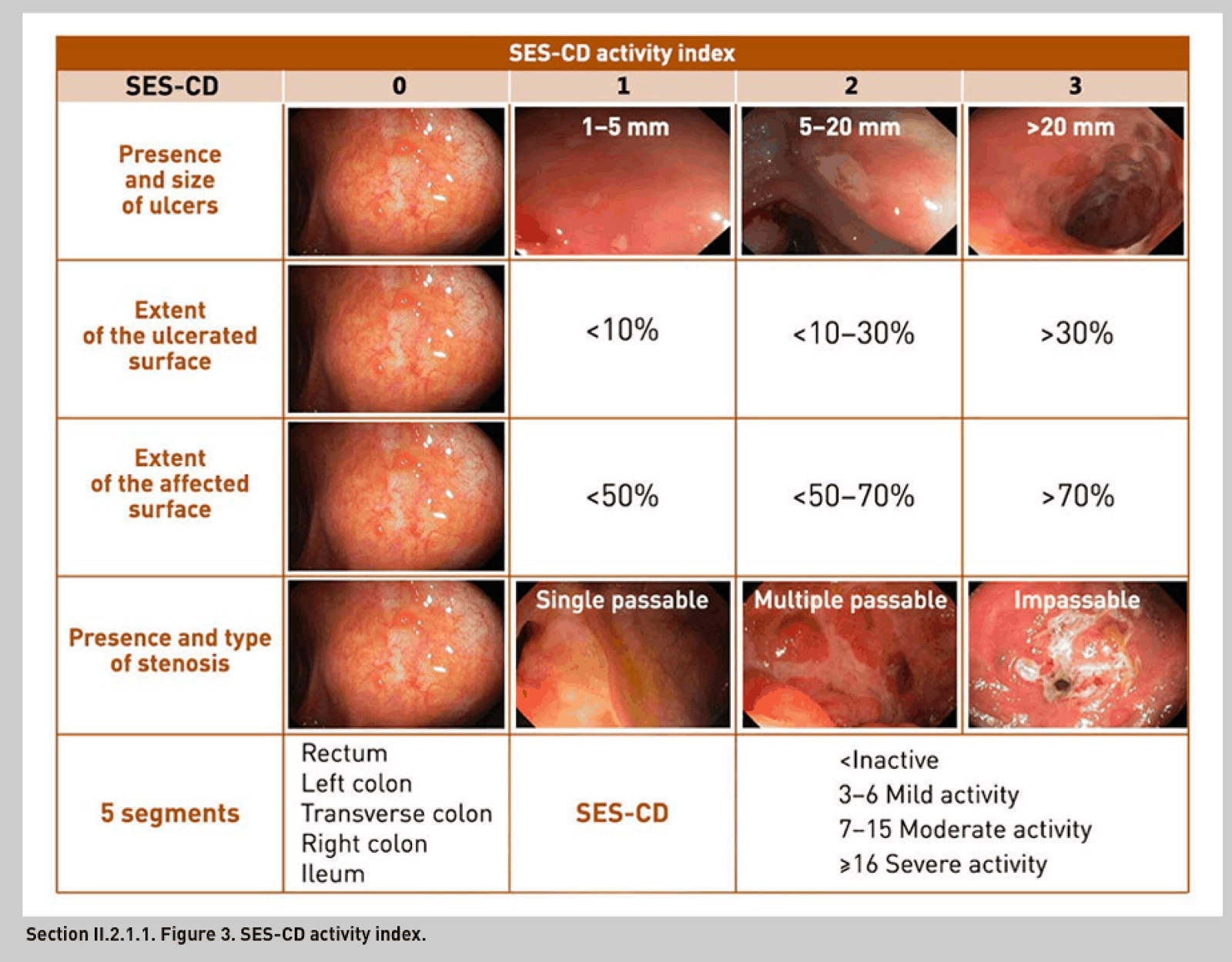
Simple Endoscopic Score for Crohn’s
- This is what is used commonly in New Zealand hospitals.
- 5 – regions – TI, right colon, transverse, left colon/sigmoid and then rectum.
- Looks at presence of ulcers, size of ulcers, extent of ulcerated surface, and presence and degree of narrowing.
- Is a good tool for assessing response to therapy.
Extraintestinal manifestations
Related to disease activity
- Aphthous ulceration (10%)
- Erythema nodosum (10%)
- Pyoderma gangrenosum (1%)
- Neutrophilic dermatitis with necrotic ulcers
- Avoid surgery as it can lead to a larger wound that does not heal
- Treat with antibiotics for superinfection, steroids and immunomodulators
- Acute peripheral arthropathy (10%)
- Eye complications EXCEPT UVEITIS (may or may not) (10%)
- Amyloidosis (1%)
Unrelated to disease activity
- Sacroileitis (10%) - with minimal sx usually
- Ankylosing spondylitis (1%)
- PSC (rare)
- Chronic active hepatitis (2%)
- Cirrhosis (2%)
- Gallstones (30%)
- Bile salt malabsorption
- Renal stones (10%)
- Steatorrhoea causes increased absorption of oxalate causing oxalate renal stones
- DVT/PE
- Metastatic Crohn’s disease
- Rare
- Nodular ulcerating skin lesions occur at distant sites including the vulva, submammary areas and extremities.
- Biopsies show non-caseating granulomas
- Clubbing of chronic disease
Pregnancy
- Active disease at conception = higher risk of spontaneous abortion, preterm labour and 50% risk of relapse of disease
- The risk of relapse is only 25% if disease is inactive at conception.
Colorectal cancer
Low risk - every 5 years
- extensive but quiescent ulcerative colitis or
- extensive but quiescent Crohn’s colitis or
- left-sided ulcerative colitis (but not proctitis alone) or Crohn’s colitis or similar extent.
Intermediate risk - every 3 years
- extensive ulcerative or Crohn’s colitis with mild active inflammation that has been confirmed histologically or
- post-inflammatory polyps or
- family history of colorectal cancer in a first-degree relative aged 50 years or over.
High risk - every 1 year
- extensive ulcerative or Crohn’s colitis with moderate or severe active inflammation that has been confirmed histologically or
- primary sclerosing cholangitis (including after liver transplant) or
- colonic stricture in the past 5 years or
- any grade of dysplasia in the past 5 years or
- family history of colorectal cancer in a first-degree relative aged under 50 years.
Baseline colonoscopy should be offered 8-10 years after diagnosis
Management
Medical management
Inducing remission
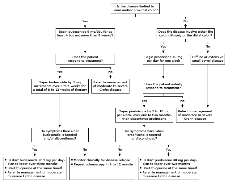
Mild to moderate
- Steroids, pred 20-40mg daily for 2-3 weeks then tapering
- Ileal and/or right colon: budesonide 9mg per day
- Crohn’s colitis: Aminosalicylates oral and/or enema
- Perianal disease: metronidazole 400mg tds or ciprofloxacin 500mg bd Moderate-Severe disease
- IV prednisone 60-80mg daily
- First line in fistulizing disease: combination therapy with an Anti-TNF plus an Immunomodulators
- First line in non-fistulizing disease: Biologic agents monotherapy
- Second line – Alternative Biologic agents +/- Immunomodulators
Maintenance of remission
Mild to moderate
- Wean steroids, if fails treat start Immunomodulators + treat as moderate-severe disease Moderate-Severe disease
- If remission with Biologic agents alone then continue indefinitely. If a Immunomodulators was also used then this should be discontinued in 1-2 years.
Mild disease
Nutrition
- Intraluminal dietary antigens may drive inflammatory response so removal of these may bring remission.
- TPN is effective in inducing remission in 60-80% of patients (similar to steroids, but combining both has no additional benefit)
- Total enteral nutrition equally as effective
- Polymeric diet is also an option for remission and maintenance.
- Enteral often first line for children with Crohn’s.
Steroids
- Good for induction
- Side effects
- Impact on surgical outcomes if used > 6 weeks and >20mg
Immunomodulators – thiopurines
- Used to reduce steroid requirements
- Inhibit cell proliferation and suppress cell-mediated events by inhibiting the activity of cytotoxic T cells and natural killer cells.
- Onset 3-6 months.
- Toxicity 20 – 30%, pancreatitis 3 – 15%. Other effects – fevers, rash, arthralgia, hepatitis, marrow-suppression
- Need to monitor drug concentration
Biologic agents – monoclonal antibodies
- TNF-a inhibitor.
- Good for inducing remission
- Possible increased risk of malignancy long term
- Need to monitor drug concentration
- Possible increase in infective complications postop
Other medications
Antibiotics
- Metronidazole and ciprofloxacin are used to treat symptoms of perianal disease.
- Long-term use of metronidazole is contraindicated because of the risk of peripheral neuropathy. Antidiarrhoeal medications and anticholinergic agents
- useful
- avoid in severe attacks NSAIDS
- should be avoided – may make disease worse. Opioids
- can increase bowel spasm Cholestyramine
- useful for treating bile salt diarrhoea
Surgical management
- Minimum amount of bowel resected to re-establish satisfactory intestinal function.
- If indication for surgery, operate early.
Principles of surgery:
- Optimising perioperative care
- MDT
- DVT prophylaxis
- Possible steroid cover if risk of adrenal suppression
- Nutrition
- Sepsis controlled
- Electrolytes corrected
Recurrence after surgery
- Smoking doubles the risk of reucrrance
- prophylactic use of 5-ASA has a modest effect to reduce recurrence by 15%
Technique
- Open or laparoscopic similar. Lap shorter LOS, fewer adhesions
- No difference between hand sewn end-to-end or stapled side-to-side anastomosis
- At first Crohn’s op, remove appendix
- Thick mesenteric pedicles – may need double-suture ligation
- Preserve bowel length
- Small-bowel and ileocolic
Indications:
- Stenosis causing obstructive symptoms (most frequent)
- Enterocutaneous or intra-abdominal fistulas to other organs
- Draining intra-abdominal or retroperitoneal abscesses
- Controlling acute or chronic bleeding
- Free perforation
- Failed medical management
Gastroduodenal disease
- Gastrojejunostomy for duodenal or pyloric stenosis
- Fistulas to duodenum - jejunal serosal patch or Roux-en-Y limb may be preferable to primary suture of duodenal defect.
Ileocolic
- At 5 years 80% of patients with ileal disease will have had an operation
- Resection with a few cm.
- Effect of Resection Margins on the Recurrence of Crohn’s Disease in the Small Bowel, page 1
- 1996 study from Cleveland clinic showed no difference in recurrance comparing limited (2cm) with extended (12cm) margins.
- Average re-op rate is around 10 years.
- Balloon dilatation sometimes possible - stricture < 4 cm without angulation and no active disease
- Isolated ileocolic disease – could favour surgery as opposed to medical therapy. LIR!C study, showed better QoL for surgery vs infliximab but comparable other parameters such as days off work etc.
- Laparoscopic ileocaecal resection versus infliximab for terminal ileitis in Crohn’s disease- a randomised controlled, open-label, multicentre trial, page 2
Stricutures
- Can be inflammatory or fibrotic.
- Inflammatory
- Usually treated with steroids followed by medical management.
- Fibrotic
- Endoscopic dilatation – good for strictures which are <5cm in length. Up to 50% of patients will avoid surgery over a 5 year period. There is a risk of perforation or bleeding.
- Surgery
- Foley catheter can be placed to identify strictures
- Strictureplasty
- Resection for longer segments of active disease
- Inflammatory
Fistulas and abscesses
Enterocutaneous fistulas and intra-abdominal abscess
- S - Sepsis
- Resuscitate the patient, correct electrolytes, drain sepsis, antibiotics
- N - Nutrition
- Establish nutrition – enteral or TPN
- A - Anatomy
- Work up with imaging to define extent of disease, exclude obstructing lesions and delineate fistula tracts
- P -Plan definitive operation:
- Likely needs complete adhesiolysis & en bloc resection of diseased bowel + fistula tract with primary anastomosis.
A. Spontaneous fistula
- General benefit from earlier surgery due to:
- Fistula won’t heal spontaneously
- No concern about recent laparotomy making surgery difficult
- Bowel perforation occurs slowly and abdominal sepsis usually localised, reducing systemic insult
- Although aim is to optimise patient’s general & nutritional state, active Crohn’s disease will limit what is achievable.
B. Postoperative fistula
- Usually close with conservative measures.
- Delay closure as many heal by 6 weeks unless:
- Originates from diseased segment
- Arises from anastomotic breakdown > 50% of circumference
- Very short tract or communication between skin and mucosa
- Bowel obstruction distal to it
- If > 12 weeks, unlikely to close spontaneously.
Intraabdominal fistulas
- Usually spontaneous.
- En bloc resection of primary defect with simple closure of secondary defect except duodenum.
Free perforation
- (1%)
- Usually need to operate < 24 hours with resection and exteriorisation of bowel ends.
Colonic and rectal Crohn’s
Emergency colectomy +/- ileostomy
- Indications:
- Toxic dilatation
- Haemorrhage
- Perforation
- Severe colitis not responding to medical management
- If does respond then completion proctectomy usually done when in good health.
- Rectal stump surveillance required
- Loop ileostomy is an option
Segmental colectomy
- Appropriate for symptomatic stricture
- If multiple segments, earlier recurrence with segmental resection
Total colectomy and ileorectal anastomosis
- Suitable in 25% with rectal sparing, normal function and sphincter.
Panproctocolectomy
- Most definitive with lowest recurrence rate.
- Rectal dissection with intersphincteric and perimuscular to avoid parasympathetic nerves.
- Delayed wound healing is frequent
Restorative proctocolectomy
- Crohn’s traditionally contraindication to pouch. However some surgeons will offer pouch to well-informed patient with isolated colonic Crohn’s who requires proctocolectomy.
Perianal disease
Clinical
- Anal fissures – most in posterior midline.
- Abscesses – may arise from deep cavitating ulcers or distorted anal glands.
- Fistulas
- Hughes’ classification of perianal lesions in Crohn’s
- Primary lesions
- Anal fissure
- Ulcerated oedematous pile
- Cavitating ulcer
- Aggressive ulceration
- Secondary lesions
- Skin tags
- Anal/rectal strictures
- Perianal abscess/fistula
- Anovaginal/rectovaginal fistula
- Carcinoma
- Incidental lesions
- Piles
- Perianal abscess or fistula
- Skin tags
- Cryptitis
- Hidradenitis suppurativa
- Primary lesions
Investigation:
- EUA,
- MRI
- Endoanal ultrasound
Medical treatment
- Ciprofloxacin, metronidazole, azathioprine, infliximab and adalimumab all effective
- Drain sepsis +/- stoma + start biologic for perianal Crohn’s
Fistulas
- Setons to achieve drainage and commence biological agent. Setons can be removed later if good response.
- When disease is in remission a standard approach can be considered
- Defunctioning ileostomy for perianal disease may be an option.
- Proctectomy in severe cases
Anal fissures
- treat as normal
Abscess
- drain
Rectovaginal fistulas
- Depending on the condition of local tissues, options include a rectal advancement flap, anocutaneous advancement flaps, vaginal flaps, a Martius graft, a gracilis interposition or sphincteroplasty
Abscess
- drain
Bile salt malabsorption
- This is common in ileal Crohn’s. Due to the fact that bile salts are not being absorbed in the inflamed terminal ileum.
- Can get associated reduced absorption of Vitamins ADEK.
- Decreased bile salt absorption can lead to precipitation of cholesterol stones.
- Decreased bile salt absorption can lead to steatorrhoea. Steatorrhoea promotes the absorption of oxalate which can cause kidney stones.
- Bile salts in the colon precipitate diarrhoea.
Prognosis
Slightly higher mortality rates (RR 1.4) High impairment in QoL.