Section: Hepatobiliary Sub-section: Liver Curriculum: Curriculum, page 85
Definition
Epidemiology
- 6th most common neoplasm, 5% of all cancers.
- 2nd most common cause of cancer-related deaths.
- 90% of all primary liver malignancy
- Incidence: 15/100,000 in men
- Related to geographic variation of prevalence of hepatitis B and hepatitis C
- SE Asia >20/100,000. Northern Europe and South central Asia <5/100,000.
Risk factors
Cirrhosis (no. 1)
- Highest risk reported for chronic viral hepatitis
- Lower risks with other forms such as primary biliary cirrhosis
- 10-20% have absence of cirrhosis (but likely have mild degree of fibrosis, steatosis or dysplasia)
Infections
Hepatitis B infection
- Chronic HBV most frequent risk factor worldwide (>50% of cases)
- HBV-DNA can integrate into genome of malignant hepatocytes and is detected in liver tissue of patients with HCC, despite absence of classical HBV serological markers.
- Risk increases with:
- Severity of hepatitis
- Age at infection
- Duration of infection
- Level of viral replication Hepatitis C infection
- Mechanism not clear, but majority have cirrhosis – likely presence of cirrhosis that is crucial for development of tumour.
- Risk increases with:
- Age > 55 years
- Male
- Diabetes
- Alcohol
- HBV co infection
- Obesity – likely risk factor
- Viral genotype or concentration has no impact on risk
HIV infection
- Higher prevalence of associated known risk factors; not only HCV and HBV, also alcohol abuse, NASH & diabetes.
- More rapidly progressive liver disease.
- Cirrhosis and HCC occur 15-20 years earlier in HIV-HCV co-infected patients than HCV alone patients.
Environmental
Alcohol
- Heavy (50-70 g/day) and prolonged ingestion is classical risk factor for cirrhosis and therefore HCC.
Non-alcoholic fatty liver disease (NAFLD)
- One of the common causes of liver disease.
- Spectrum from simple steatosis to more severe forms of Nonalcoholic steatohepatitis (NASH), including cirrhosis.
- Associated with T2DM, central obesity and dyslipidaemia as part of metabolic syndrome.
- Obesity and diabetes increase the risk of HCC
Aflatoxin B1
- Areas of Africa.
- Aflatoxin is ingested in food due to contamination of poorly stored staple crops by Aspergillus flavus.
- Thought to induce HCC through mutation of tumour suppressor gene p53.
Genetic
Hereditary haemochromatosis (HH)
- Autosomal recessive
- Associated with homozygosity for C282Y mutation in the haemochromatosis gene, characterised by excessive gastrointestinal absorption of iron.
- Iron overload generates oxygen/nitrogen species that cause mutations of p53 tumour suppressor gene.
Other causes
- Primary biliary cirrhosis – low risk
- Autoimmune hepatitis – low risk
- Metabolic liver diseases – increased risk
- Porphyria cutanea tarda,
- Tyrosinaemia
- Hypercitrullinaemia
- Glycogenosis type IV, hereditary fructose intolerance and Wilson disease may also develop HCC, but with lower risk.
- Copper overload generates unsaturated aldehydes that cause mutations of p53 tumour suppressor gene.
Adenoma
- Risk of malignant transformation:
- 50% risk in male; any size needs resection
- HCA > 4 cm in women
- Presence of β-catenin mutation
Pathogenesis
- 90% of patients will have cirrhosis
- Repeated rounds of cellular injury followed by regeneration in the setting of chronic inflammation contributes to the development of liver fibrosis, cirrhosis and eventually HCC
- Multi-step process estimated to take approximately 20 to 40 years.
Preneoplastic lesions
Preneoplastic lesions are
- Morphologically characterised by:
- Microscopic dysplastic foci
- Macroscopic dysplastic nodules (DNs)
- Microscopic dysplastic foci
- Lesions made of dysplastic hepatocytes < 1 mm in size and occur in chronic liver disease, particularly cirrhosis
- Dysplastic nodules
- Nodular regions < 2 cm in diameter with dysplasia but without definite histological criteria of malignancy.
- Low grade DNs
- <1 cm in diameter, slightly yellowish and have a very low probability of becoming malignant.
- High grade DNs
- Less common
- Slightly larger nodules (up to 2 cm)
- Characterised by increased cell density with irregular thin-trabecular pattern and occasionally unpaired arteries.
- May be difficult to differentiate from highly differentiated HCC, and may contain foci of well-differentiated HCC therefore considered as precancerous and 1/3 become malignant.
- Low grade DNs
- Nodular regions < 2 cm in diameter with dysplasia but without definite histological criteria of malignancy.
- Vascularisation
- Key parameter in differentiating HCC from Regenerating hepatic nodules
- Progression from nodule to low grade DN, high grade DN and frank HCC is characterised by loss of visualisation or portal tracts and development of new non-triadal arterial vessels which become the dominant blood supply in overt HCC lesions.
- This arterial neoangiogenesis is the hallmark of HCC diagnosis and rationale for chemoembolization and anti-angiogenic treatment.
Gross morphology
- HCC can be subdivided according to: gross morphology, degree of differentiation, vascularity, presence of surrounding capsule and presence of vascular invasion.
- Gross morphology
- Solitary
- Multinodular
- Multicentric - consisting of either a collection of discrete lesions in different segments developing synchronously
- Intrahepatic metastases – one dominant mass and a number of ‘daughter’ nodules located in adjacent segments
- Infiltrating – typically less differentiated with ill-defined margins
- Gross morphology
Histology
-
Degree of differentiation
- Edmondson grade 1-4
- Well-differentiated
- Moderately differentiated
- Poorly differentiated
- Undifferentiated
- Edmondson grade 1-4
-
Immunomarkers may selectively identify malignancy
- Glypican 3 (GPC3)
- Heat Shock Protein 70 (HSP70)
- Glutamine Synthetase (GS).
- Positive staining for any two can detect early and well differentiated HCC in 50–73%
-
TERT promoter – most frequent mutation
-
Increase in telomerase expression is determinant of malignant transformation.
-
Use of these markers may help determine prognosis, select treatment and assess response to chemotherapy
-
Presence of fibrous capsule
- Present in 80% of resected HCC.
- Variable thickness, may not be complete and frequently infiltrated by tumour cells.
-
Vascular invasion
- HCC has tendency to spread locally and invade blood vessels.
- Presence of portal invasion is the most important predictive factor associated with recurrence.
- Rate of portal invasion is higher in:
- Expansive type
- Poorly differentiated
- Large tumours
- Once HCC invades portal vein, tumour thrombi grow rapidly in both directions and towards the main portal vein. Tumour fragments spread throughout the liver as thrombus crosses segmental branches. Once tumour thrombus has extended into main portal vein, high risk of complete thrombosis and portal hypertension. This accounts for frequent presentation of fatal rupture of oesophageal varices or liver decompensation including ascites, jaundice and encephalopathy.
- Thrombus may extend into suprahepatic vena cava or right atrium
- High risk of lung metastases.
-
-
Rarely HCC may invade biliary tract by:
- Intraductal tumour extension
- Obstruction by fragment of necrotic tumour debris
- Haemorrhage of tumour resulting in haemobilia
- Metastatic lymph node compression of major bile ducts in porta hepatis
-
Metastases most frequently in lung. Locations in decreasing order of frequency are:
- Lung, adrenal, bones, lymph nodes, meninges, pancreas, brain and kidney
Clinical presentation
- Rarely before age 40, peak age 70.
- Male 2-4x more than female. Difference in exposure risk factors, higher BMI and higher levels of androgenic hormones
- Circumstances of diagnosis:
- Incidental during routine screening
- Incidental during assessment for deranged LFTs or another pathological condition
- Presence of liver or cancer related symptoms:
- Abdominal pain
- Malaise
- Weight loss
- Asthenia (weakness, lack of energy)
- Anorexia
- Fever
- Worsening ascites or liver decompensation in patients with underlying liver disease
- Clinical exam: signs of cirrhosis, ascites, collateral circulation, umbilical hernia, hepatomegaly and splenomegaly
- Complications:
- Rupture (5-15%)
- Superficial or protruding tumours
- Abdominal pain, haemorrhagic ascites and hypovolaemic shock (50% of patients)
- Portal vein invasion
- Upper GI bleeding, acute ascites
- Hepatic vein or IVC invasion
- PE or sudden death
- Biliary invasion (2%)
- Paraneoplastic syndromes – polyglobulia (pathologically elevated Hb), hypercalcaemia and hypoglycaemia
- Rupture (5-15%)
Investigations
Liver function tests and tumour markers
- LFTs
- Nonspecific, reflects underlying liver pathology or presence of space occupying lesion
- Jaundice most frequently due to liver decompensation
- Alpha-fetoprotein
- Most widely recognised serum marker of HCC. Secreted during foetal life but residual levels very low in adult (0-20 ng/mL).
- Levels >400 ng/ml diagnostic for HCC.
- Very high levels (>10,000 ng/mL) usually correlate with poor differentiation, tumour aggressiveness and vascular invasion.
- Other tumours can have increased levels e.g. non-seminal germinal tumours, hepatoid gastric tumours, neuroendocrine tumours
Imaging
- Aim is to screen high risk patients (best done with USS), differentiate HCC from other space-occupying lesions and select an appropriate treatment.
- Differentiation relies on vascularisation. Most reliable imaging features of HCC are:
- Presence of hyperarterialisation of nodule in early (arterial) phase, and
- Non-peripheral wash-out during portal or late phase following injection (tumour becomes hypovascular compared to adjacent parenchyma)
- Choosing treatment depends on number of lesions, size and extent, presence of daughter nodules, vascular invasion, extrahepatic spread and underlying liver disease.
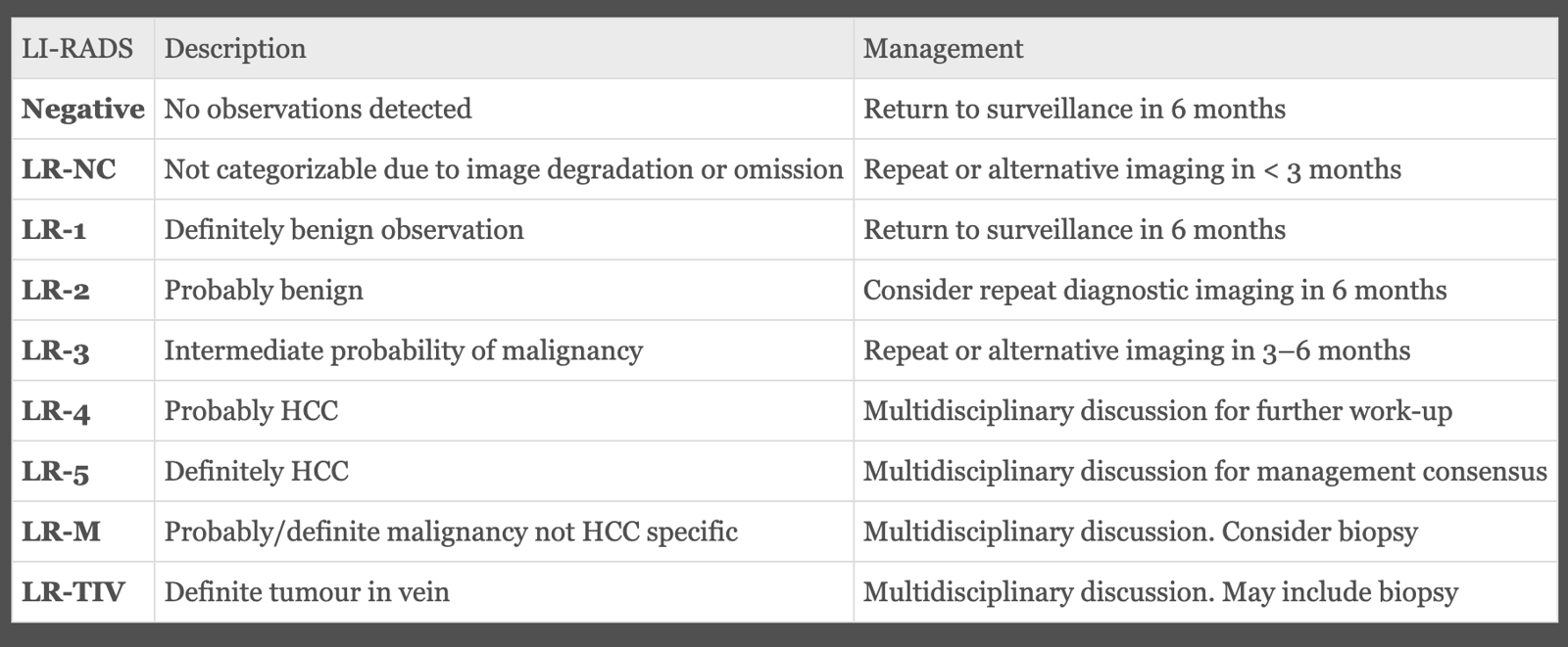
LI-RADs
Adopted in 2011 by the American College of Radiology, the Liver Imaging Reporting and Data System (LI-RADS) provides a set of standardised terminology and establishes a framework for the classification of liver lesions in the setting of cirrhosis, ranging from LR-1 (definitely benign) to LR-5 (definitely HCC)
Ultrasound:
- First line investigation for screening – low cost, high availability, high sensitivity in identifying focal liver mass. Accuracy depends on dimension, location of tumour and operator experience.
- Typically, small HCCs are hypoechoic and homogenous, cannot be differentiated from regenerating or dysplastic nodules.
- With increasing size, may become hypo or hyperechoic but importantly, heterogenous. A hypoechoic peripheral rim corresponds to capsule.
- Infiltrating type difficult to identify in heterogenous cirrhotic liver.
- Doppler may demonstrate feeding artery or draining veins.
- Also accurate in identifying vascular or biliary invasion and indirect evidence of cirrhosis such as segmental atrophy, splenomegaly, ascites or collateral veins.
- Limitations:
- Operator experience
- Location on surface, left lateral segment, upper segments.
- Obesity (thickened abdo wall, steatotic liver)
CT:
- More accurate than US in identifying HCC and their lobar/segmental distribution.
- Useful for identifying features of underlying cirrhosis, accurately measuring liver and tumour volumes and assessing extrahepatic tumour spread.
- HCCs usually hypodense, early uptake of contrast and a mosaic shape pattern. Portal phase washout.
- Variable vascularity depending on grade.
- Capsule (if present) best seen during portal or late phase - enhanced thickening
- Vascular invasion of segmental branches may be identified. Intratumoural arterioportal fistula may occur.
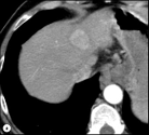
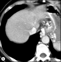
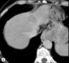 Figure: (a) arterial – early uptake (b) portal – isodense (c) late – washout
MRI:
- More accurate in differentiating HCC from other liver tumours
- Characteristics are:
- Mosaic shape structure
- Presence of capsule
- Usually hypointense on T1, hyperintense on T2, but can vary.
- Contrast enhanced ultrasound
- Primovist
- Hepatospecific paramagnetic gadolinium-based contrast agent, used exclusively in MRI liver imagingAngiography
- Used for arterial chemoembolization PET
- Limited use, low sensitivity (60%)
Histology
- Pathological confirmation of HCC can be cytology, histology or combination
- Accuracy increased if non-tumourous tissue available for comparison
- Liver biopsy limited by risk of haemorrhage, pain, seeding and vascular spread. Incidence of needle tract seeding is 1-5%.
- Risk of seeding balanced by risk of pursuing an aggressive treatment such as resection or transplant in patient without malignancy
- Should access nodule through thick area of normal liver rather than puncture directly.
- Diagnosis reinforced by staining for GPC3, HSP70 and GS.
- High false-negative rate for FNA esp small lesions
Diagnosis
- Current guidelines for ndoules 0-2cm
- For nodules < 1 cm on US, considered unlikely other imaging would reliably confirm diagnosis.
- For repeat US at 3-6 months until lesion disappeared, enlarged or displayed characteristics of HCC.
- If no growth for 2 years, then continue routine surveillance.
- For nodules > 1 cm on US, diagnosis can be established by contrast CT or MRI.
- If findings not characteristic or vascular features atypical or absence of cirrhosis, a diagnostic biopsy recommended
- Biopsy of small lesions evaluated by expert pathologist. Tissue not clearly HCC should be stained with CD34, CK7, GPC3, HSP70 and GS to improve diagnostic accuracy.
- If negative, does not exclude diagnosis. Should have repeat imaging at 3-6 monthly intervals – if enlarges but remains atypical for HCC, repeat biopsy recommended.
- For nodules < 1 cm on US, considered unlikely other imaging would reliably confirm diagnosis.
- After initial recommendations, contrast-enhanced US may provide false positive HCC and cannot selectively differentiate intrahepatic cholangiocarcinoma from HCC, so technique has been withdrawn from algorithm.
Natural history
- Traditionally medial survival of 6 months in symptomatic patients, may be 3 years in asymptomatic untreated patients who are not end-stage.
- Survival is influenced by:
- Morphological spread of tumour
- Presence and severity of cancer-related symptoms
- Severity and evolution of underlying cirrhosis.
- Multiple different staging classifications
Staging
AJCC 8th stage
| T1 | Single tumour < 2 cm, or > 2-5 cm without vascular invasion |
|---|---|
| T2 | Single tumour with vascular invasion or multiple tumours, none > 5 cm |
| T3A | Multiple tumours, any > 5 cm |
| T3B | Single tumour or multiple tumours of any size involving a major branch of the portal or hepatic vein |
| T4 | Tumour(s) with direct invasion of adjacent organs other than gallbladder or with visceral peritoneum |
| N1 | Regional lymph nodes |
| M1 | Distant metastatic disease |
- Cancer related symptoms have detrimental impact on outcome – assessed by WHO performance status or Karnofsky index
- Liver damage traditionally assessed by Child-Pugh score. This was designed to assess functional reserve of cirrhotic patients undergoing portocaval shunt; not entirely appropriate for HCC where therapeutic options include liver transplant and resection.
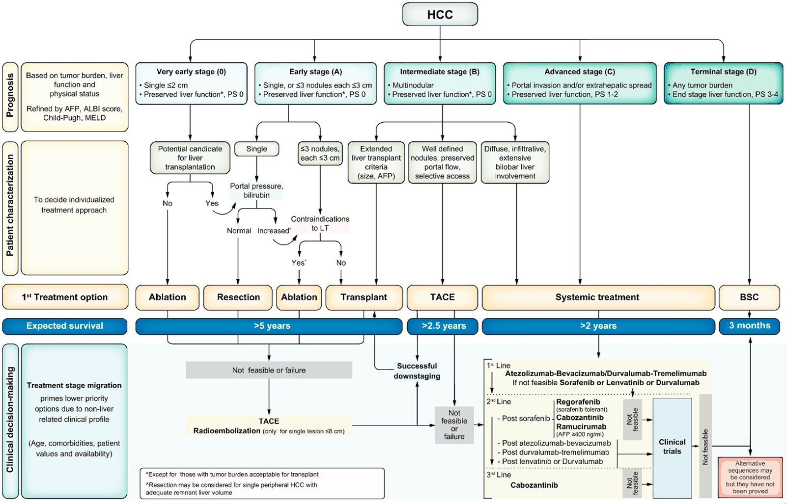
BCLC algorithm
Stage 0-D
 Milan Criteria
University of California San Francisco Criteria
Milan Criteria
University of California San Francisco Criteria
CLIP (Cancer of the Liver Italian Program Score) “CTAP”
- Child-Pugh Score
- Tumour morphology (uninodular <50% of liver, multinodular <50%, extension >50%)
- AFP level (< or > than 400ng/dL)
Tumour Burden Score
- Takes into account largest tumour size and number of tumour nodules in a continuous score
- <3.36 = low
- 3.36 – 13.74 = medium
-
13.74 = high
Correlates well with OS after resection (5YS for low = 78%, medium = 61%, high = 39%)
Can provide rationale for resection of HCC that might not meet BCLC criteria (ie BCLC-B).
ALBI score
Includes bilirubin and albumin to predict survival in HCC pts Avoids subjective criteria used in CP score (ascites, encephalopathy)
Makuuchi criteria
- To select patients for resection, using ICG clearance rate at 15 mins (ICG-R15)
- 0.5mg/kg ICG given IV, then serum ICG tested at 15 mins
- ICG binds to serum proteins and is taken up by first pass metabolism by hepatocytes and excreted unchanged in bile. So rate of clearance reflects liver function
Criteria (“ABI”):
- Ascites
- Bilirubin
- ICG-R15
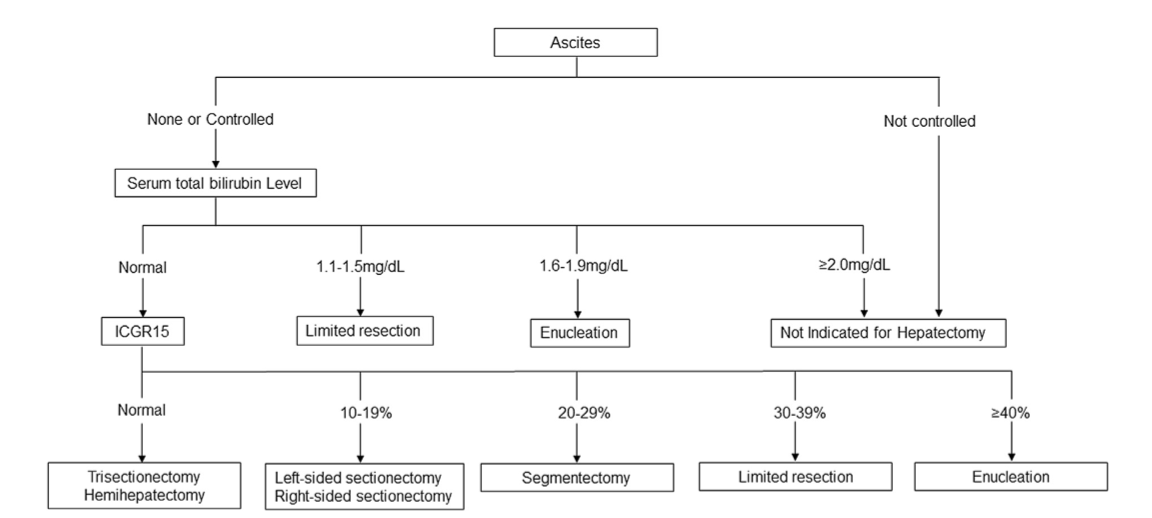
ICG-R15 can be used as a surrogate marker of elevated HVPG and CSPH
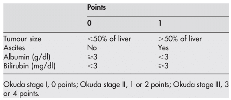
Okuda staging
Portal Vein Tumour Thrombus (PVTT)
- Confers BCLC stage C and typically associated with poor prognosis – conventional treatment is Sorafenib, median survival only a few months
- Liver Cancer Study Group of Japan classification:
- Vp1 = thrombus in third order branches or higher
- Vp2 = second order branches
- Vp3 = first order
- Vp4 = main PV (or contralateral to side of tumour, or both)
- Up to Vp3 has some survival benefit with resection. Increasing Vp closely correlated with OS - Higaki bioscience trends 2017
Screening
- HCC fulfils most of the WHO principles of population screening to be justified.
- High mortality in symptomatic stage.
- At risk population clearly defined (HCV & HBV related cirrhosis, esp male and > 60)
- Acceptable screening tests with low morbidity and high efficacy exist which pick up tumour in early/latent stage
- Effective treatments exist
- Two most common tests are US and serum AFP (although many consider this of little value)
- Usually 6 monthly intervals.
- Need to ensure patient has appropriate treatment option available. E.g. Child-Pugh C and if not potential liver transplant candidate, screening is inappropriate.
- Limitations:
- 20-50% of HCC have previously undiagnosed cirrhosis, therefore escaped surveillance.
- Limited access to medical care and compliance (esp alcohol cirrhosis)
- US operator dependent
Assessment
- Cancer stage – number/size of nodules, relation to pedicles, bilateral disease, macrovascular invasion, metastases
- Underlying liver disease – Child-Pugh Score, MELD, platelets
- Blood tests, CT, MRI
- Fibroscan +/- liver biopsy.
- Liver Function: ICG clearance, hepatobiliary scintigraphy
- FLR as a ratio of the total liver volume (minus the tumour volume). Ratios of at least 25% are considered safe in healthy, non-cirrhotic livers. An FLR of at least 30–40% is recommended in patients with chronic liver disease and a Child-Pugh A score
- Portal hypertension:
- Non-invasive assessment of PC gradient: fibroscan, plt count, CT varices, gastroscopy
- Invasive: right heart catheter or EUS into portal vein
- High risk features: preop ascites, MELD>11, Child>B7, bili >34
- Performance status & non-liver comorbidities – ADLs, ECOG, albumin
Treatment option
Broadly:
- If Child-Pugh A and resectable → resect
- If B-C, portal HTN or otherwise unresectable → transplant if transplant candidate (Milan or UCSF criteria)
- If CP C → palliate
- If metastatic disease → systemic treatment
- If unresectable and not transplant candidate →options include TACE, RFA, radioembolization and stereotactic radiotherapy
Liver resection
In normal liver
- Partial liver resection.
- Treatment of choice in non-cirrhotic patient’s
- 5 year survival can reach 70% to 80%
- Lymphadenectomy recommended (prevalence of LN mets ~15%, compared to < 5% in cirrhosis)
- Adjuvant chemotherapy not recommended.
- Regular follow-up with CT 6 monthly, as early detection and treatment of recurrence may improve survival.
In patients with cirrhosis
- Main limitations:
- Tumour multifocal in 20-60% at diagnosis
- Cirrhosis is high risk factor for postoperative complications
- Oncological resection require wide margins where diseased underlying liver needs parenchymal sparing.
- Risk of recurrence high because cirrhosis persists
- Risk of surgery increased due to coagulation defects, portal hypertension, liver failure and impaired regeneration. Mortality ~5%.
- Hepatectomy should only be performed if well preserved liver function, with a normal bilirubin and hepatic venous pressure gradient < 10 mmHg.
- In Child-Pugh A, without clinically significant portal hypertension (hepatic venous pressure gradient > 10 mmHg, oesophageal varices, splenomegaly > 12 cm and thrombocytopenia < 100,000/mm3).
- Remnant of 30-40% required
- PVE should be undertaken to increase remnant volume and preoperatively test ability to regenerate.
- Technique
- Anatomical resection rather than tumourectomies should be performed.
- To remove tumour and adjacent segments that have same portal tributaries.
- 20% improvement in OS and DFS.
- Margins should be > 1-2cm
- To ensure potential satellite nodules also resected.
- 75% 5-year survival for 2 cm vs 49% for 1 cm.
- Laparoscopic resection
- Not proven as better but may have less intraoperative bleeding, fewer postop complications, reduced postop analgesia and shorter hospital stay.
- May reduce risk of postop ascites and facilitate subsequent transplant with fewer adhesions.
- Intraoperative ultrasound
- to detect potential unrecognised nodules between 0.5 and 1 cm and optimize the surgical resection efficacy, while at the same time being instrumental to perform anatomical resections
- Anatomical resection rather than tumourectomies should be performed.
- Outcomes
- 5-year survival 44%
- Predictors of survival are:
- Age
- Degree of liver damage
- AFP level
- Tumour diameter
- Number of nodules
- Vascular invasion
- Surgical margins
- Treatment of recurrence
- Two methods:
- Management of underlying chronic liver disease
- Actively screen operated patients and treat recurrences if confined to liver by repeat resection or transplant.
- Lack of evidence for neoadjuvant or adjuvant therapy.
- Most effective way to prevent recurrence is transplant
- Two methods:
Liver transplant
- It removes both detectable and undetectable tumour nodules with preneoplastic lesions present in cirrhotic liver.
- Simultaneously treats underlying cirrhosis and prevents development of postoperative or distant complications associated with portal hypertension and liver failure.
- Patient selection:
- No portal invasion
- No extrahepatic disease
- Plus:
- Milan criteria
- 1 nodule ≤ 5 cm or even 3 nodules ≤ 3 cm
- University of California San Francisco (UCSF) expanded criteria
- Limited tumour burden. UCSF criteria (more inclusive than Milan criteria):
- Single tumour < 6.5 cm
- Or ≤ 3, largest < 4.5 cm with sum of diameters < 8 cm.
- Milan criteria
- Treatment on waiting list
- Average > 12 months on list. Up to 25% may be excluded while waiting. Approaches developed to avoid drop-outs:
- Living-donor liver transplantation (LDLT)
- Drawbacks:
- Inherent risk for donor
- Small for size grafts
- Only 25-30% of candidates have potential donor
- Drawbacks:
- Rules for graft allocation. MELD and Milan criteria
- Treatment by resection, ablation or chemoembolization while on waiting list to avoid tumour progression beyond criteria.
- Living-donor liver transplantation (LDLT)
- Average > 12 months on list. Up to 25% may be excluded while waiting. Approaches developed to avoid drop-outs:
Liver Ablation
Four techniques:
- Chemical ablation (percutaneous ethanol injection, PEI)
- Thermal ablation (radiofrequency, RFA)
- Microwave ablation (MWA)
- Cryoablation
Radio-frequency ablation
- RFA first line ablation technique. RFA > perc ethanol injection, with lower recurrence and fewer treatment sessions required.
- Advantages:
- Minimally invasive
- Low waiting time
- Preserve uninvolved parenchyma
- No systemic side effects
- Avoid mortality/morbidity risks of major hepatic surgery
- Disadvantages:
- Only small tumours < 3-5 cm likely to be treated successfully
- Technical limitations:
- Multiple tumours (>3) limitation because need for repeat punctures.
- Need to clearly visualise tumour by US; isoechoic tumours in upper segment 4, 7 and 8 or edge of left lateral section may be unsuitable.
- Needle needs to enter tumour via hepatic parenchyma due to risk of intraperitoneal bleeding or seeding – not possible for superficial or protruding tumours
- Does not treat underlying liver disease.
- Recurrence common
- Indications:
- Previously used if patient unsuitable for resectional surgery. However now being considered first-line treatment for single nodules < 2 cm. (RCTs have shown no difference in 5-year survival for HCC < 2 cm)
- Neoadjuvant treatment in liver transplant candidates, and for treatment of recurrence after liver resection.
- Contraindications:
- Gross ascites (risk of intraperitoneal bleeding)
- Uncorrectable coagulopathy
- Previous bilioenteric anastomosis or endoscopic sphincterotomy associated with bile bacterial contamination – risk of abscess
- Proximity to colon, duodenum, stomach or biliary confluence – risk of injury or perforation by heating
- RFA (not microwave) contraindicated with pacemaker
- Methods and margins:
- Ablation should aim to achieve a safety margin to control satellite nodules. 5mm margin.
- Can combine with TACE.
- Treatment response assessed 1 month post procedure.
- Advantages:
Transarterial chemoembolization
- TACE - Selective infusion of embolizing particles coupled or not with cytotoxic agents or for the intraarterial infusion of radioactive isotopes
- doxorubicin or epirubicin, and/or cisplatin or miriplatin
- Emulsion of cytotoxic agents with lipiodol followed by spongostan
- HCC receives 100% of blood supply from the artery
- Occasionally need to embolise diaphragmatic or mammary arteries to achieve adequate control.
- Iodised oil injection combined to improve efficacy. Retained in malignant tumours (cleared from normal hepatic parenchyma), may be used for targeting cytotoxic drugs.
- Contraindications:
- Liver decompensation
- Biliary obstruction
- Bilioenteric anastomosis
- Impaired kidney function
- Portal vein thrombosis (unless limited to section of liver only)
- Morbidity:
- 75% develop post embolization syndrome – fever, abdominal pain, nausea and raised serum transaminase levels. Self-limiting.
- More severe complications: cholecystitis or GB infarction, gastric/duodenal wall necrosis & pancreatitis. Hepatic abscess rare.
- Monitoring:
- CT 1 month – can see disappearance of arterial vascular supply to tumour and decrease in diameter.
- Efficacy:
- Improves survival. Median 34 months Japanese study.
- Predictors of survival:
- Degree of liver damage
- Portal vein invasion
- Maximum tumour size
- Number of lesions
- AFP levels
- Predictors of survival:
- Improves survival. Median 34 months Japanese study.
- Contraindications:
- Patients with liver-only HCC without vascular invasion who are not amenable to resection, transplantation, or ablation should be considered for intra-arterial therapies
Systemic chemotherapy
- Limited role
- Recommended for patients with preserved liver function who present with extrahepatic spread, have macrovascular invasion, or are ineligible for liver-directed modalities because of refractoriness or contraindication
- IMbrave150 Trial
- Unresectable hepatocellular carcinoma who had not previously received systemic treatment were randomly assigned in a 2:1 ratio to receive either Atezolizumab plus Bevacizumab or Sorafenib
- Overall survival at 12 months was 67.2% with atezolizumab–bevacizumab and 54.6% with sorafenib
- Sorafenib recommended as second-line palliative option in patients not eligible for resection, transplant, perc ablation or TACE if they still have preserved liver function.
- Targets tyrosine kinases vascular endothelial growth factor (VEGR) receptors 2 and 3, and the platelet-derived growth factor receptor β.
Selective internal radiation treatment
- Injecting radioisotopes of iodine-iodised oil or Yttrium labelled microspheres directly into hepatic artery (radioembolization) – increased delivery within tumour and decreased toxicity.
- May be possible in portal vein thrombosis (where TACE is contraindicated).
Stereotactic body radiotherapy (SABR/SBRT)
- Indicated for unresectable or medically comorbid. Alternative to ablation or embolization, or when these therapies have failed or contraindicated.
- SBRT – hypofractionated RT with photons that deliver large ablative doses of radiation
BCLC algorithm
Eastern Cooperative Oncology Group (ECOG) scale:
- PS 0 – you are fully active, more or less as you were before your illness
- PS 1 – you can’t carry out heavy physical work, but can do anything else
- PS 2 – you are up and about more than half the day. You can look after yourself but can’t work
- PS 3 – you are in bed or a chair for more than half the day. You need help to look after yourself
- PS 4 – you are in bed or a chair all the time and need complete care
Uptodate treatment algorithm
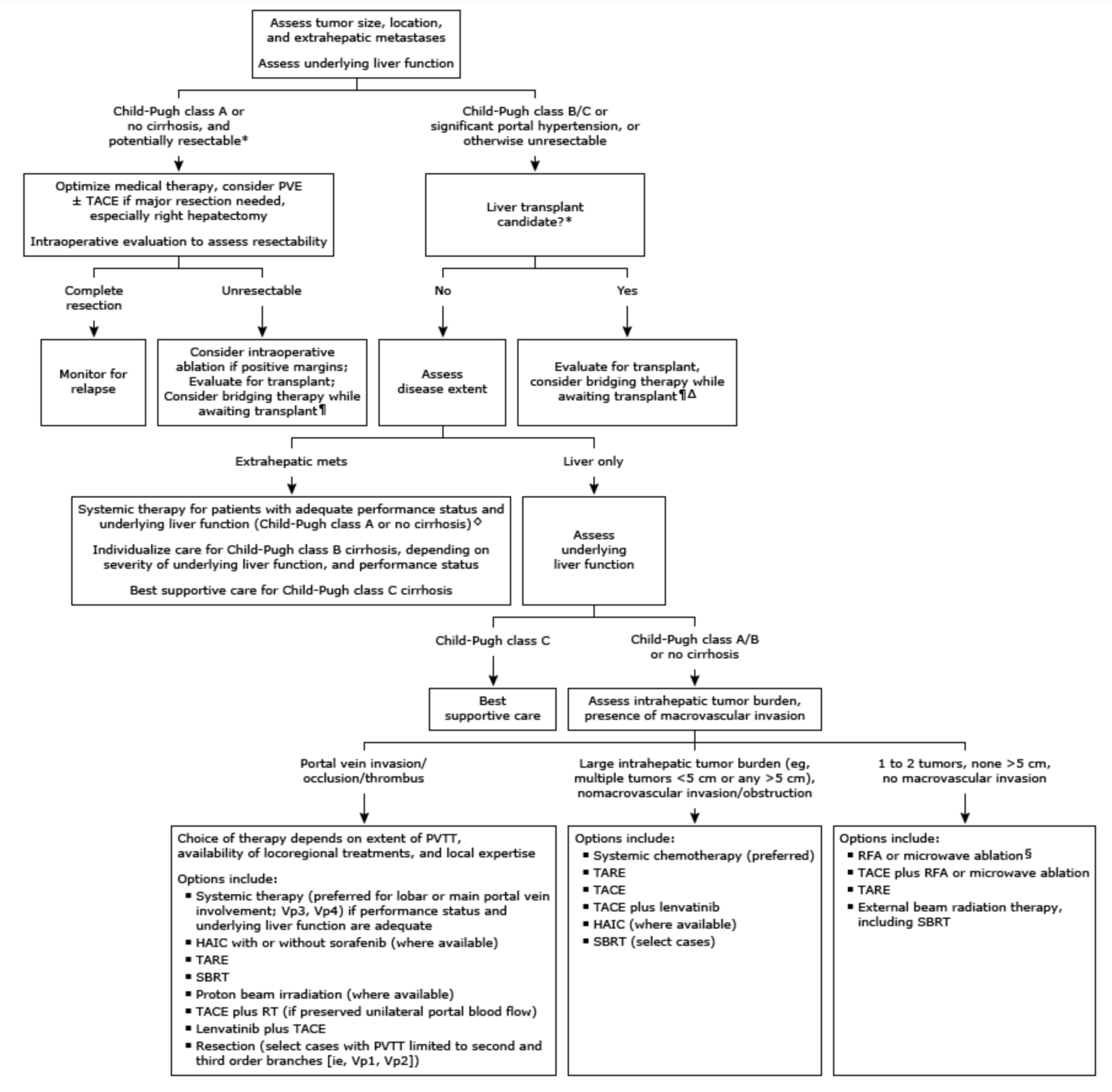
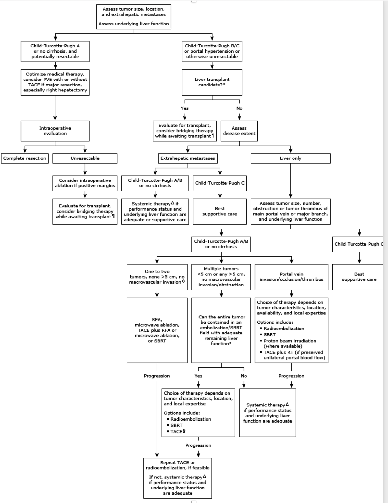
Treatment of complicated HCC
- HCC with macroscopic portal vein invasion:
- Contraindication for transplant and ablation
- Surgical resection considered if thrombus not into main portal vein.
- TACE can be used if thrombus limited to a section of liver and embolization is highly selective and partial arterial occlusion.
- Radioembolisation and anti-angiogenic therapy indicated
- HCC with macroscopic invasion of hepatic veins:
- Worse prognosis
- If confined to hepatic vein, can consider resection.
- High risk of pulmonary mets within 6-12 months of surgery
- Extension to IVC or right atrium beyond any treatment
- Ruptured HCC:
- Actively treated unless terminal presentation
- Aim is to stop bleeding, ideally by arterial embolization.
- Subsequent hepatectomy can be associated with long-term survival.
- Bleeding not necessarily due to tumour rupture, but occasionally due to rupture of artery at junction of tumour and adjacent parenchyma
- Even if tumour ruptured, not always associated with peritoneal seeding of tumour cells.
Fibrolamellar carcinoma (FLC)
- Definition:
- Rare variant of HCC.
- Defined as well-differentiated polygonal hepatic tumour cells with an eosinophilic granular cytoplasm surrounded by a fibrous lamellar stroma
- Epidemiology:
- 1% of HCC.
- Younger age (20-35 years).
- Not due to chronic liver disease.
- Women > men.
- Presentation:
- Usually large at time of diagnosis (8-10 cm)
- Symptoms: palpable mass, abdominal pain, weight loss, malaise, anorexia
- Imaging:
- Large solitary hypervascular heterogenous liver mass with central hypodense region due to central necrosis or fibrosis.
- On MRI, central scar has low attenuation on T2 (as opposed to high attenuation in FNH).
- Well defined margins and calcification in 68%
- Pathology:
- Histology: Deeply eosinophilic, polygonal neoplastic cells surrounded by dense, layered fibrous stroma.
- Lymph node invasion within hepatic pedicle is frequent (60%).
- Treatment:
- Resection. Transplant has little or no place.
- Lymphadenectomy recommended.
- Prognosis:
- Better than HCC. 5-year survival after resection 50-75%
- Recurrence:
- High risk within liver, LNs or distant mets
- Repeat surgery reasonable option due to relatively indolent course of disease and inefficacy of non-surgical treatments
- Follow-up
- Close long-term follow-up mandatory. Recurrence and death beyond 5 years common.