Section: Hepatobiliary Sub-section: Pancreas Curriculum: Curriculum, page 90
Definition
Benign inflammatory disease, characterised by chronic pancreatic inflammation, atrophy and scarring/fibrosis, irreversibly damaging the gland, and resulting in loss of pancreatic exocrine and endocrine function.
- Multiple ways to classify:
- Aetiology (EtOH, genetic or idiopathic)
- Morphology (large-duct disease, small-duct disease and minimal change)
- Temporal (early-stage, late stage)
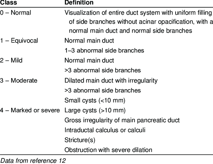
Cambridge classification (morphology based)
Epidemiology
- Incidence – 4/100,000/year. Prevalence 40-50/100,000
Aetiology
- TIGAR-O
- Toxic-metabolic – alcohol, smoking, hypercalcaemia, hyperlipidaemia, chronic renal failure, medications, toxins
- Idiopathic – associated with early-onset and late-onset CP, tropical CP
- Genetic mutations – PRSS1, CFTR, SPINK1, others
- Autoimmune - IgG4
- Recurrent and severe AP-associated CP – post-necrotic severe AP, vascular disease/ischaemic, post-irradiation
- Obstructive – Pancreatic divisum, SOOD, duct-obstruction e.g. tumour, post-traumatic pancreatic duct scars
Pathology
- Multiple theories.
- Incompletely understood
- Two hit theory– initial hit which initiates acute pancreatitis followed by an abnormal response which leads to inflammation becoming chronic and chronic pancreatitis.
- initial insult with iոϳսry, followed by an attempt at healing through fibrosis and regeneration
- Initial event
- Alcohol:
- Increases total protein concentration in pancreatic juice, promotes synthesis and secretion of lithostathine by acinar cells and increases GLP2 secretion. These cause protein precipitation with subsequent formation of protein plugs and stones within pancreatic duct (leading to obstruction with autodigestion by pancreatic enzymes).
- Also EtOH stimulates PSCs (pancreatic stellate cells; type of resident fibroblast) to differentiate and synthesise proteins such as collagens, fibronectin, MMPs, causing fibrosis.
- Autoimmune
- Lymphocytes cause immune mediated destruction of pancreatic duct epithelium
- Obstructive
- Blockage of main duct due to underlying aetiology
- Alcohol:
- Second hit
- General: persistent inflammation causes irreversible fibrosis and atrophy with chronic pain, and endocrine & exocrine insufficiency. Release of inflammatory mediators such as PDGF, TGF-β, TNF-α, IL-1 and IL-6. Calcifications caused by dystrophic calcification.
- Genetic:
- Protease serine 1 (PRSS1) mutation causes increased trypsinogen activation within acinar cells.
- SPINK1 regulates premature activation of trypsinogen.
- CFTR – bicarbonate and chloride secretion regulation. Decreased pancreatic juice volume, increasing concentration of pancreatic enzymes within duct.
Clinical
Pathogenesis of pain
- Complex and multifactorial.
- Pancreatic pain
- relates to inflammation of gland & obstruction of main pancreatic duct
- Extra-pancreatic
- relates to abnormal neural pathways and aberrant central nervous system perception of pain
- Objective evidence from studies showing the number and diameter of non-myelinated type C pain fibres significantly increased in patients with CP.
- relates to abnormal neural pathways and aberrant central nervous system perception of pain
- May be treatment related side-effects, e.g. opioid-induced gut dysmotility.
- Pancreatic pain
Clinical presentations
- Acute presentation of chronic pancreatitis
- Acute abdominal pain, high lipase or as per Atlanta 2012 diagnostic criteria – treated as acute pancreatitis with adequate initial resuscitation and treat avoidable causes of recurrent pancreatitis.
- Presence of parenchymal calcification on CT or dilated main pancreatic duct may suggests underlying diagnosis of chronic pancreatitis
- Chronic presentation of chronic pancreatitis
- Abdominal pain with normal or slight hyperlipasaemia
- May only need temporary escalation of analgesia and maintenance fluids.
- Counselling to reduce EtOH consumption
- Considered for endoscopic or surgical management
- Index presentation with complications of chronic pancreatitis
- Pseudocyst, bleed from false aneurysm, biliary or duodenal obstruction from chronic fibrosis
- Asymptomatic incidental finding
- CT may find parenchymal calcification without symptoms.
- Excluded diabetes.
- No role for intervention of asymptomatic
Clinical course
- Typically relapsing with intermittent abdominal pain.
- Endocrine and exocrine function become compromised during clinical course.
- Diabetes more frequent in long standing chronic pancreatitis
Diagnosis
- American guidelines:
- Definitive evidence – intraductal calcification most specific.
- Moderate – calcification on CT or histology
- Probable – abnormal imaging + history suggestive and abnormal physiology
- Insufficient – abdominal pain with no imaging changes, prior ERCP, stenting
Differential diagnoses of pancreatic mass
- Pancreatic cancer
- Focal mass on bg of chronic pancreatitis.
- Ca 19-9 may not be accurate.
- EUS/FNA may confirm cancer, but cannot exclude even if negative cytology. Surgical resection should be considered.
- Autoimmune pancreatitis
- Chronic inflammatory condition, may present with mass, pain and jaundice.
- Associated with extra-pancreatic stricture of upper and intrahepatic bile ducts, not typically with pancreatic calcification. Classic sausage-shaped swollen pancreas
- Associated with IgG4 elevation
- Ideally tissue diagnosis obtained
- IPNM
- Typically main-duct IPMN has intraductal mucin seen extruding from ampulla, whereas duct dilatation from CP associated with stricture formation and parenchymal calcification.
Investigations
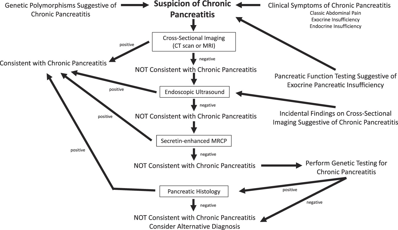 American College of Gastroenterology Guidline
Baseline assessment:
- History – nature, duration, age of first onset, associated factors e.g. jaundice or vomiting. EtOH history. Smoking. FHx.
- Exam – BMI, abdominal exam, urinalysis.
- Bloods – FBC, U&Es, LFTs, glucose (+HbA1c), CRP, Ca 19-9. IgG4
- Stool – faecal elastase
- Imaging:
- Abdo USS – presence of gallstones, coexistent liver disease, dilated duct, change in size of gland
- CT – dilatation of pancreatic duct, pancreatic calcification, parenchymal atrophy (all late signs)
- MRI – occasionally used. Better for ductal assessment.
- EUS – complements cross sectional imaging, more sensitive than CT for early CP.
- ERCP – not used for diagnosis
- Exocrine function – faecal elastase, serum trypsinogen, chymotrypsin – usually all in late stages when steatorrhoea already present.
Management
Medical
- MDT approach, chronic/complex pain team, dietician, surgeons, gastro, endocrine, drug & alcohol
- Analgesia – WHO ladder
- Lifestyle – EtOH avoidance and smoking cessation
- Exocrine replacement and nutrition
- Endocrine management - diabetes
Endoscopic management
- Endoscopic drainage of main pancreatic duct
- Surgical drainage more effective, with better relief of pain long term and fewer procedures.
- ESCAPE Trial — JAMA 2020 + JAMA Surgery 2024 (Long-term)
- Early surgery superior to endoscopy-first approach for pain relief at 18 months
- Long-term follow-up (JAMA Surgery 2024): superior outcomes maintained with early surgery vs endoscopy-first strategy
- 51% of patients in the endoscopy-first group ultimately crossed over to surgery, demonstrating high endoscopy failure rate
- ESCAPE Trial — JAMA 2020 + JAMA Surgery 2024 (Long-term)
- Indicated for dilated main pancreatic duct without pancreatic head mass.
- Principles: Access pancreatic duct by sphincterotomy, remove intraductal stones and leave stent to facilitate prolonged drainage.
- For large calculi, ESWL should be used in combination with endoscopic clearance
- Surgical drainage more effective, with better relief of pain long term and fewer procedures.
- Endoscopic coeliac plexus block
- If good response by local, can consider destruction of plexus by injection of alcohol.
- Treatment of complications of CP
- Distal bile stricture, pancreatic pseudocyst, short term duodenal stenting
Surgical management
Indications:
- Pain control
- Complications of chronic pancreatitis e.g.
- Distal bile duct stricture in younger patients where long-term stenting not optimal
- Gastric outlet obstruction secondary to duodenal stricture
- Pseudocysts in tail may require distal pancreatectomy
- Case selection:
- MDT approach
- Depends on age, comorbidity. Type of surgery depends on these and morphology.
- Ideally alcohol avoidance for >6 months prior to surgery
Timing
- No clear consensus on optimal timing.
- Earlier intervention may avoid long term pain and opioid dependence.
- Contrary, patients may have unnecessary surgery.
Preoperative optimisation:
- Cardiopulmonary exercise testing
- Dietician
- Physiotherapy
- Pain management
- Diabetes management
Type of surgery
- Head of pancreas mass concerning for cancer
- Pancreatoduodenectomy
- Head of pancreas mass with low index of concern
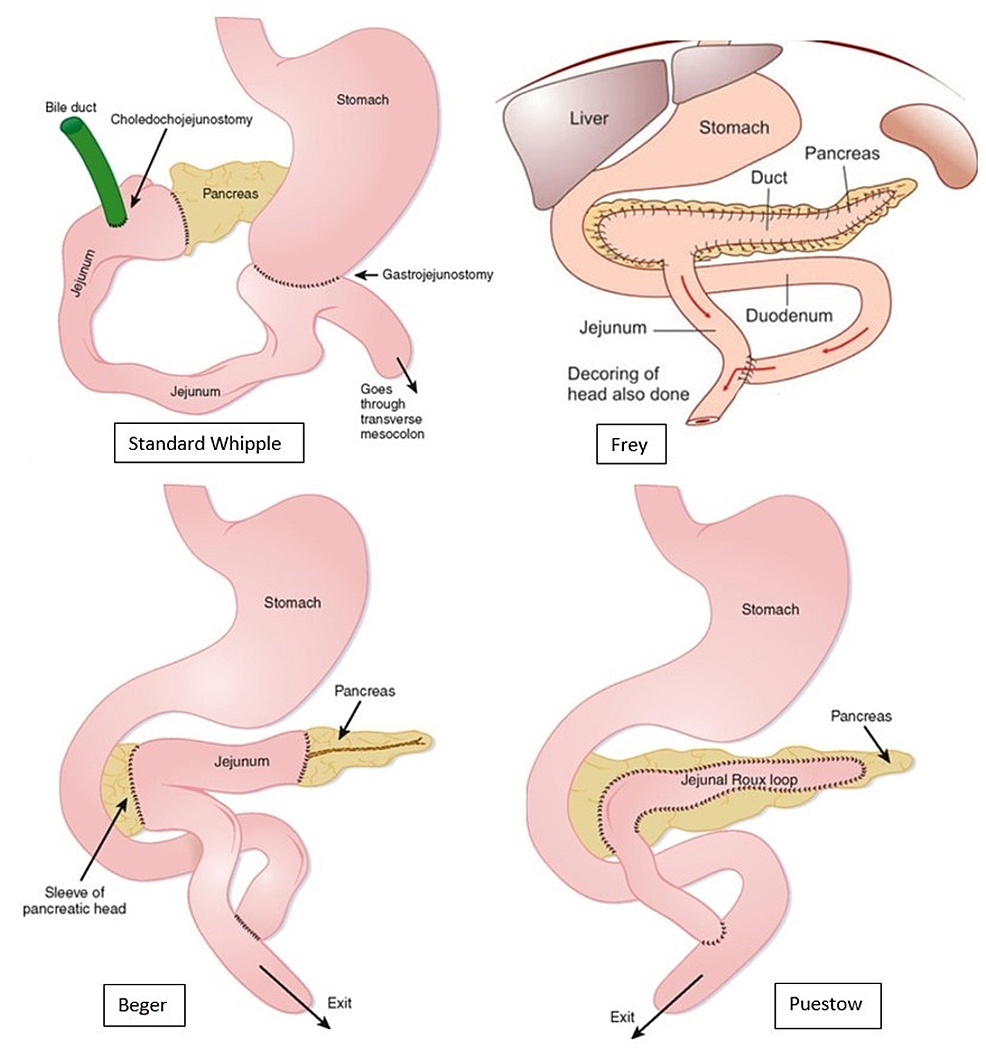
- Longitudinal pancreatojejunostomy with partial pancreatic head resection (Frey)
- Duodenum-preserving partial pancreatic head resection (Beger procedure)
- No head of pancreas mass and large duct disease
- Longitudinal pancreatojejunostomy (Puestow)
- Longitudinal pancreatojejunostomy with partial pancreatic head resection (Frey)
- Total pancreatectomy
- Not widely used
- Many supporters of total pancreatectomy with islet autotransplantation (TPIAT)
- Need facility for islet isolation.
- Reserved for chronic pancreatitis with low risk of cancer, e.g. younger patients with genetic chronic pancreatitis
Drainage Procedures
- Puestow procedure (lateral pancreaticojejunostomy) = drainage procedure
- Main PD needs to be >6mm
- Doesn’t drain head so pain may persist, as well as biliary obstruction Mixed Procedures
- Frey procedure
- Core out pancreatic head and lateral panc-jej
- Simpler to perform
- Avoids transection of pancreas and dissection of SMV-PV, which may be difficult in chronic pancreatitis
- Beger procedure — duodenum-preserving pancreatic head resection
- Core out pancreatic head as much as possible, and divide neck of pancreas over/beyond SMV
- Drains distal pancreas and proximal head stump by 2 panc-jejs
- Berne procedure — also duodenum-preserving pancreatic head resection
- Core out pancreatic head but don’t divide pancreas
- Single PJ over top of pancreatic head capsule Resectional Procedures
- Distal pancreatectomy – for distal disease
- Whipple’s
- Total pancreatectomy +/- islet cell transplantation (TPIAT)
Complications of longstanding chronic pancreatitis
- Biliary stricture
- Initial management
- Exclusion of neoplasia
- Cross-sectional imaging, EUS/FNA
- ERCP drainage.
- Biliary bypass considered for patients stent dependent with minimal comorbidity but persistent symptomatic stricture.
- Roux-en-Y hepaticojejunostomy preferred.
- Choledochoduodenostomy & cholecystoduodenostomy now obsolete, high rates of failure
- Initial management
- Duodenal stenosis
- Short term placement of removable duodenal stent.
- Preferred tx is laparoscopic gastro-jejunostomy
- Pancreatic ascites
- Rare. Defined as massive accumulation of pancreatic fluid in the peritoneal cavity.
- Amylase in ascitic fluid 3x plasma levels.
- Occasionally fluid can track through diaphragmatic hiatus into mediastinum, occasionally reaching pleura or bronchus.
- Initial management by perc drain and nutritional support, followed by ERCP to locate site of leakage + pancreatic duct stent insertion.
- Surgery rarely required.
- Somatostatin or octreotide + diuretics and repeated paracentesis may be beneficial for some patients.
- Pseudocyst
- Persistent fluid collection > 4 weeks from episode of acute pancreatitis with little or no necrosis
- Endoscopic drainage preferred.
- Stent can be left is situ to prevent closure of the endoscopic cystgastrostomy.
- Transpapillary may be effective if duct stricture can be negotiated
- Pseudocyst secondary to main duct disruption
- Disconnected duct syndrome c
- Endoscopic drainage but is one of the rare indications for distal pancreatectomy in chronic pancreatitis.
- False aneurysm
- GI haemorrhage due to false aneurysm. Splenic artery and GDA.
- Optimal intervention is embolization.
- Extrahepatic portal hypertension
- Chronic inflammation and swelling around head can lead to PV occlusion, with development of cavernous transformation.
- Unclear benefit/risk of anticoagulation in occlusion (favoured in thrombosis)
- Can lead to variceal collateral circulation through wall of stomach.
- TIPSS not effective due to portal vein thrombosis.
- GI bleeding rare.
- SMV/PV thrombosis with collaterals restricts subsequent surgical approaches.
- Pancreatic cancer complicating CP
- Lifetime risk is RR 13 for CP, with 10-20 year lag between incidence of pancreatitis and pancreatic malignancy.