Section: Hepatobiliary Sub-section: Spleen Curriculum: Curriculum, page 92
Anatomy
- 300-500 LN in the body; approximately 1/3 of them are in the head & neck
- Normal lymphocytes circulate, passing from blood into & though the lymph nodes then into efferent lymphatics surveying for antigen for which they have a receptor; if this Ag is not present, then they leave the LN
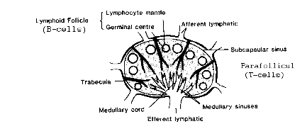
- LN structure
- Capsule
- Afferent lymphatics → lymphatic fluid enters subcapsular sinus.
- Cortex
- In the absence of immune stimulation: the cortex contains “Primary follicles” (composed of B cells – virgin B cells or memory B cells)
- With antigenic stimulation, the B cells are stimulated & this converts the primary follicle into a “secondary follicle” or “germinal centre”, surrounded by a mantle zone of transient small lymphocytes & a central replicating area
- Paracortex = region surrounding & beneath germinal centres
- Site of T cell homing; T cells replicate in response to antigen
- Medulla = deep to cortex & paracortex – composed of medullary cords & sinuses,
- Plasma cells aggregate at the cords and release their Immunoglobulins into efferent lymph
- Medullary vessels: artery & vein @hilum
- Efferent lymphatic(s) @hilum
Biopsy
- Diagnosis
- Need adequate tissue for histologic, immunologic, and molecular biologic assessment.
- Excisional or incisional biopsy
- Multiple core needle
- FNA
- FNΑ is neither adequate to exclude a diagnosis of lymphοma (eg, due to limited diagnostic material, sampling issues, loss of fragile cells during processing) nor does it enable definitive classification (because it disrupts the nodal architecture)
- Need adequate tissue for histologic, immunologic, and molecular biologic assessment.
- Send
- Fixed in Formalin
- Histology
- Can also do if required
- Immunohistochemistry - can get information that you get form flow cytometry
- Fluorescence in situ hybridization (FIЅH) - can get information detected by conventional karyotype
- Fresh
- Flow cytometry
- Conventional Karyotyping
- Fixed in Formalin
- Correlate with blood tests: PSA, Ca-125
- USS:
- presence of echogenic hilum reflects nodal architecture; it’s absence is predictive of pathology
- USS can be useful in identifying abnormal tissue & avoiding necrotic nodes
- presence of echogenic hilum reflects nodal architecture; it’s absence is predictive of pathology
- If searching for a primary: (also see Axillary nodes Unknown Primary in Breast section)
- PR
- CT
- TV USS
- ± endoscopy if anaemic or symptomatic
- ± PET
- Of all lymphadenopathy of unknown primary, adenocarcinoma is most common
Castleman disease
- aka: giant lymph node hyperplasia, angiofollicular hyperplasia, lymph node harmatoma, follicular lymphoreticuloma or benign giant lymphoma
- a rare and usually benign lymphoproliferative disease, cured by surgical resection