Section: Hepatobiliary Sub-section: Spleen Curriculum: Curriculum, page 92
Overview
- Largest lymphoid organ
- Size: 1 x 3 x 5 inches
- Weight: 7 ounces (200 grams)
- Location: Deep to left ribs 9 to 11
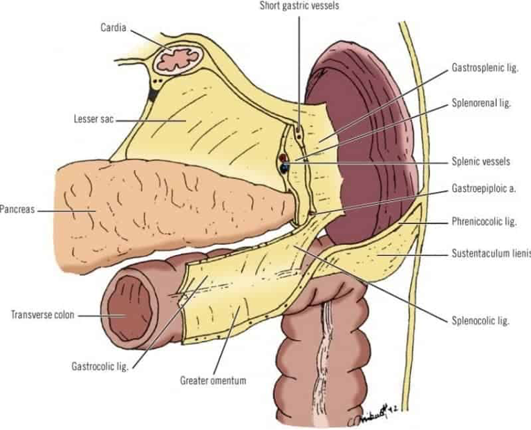
- Lies at the left margin of the Lesser Sac
- NB: Spleen does NOT project into the Lesser Sac
- NB: Spleen projects into the Greater Sac
- NB: Spleen is separated from the stomach by the peritoneum of the Greater Sac
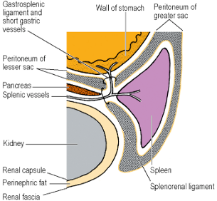
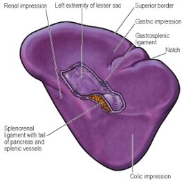
- Hilum lies at the angle between the Stomach and Left Kidney
- Each impresses a concavity alongside the attached splenic vessels
- Long axis – lies along the line of the 10th rib
- Lower pole – does not normally project beyond the Midaxillary Line
- Small colic area lies in contact with the Splenic Flexure and the Phrenicocolic Ligament
- Visceral surface invests all surfaces – gastric, diaphragmatic, colic and renal
- Convex surface related entirely to the diaphragm
- Gastrosplenic Ligament
- Two leaves of the Greater Omentum that pass from the Hilum forwards to the Greater Curvature of the Stomach
- Contains the Short Gastric Vessels
- Splenorenal Ligament
- Two leaves of Greater Omentum that pass backwards to the front of the Kidneys
- Hilum makes contact with the Tail of the Pancreas
- Contains Tail of Pancreas and Splenic Vessels
- Phrenicocolic Ligament
- Connects Splenic Flexure to the Diaphragm
- Small colic area of Spleen lies in contact with this ligament
- Phrenicosplenic Ligament
- Attaches Spleen to the Diaphragm
- Spleen must double in size before its anterior border passes beyond the left costal margin
Relations
Closely Related Structures
- Diaphragm
- Splenic Artery and Vein
- Short Gastric Arteries
Closely Related Organs
- Stomach
- Left Kidney
- Splenic Flexure of the Colon
- Pancreas
Arterial supply
- Splenic artery – which is a tortuous vessel at superior border of pancreas.
- Gives off left gastroepiploic and short gastrics prior to hilum.
- Brnachs to the pancras
- Dorsal pancreatic artery
- Anastomoses with the pancreatico-duodenal arcade
- Pancreatica magna artery
- Caudal pancreatic artery
- Dorsal pancreatic artery
- Divides into multiple branches that enter hilum of spleen.
- Distributive pattern - can be multiple branches which come off the main trunk 2-3cm from the Hilum.
- Magistral type – single pedicle enters into the hilum before branching off.
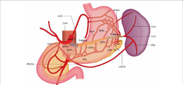 Fig. 1 Arterial splenic supply. Arteries of the tail of the pancreas (AsTP), common hepatic artery (CHA), caudal pancreatic artery (CPA), great pancreatic artery (GPA), inferior polar branch (IPB), left gastric artery (LGA), left gastro-omental artery (LGOA), lower lobar artery (LLA), posterior gastric artery (PGA), right gastro-omental artery (RGOA), short gastric arteries (SGAs), short pancreatic arteries (SPAs), superior polar Branch (SPB), splenic artery (SPLA), transverse pancreatic artery (TPA), upper lobar artery (ULA)
Venous drainage
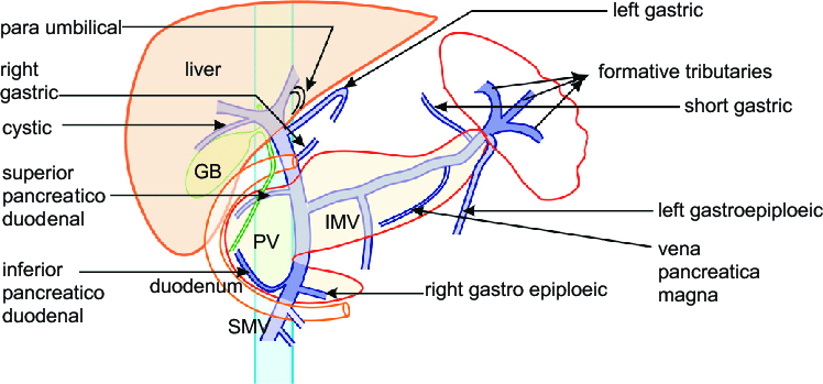
- Coalescence of open sinuses via reticular trabeculae into the splenic vein, exits hilum and courses medially to converge with the inferior and superior mesenteric veins to form the portal vein.
Lymphatic Drainage
- Drains into several nodes at the hilum ⇒ Pancreaticosplenic Nodes ⇒ Coeliac Nodes
Nerve Supply
- Sympathetic fibres only via the Coeliac Plexus
Function
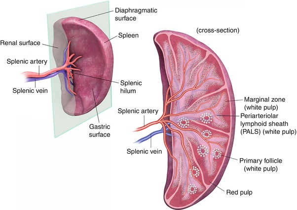
- Red pulp – site of blood filtration and haemolysis
- White pulp – opsonisation of extracellular organisms e.g. encapsulated bacteria, by B lymphocytes
- Marginal zone – antigen presenting cells to initiate lymphocyte activation
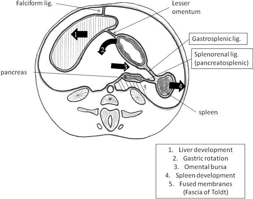
Spleen embryology
- Begins development in the 6th week
- Starts as several mesodermal cells in the Dorsal Mesogastrium
- Dorsal Mesogastrium, due to the splenic presence, becomes divided into the Splenorenal and Gastrosplenic Ligaments.
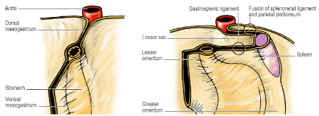
- Spleen, thus, comes to lie at the left margin of the Lesser Sac
- Original condensations become aggregated into a single organ
- Mesenchymal budding
- Splenic notch may represent a region where there is incomplete fusion
Accessory spleens/splenunculi
- Locations include the hilum, the gastrosplenic ligament, and the pancreatic tail
- Usual location – along the Splenic Vessels or in the Peritoneal Attachments
- Rarely larger than 2cm
- Can also be found in the greater omentum or even down in the pelvis.
- Develops from a mesenchymal condensation from the dorsal mesogastrium during the 5th week of development.
Surgical Approach
- Splenectomy
- Essentially involves cutting its 2 pedicles – the Splenorenal and Gastrosplenic Ligaments
- In an emergency, the Left or Posterior layer of the Splenorenal Ligament is incised and the Spleen turned medially so that Splenic Vessels can be dissected away from the Tail of the Pancreas and ligated.
- Arteries should be ligated before the veins
- Short gastric vessels in the Gastrosplenic Ligament are then divided and removal completed
- In elective setting, it is usual to enter the Lesser Sac by dividing the Gastrosplenic Ligament and its vessels and then deal with the Splenic Vessels and Splenorenal Ligament
- Caution:
- Be careful not to perforate the stomach when ligating the Short Gastric Vessels
- Be careful not to damage the Tail of the Pancreas and Splenic Flexure of the Colon