Section: Head and neck Curriculum: Curriculum, page 43
General
- Deep space neck infections following the multiple fascial planes
- Potentially airway and life threatening
Fascial Spaces
- Summary
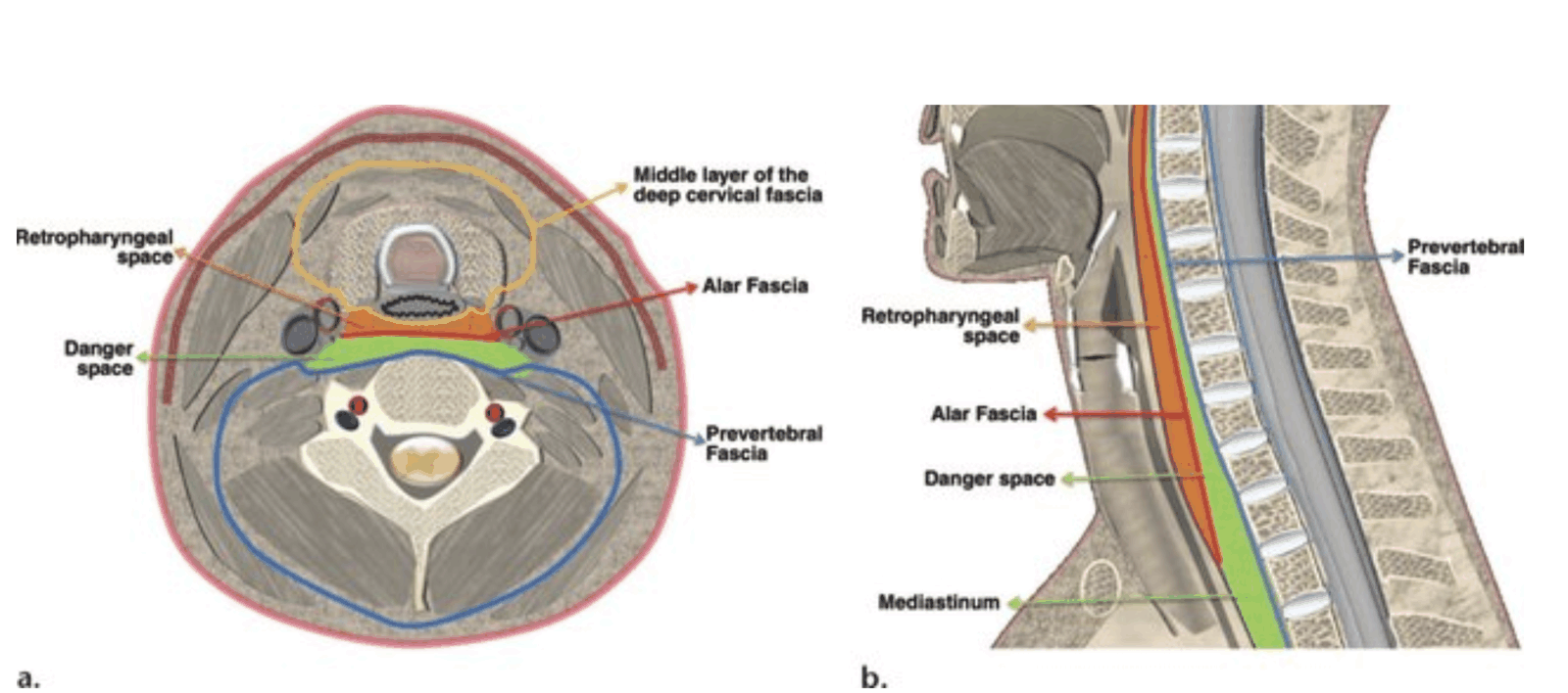
- Retropharyngeal space (between buccopharyngeal fascia and alar fascia (anterior PVF)
- Runs from skull to tracheal bifurcation (superior/post mediastinum)
- Alar fascia (anterior leaf PVF)
- Runs from skull to tracheal bifurcation (superior/post mediastinum)
- Danger space (between ant alar and post PVF of PVF
- Runs from skull to diaphragm
- Prevertebral space between post PVF leaflet and VC
- Can dissect along spinal column to coccyx
- Continuous with psoas fascia
- Retropharyngeal space (between buccopharyngeal fascia and alar fascia (anterior PVF)
Submandibular and Sublingual Space
- Borders
- Anterior and lateral - mandible
- Superior - mylohyoid
- Inferior - hyoid bone
- Medial - anterior belly of digastric
- Sublingual space is superior to the submandibular space (above mylohyoid) but continuous with it posteriorly
- Infection can cause
- Tongue swelling and airway obstruction
- Ludwig’s angina- potentially life threatening and rapidly spreading cellulitis originating from dental infection and causing infection of the floor of the mouth (all three spaces - submandibular, submental and sublingual)
Parapharyngeal Space
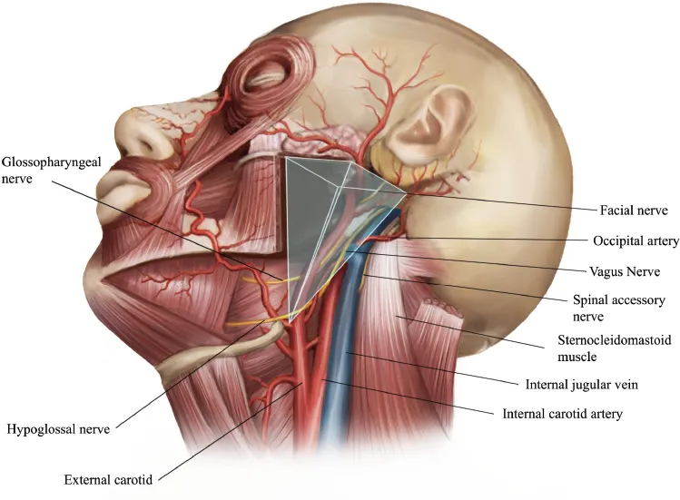
- Inverted cone - base of skull to hyoid
- Boundaries
- Lateral
- Superficial layer of deep cervical fascia extending between styloid process and mandibular ramus
- Mandible
- Muscles of mastication
- Medial
- Superior pharyngeal constrictor
- Inferior
- Hypoid
- Superior
- Skull base
- Lateral
- Communicates with retropharyngeal space laterally at carotid sheath area
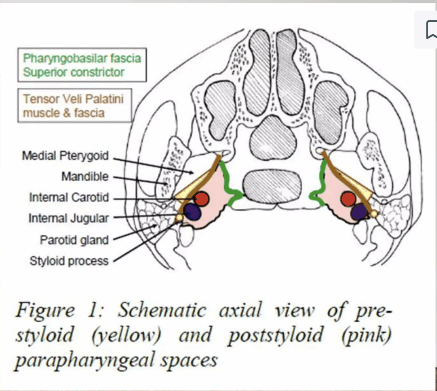
- Divided into 2 compartments by styloid process + muscles, stylomandibular ligament and their attachment to hyoid bone
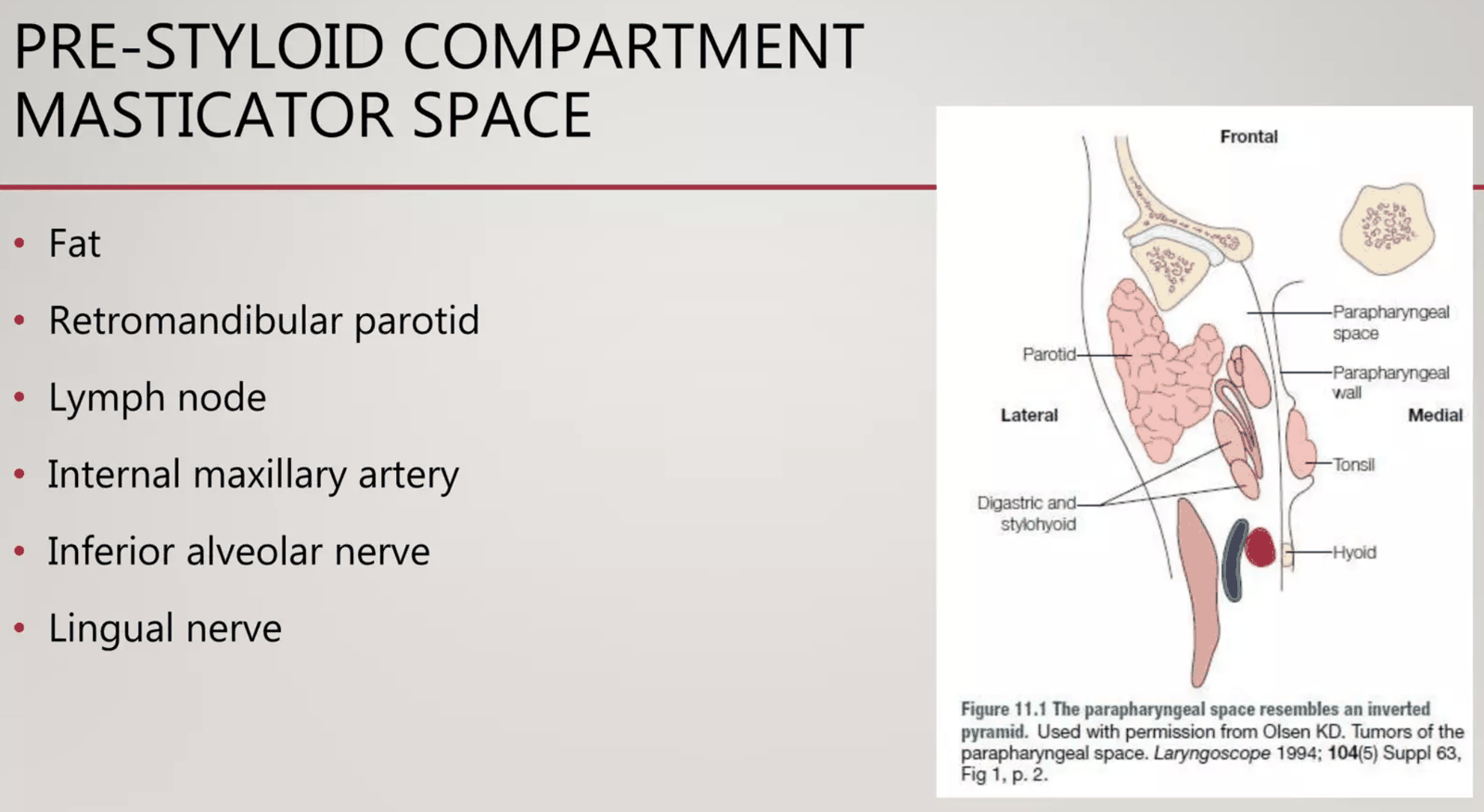
- Anterior (muscular)
- No important contents
- Closely related to tonsillar fossa
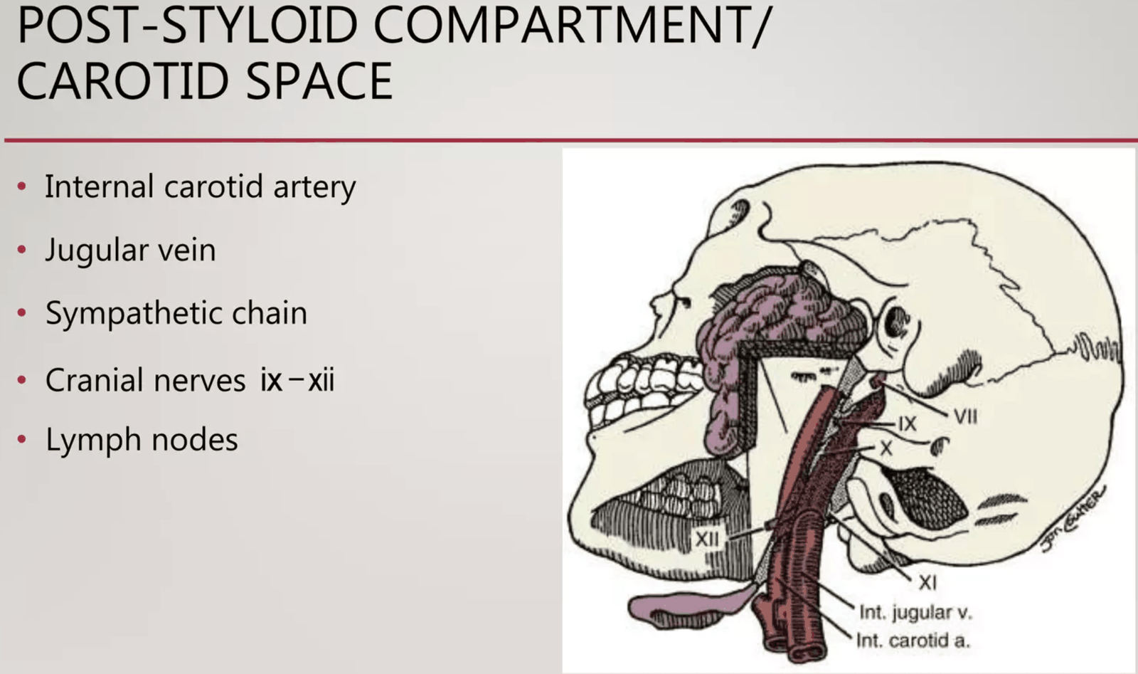
- Posterior (neurovascular)
- CN 9 and 12 superiorly
- CN 10 inferiorly
- Carotid sheath and contents
- Sheath runs in posterior aspect of parapharyngeal space
- Pierces apex of cone to enter mediastinum
- Sympathetic trunk
- Anterior (muscular)
- Infection - from pharyngitis, tonsillitis, parotitis, otitis or mastoiditis, odontogenic infection and masticator space infection
Retropharyngeal Space
- Behind hypopharynx and oesophagus
- Between pretracheal fascia anteriorly and alar fascia posteriorly
- Anteriorly - buccopharyngeal fascia which surrounds the constrictor muscles
- Posteriorly - alar fascia which is a delamination of the prevertabral fascia that attaches to the buccopharyngeal fascia at about C7
- Communicates with parapharyngeal space laterally
Danger Space
- (MOST important FOR INFECTION SPREAD FROM NECK TO CHEST)
- Located behind the retropharyngeal space
- Behind alar fascia and in front of the prevertebral fascia
- From base of skull to diaphragm through POSTERIOR MEDIASTINUM
Prevertebral Space
- Base of skull to coccyx and continuous with psoas fascia
- Posterior to the danger space
- Anterior = prevertebral fascia
- Posterior = Vertebral column
- Infection can dissect all the way down the spinal column
Other spaces
- Pretracheal Space
- Peritonsillar Space
- Parotid Space
Lymph Nodes
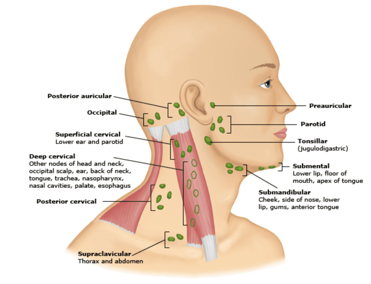
- Lateral cervical chain = common root for drainage
- Large deep chain = all H+N final drainage
- Along carotid sheath
- When infected → enlarge → can cause bacteraemia
- MALT - mucosa associated lymphoid tissue (like GALT)
- Waldeyers ring
- Nasopharyngeal lymphoid tissue aggregation
- Palatine, lingual, adenoid and tonsillar lymphoid tissue
- When inflamed can cause acute AW obstruction
- Waldeyers ring
Pathogenesis
- Arise secondary to primary infections of
- Mandibular teeth
- Tonsils
- Parotid
- Deep cervical LN
- Middle ear
- Paranasal sinuses
Microbiology
- Polymicrobial
- Normal flora of upper respi tract and its all interconnected
- Can vary based on location
- Odontogenic
- G+ - strep viridans, strep anginosus
- Anaerobes - peptostrep, bacteroides, actinomyces
- Oropharyngeal
- Strep pyogenes, haemophilus, fusobacterium
- Otogenic
- Staph aureus, pseudomonas
- Sinogenic
- Strep pneumoniae, haemophilus, moraxella, staph aureus
- Prevertebral
- Staph aureus, pseudomonas, mycobacteria, fungi
- Less common bugs in susceptible (imm comp, DM, trauma)
- Pseudomonas
- Enterobacter
- Klebsiella pneumoniae
- Odontogenic
Clinical
- Sepsis
- Sore throat
- Trismus - inability to open jaw
- → pressure/infection on muscles of mastication (masseter/pterygoids) or
- CN 5 motor branch
- Dysphagia/odynophagia
- Stridor/SOB
- Dysphonia/hoarseness - CN10
- Unilateral tongue paresis - CN12
- External palpation not helpful but oral cavity palpation may be
- Bulging pharyngeal wall (unilateral)
- Torticollis
- Stiff neck
- Crepitus
- Examination includes - full ENT exam
Investigations
- If AW compromised → SORT AW FIRST
- Bloods and cultures
- CT head, neck and maybe chest with IV contrast
- MRI
- X-rays - thickened prevertebral soft tissues, air-fluid levels, FB, cervical lordosis, tracheolaryngeal forward displacement
Management
- Concepts
- Early ICU/anaesthetics/ENT involvement
- AIRWAY
- Assess and secure early
Medical
- IV abx - maximum doses of broad coverage and narrow spectrum with culture results
Radiological
- USS or CT drainage
- Consider in the stable pt with no airway compromise and infection confined to the neck
- Low threshold to proceed to operative management
Surgical
- Considerations
- Only do when there is evidence of pus/collection
- Doing it too early can destroy the natural tissue planes and hasten the spread of infection
- AIRWAY
- Indications
- Unstable patient
- Airway compromise
- Failed non-operative mx
- Complications
- Facilitating infection spread - disruption of natural tissue planes
- Nerve injury - CN 5, 7, 9, 10, 11, 12
- Vascular injury - carotid sheath
- Haematoma
- Tracheoesophageal injury and fistula How To Do
Incision and Drainage
- Transoral I+D
- Transcervical I+D
Summary of Abscess Types
- Parapharyngeal Space
- Cause
- Dental, peristonsillar abscess, parotitis, otitis, mastoiditis
- Clinical
- Trismus, swelling below angle of mandible, medial pharyngeal wall bulging, sepsis
- Anterior compartment - std, posterior compartment - Horners or the like for CN 9, 10 and 12
- Complications
- Carotid sheath involvement (carotid mycotic aneurysm, IJV septic emboli), AW obstruction, sepsis
- Cause
- Retropharyngeal Space
- Clinical - as above
- Complications
- Can track into danger space - entire length of posterior mediastinum
- Mediastinitis and empyema
- Can track into anterior and posterior regions of superior mediastinum
- Aspiration pneumonia
- Mx for para and retro
- CT/MRI
- IV abx
- Source control
- Remove causative tooth
- Percutaneous drainage if confined to neck and no AW compromise
- Surgical drainage if pus and going into chest
- Cervical and thoracic approaches
- SLT to clear the swallow prior to reinstituting diet
- Prevertebral Space
- Cause
- Discitis/ostemyelitis - DM, immune comp, IVDU, EtOH
- Clinical
- As above
- Plus cord compression - spinal epidural abscess
- Mx
- Same as above - dx, abx, drain if pus either USG or surgically
- Cause
- Other Considerations
- Ludwig’s Angina
- Bilateral infection of floor of mouth - submandibular, sublingual and submental spaces
- Usually secondary to dental infection - mandibular molars
- Polymicrobial cellulitis - aggressive and rapidly spreading
- Can compromise AW
- Ludwig’s Angina