Emergency Cricothyroidotomy
- Horizontal stab incision between the Cricoid and Thyroid Cartilages
- Structures encountered: Skin, platysma, investing layer of deep cervical fascia and Cricothyroid Membrane
- Principles
- Aseptic equipment if possible
- Quick prep of the neck and some sterile gloves.
- Positioned with their neck extended as much as possible
- Open procedure
- 2-3 cm midline vertical skinned incision
- centered over the cricothyroid membrane
- midline incision avoids the anterior jugular veins and bleeding from those.
- Dissecting sharply to the cricothyroid membrane and pierce this with my scalpel
- Artery clip or similar clamp to insert it into the cricothyroid membrane and dilate
- Place a small ET tube of size 5 or 6 directly into the airway.
- 2-3 cm midline vertical skinned incision
- Scalpel bugie technique
- Identify cricothyroid membrane and stabilise with nondominant (ND) hand
- With scalpel in dominant (D) hand make a horizontal stab incision through cricothyroid
- membrane
- Rotate blade through 90 so that the blade points caudally
- Pull scalpel towards you, maintain perpendicularity producing a triangular hole
- Switch hands so that the ND hand now stabilises the scalpel
- With the bougie pointing away and parallel to the floor, insert tip into trachea using the blade as a guide
- Rotate and align bougie to allow insertion along the line of the trachea
- Reoxygenate via bougie with jet ventilator
- Railroad lubricated 6.0 ETT (remove 15-mm connector to aid passage over bougie).
- Continually rotate tube to facilitate placement
- Remove bougie
- Reattach 15-mm connector and ventilate via circuit
- Secure tube and check bilateral ventilation
Tracheostomy
Preparation
- Review indications and imaging
- Consent patient
- GA, ivabx
- Position supine on shoulder roll with head ring, neck + head extended
- Mark thyroid and cricoid cartilages, midline, suprasternal notch
Surgery
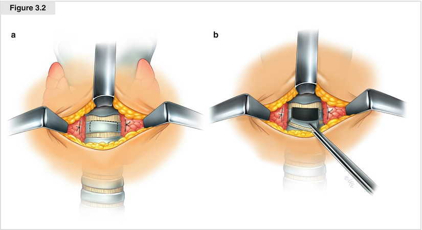
- Horizontal incision over trachea
- Raise subplatysmal flaps and retract with 2x Joll’s retractors
- Separate straps in midline
- Divide isthmus of thyroid and ligate with 3-0 vicryl
- Use tracheal tenaculum hook in cricoid cartilage to retract superiorly to better expose trachea Identify 2nd and 3rd tracheal rings
- Ask anaesthetics to turn down O2 as much as possible and deflate ETT balloon
- Incise tracheal rings vertically and horizontal above to raise inferior Bjork flap–hold up and open with a hook retractor.
- Suture flap to skin with silk sutures
- Ask anaesthesia to withdraw ETT until end can be seen just above level of tracheotomy
- Immediately place tracheostomy tube with obturator
- Inflate cuff, and swap ventilator tubing onto tube
- Check misting + CO2
- Haemostasis
- Layered closure with absorbable sutures, suture tracheostomy cuff to neck and secure with tie around neck
Postop
-
Confirm adequacy of ventilation by checking CO2, chest rise, auscultate
-
Haemostasis
-
Regular suction
-
Plan for trache removal/permanence
-
Indications for Surgical Airway in trauma
- Laryngeal #
- Penetrating throat and neck injuries
- Severe maxfax injuries with bleeding, deformity, oedema
- Airway burns/inhalation injuries
- Can’t intubate can’t ventilate
-
Complications
-
Immediate complications
- Airway loss
- Bleeding
- Can be anterior jugular, carotid, IJV, innominate, isthmus of the thyroid.
- Air leak
- subcutaneous emphysema
- Tube falling ou
-
Late complications
- Lack of spontaneous closure or healing of the tracheostomy once it’s removed
- Requiring surgical repair
- Infection
- Scarring
- Erode into large veins
- Inominate vein
- Can have herald bleed just prior to life-threatening hemorrhage.
- Iinsert a gloved finger into the tracheostomy stoma and occlude the vessel against the sternum and call for help.
- Inominate vein
- Tracheosophageal fistula
- Tracheal stenosis.
- Lack of spontaneous closure or healing of the tracheostomy once it’s removed