Section: Trauma Curriculum: Curriculum, page 75
(AT) MIST
- A - Age - name, age, sex
- T - Time of incident
- M - Mechanism of injury and Medical Complaint
- I - Injuries sustained and Illness
- S - Signs and Sx
- T - Treatment
Principles
- Simultaneous assessment and resuscitation
- Life-saving surgery
- Complete physical examination
- Diagnostic studies if pt becomes hemodynamically stable
- Broad Concepts
- 60% of severe trauma pts die within 1st 4 hrs in hospital
- Haemorrhage or CNS damage
- Require rapid restoration of adequate tissue perfusion and haemorrhage control
- 60% of severe trauma pts die within 1st 4 hrs in hospital
- Goals
- Unstable pt → Theatre or ICU within 30 mins (unless emergency ED surgery)
- Stable pt → ED time 30-60 mins
- In CT or ICU within 60 mins
- Can only move onto secondary survey from primary once H/D stable
- If not, needs OT or ICU
Primary Survey
- Goals
- Identify and treat life threatening injuries
- A→E
- Identified in the order that the injuries will cause death
- Establish patent airway and c-spine control
- Adequate ventilation
- Maintaining circulation (including intravascular control volume and cardiac function)
- Assessing the global neurological status
ABCDE
- Systematic and simultaneously assess and treat
AIRWAY and C-spine
- Early ETT with cervical spine protection
- In severe trauma when unconscious or in shock
- Indications for Surgical Airway Cricothyroidotomy
- Laryngeal #
- Penetrating throat and neck injuries
- Severe maxfax injuries with bleeding, deformity, oedema
- Airway burns/inhalation injuries
- Can’t intubate can’t ventilate
- Options
- Emergency cricothyroidotomy
- Elective tracheostomy
- Priorities
- Clear upper AW
- Give high flow O2
- Obtain definitive airway → ETT (cuffed tube) or surgical
BREATHING
- Midline trachea
- RR/Sats/adequacy of breathing
- Tension PTX
- Finger thoracostomy and 3 sided occlusive dressing
- Then chest drain
- Massive HTX/flail chest/contusion/tamponade/tracheobronchial injury
- Chest drain
CIRCULATION
- = assessment for shock, IVL large bore, bloods (FBC, G+H, coags, gas), resus fluids, MTP, prevent and treat lethal triad
- Shock
- Cool/pale peripheries, prolonged CRT, thready pulse, reduced LOC
- Blood on the floor and 4 more
- Floor - External bleeding
- Chest - CXR
- Abdomen - eFAST
- Pelvis – Pelvic XR
- Extremities/long bones - Clinical exam and XR
- Neck veins and shock
- Assume hypovolaemic hemorrhagic shock if flat
- If dilated then assess for:
- Tension Pneumothorax
- Tamponade
- Myocardial contusion (cardiogenic)
- MI (cardiogenic)
- Air Embolism
- Tension PTX - CXR/clinical dx → Chest drain
- Tamponade
- Penetrating torso injuries → Cardiac box (nipples, clavs and costal margin)
- Distended neck veins, peripheral shut down, pulsus paradoxus
- Drop in BP on inspiration by > 10mmHg
- USS dx
- Options for tx
- Needle pericardiocentesis (not preferred in trauma)
- Guided with USS
- Subxiphoid pericardial window via emergency thoracotomy
- Needle pericardiocentesis (not preferred in trauma)
- Myocardial contusion - rare
- MI - common in elderly and may have caused the trauma
- Air Embolism
- Air in systemic system due to bronchopulmonary fistula
- Causes - blunt trauma (rib #) or gunshot/stab
- Can have neurology without hx of head injury - air bubble occlusion
- Check fundoscopy - air bubbles in retinal vessels
- Surgery
- Thoracotomy and hilar clamping
- Resuscitate (expand IV volume) and aspirate air from heart
- Repair or lobar resection
- Haemorrhagic shock
- Goal
- Stop the bleeding - if not possible
- IV access, blood sample and appropriate resus
- Identify cause/location of bleed
- Prevent and treat coagulopathy
- Prevent and treat hypothermia
- Stop the bleeding - if not possible
- Access - Preferably central
- Subclavian/jugular/femoral
- Seldinger vs open cut down
- Bloods - Routine and blood gas
- Assess for occult blood loss
- Pleural cavity → CXR/USS
- Thigh → Clinical and X-ray
- Abdomen (incl retro and pelvis)
- If CXR/USS and leg ok → laparotomy
- Resuscitation fluids
- Crystalloid to start but quick change to blood products
- MTP activation
- Use of 2 units
- Ongoing bleeding requiring transfusion
- Local protocol for ratios
- Monitoring with bloods (incl. goal directed TEG/ROTEM)
- TXA when indicated
- Permissive hypotension in unstable bleeders
- Criteria for adequate resuscitation
- Maintain peripheral perfusion
- UO 0.5mL/kg/hr (1mL/kg/hr child)
- Keep atrial filling P at normal level
- Goal
- Don’t give 2L crystalloid bolus
- Early haemostatic resuscitation
- Guided by TEG
- 1:1:1 ratio early and minimize crystalloid in hypotensive shock
DISABILITY
- GCS, pupils, gross neuro (lateralizing signs)
- Key components
- Determine LOC
- Assess pupil size and reactivity
- Check eye movements and oculovestibular responses
- Document skeletal and spontaneous movements
- Determine pattern of breathing
- Peripheral sensory exam
- GCS
- Consciousness made up of:
- Awareness - Goal directed/purposeful behaviour
- Use of language
- Protection from painful stimulus
- Arousal - Simple wakefulness
- Brainstem function - Any eye opening
- Coma - Loss of awareness and arousal
- Awareness - Goal directed/purposeful behaviour
- Consciousness made up of:
- Key marker is deterioration in GCS - prompts emergent CT head
ENVIRONMENT/ EXPOSURE
-
Remove clothes and log roll
-
Check temperature and warming measures
-
Body temperature fluids via rapid infusers, warmed mattresses, warm air blankets
-
Prevent heat loss - blanket, Bair hugger, environment
-
Hypothermia shifts O2 curve left → Reduced oxygen delivery
- Reduce ability for liver to metab citrate/lactic acid
- Arrhythmia
-
Primary Survey Diagnostic Studies for unstable pt ADJUNCTS
- CXR
- Pelvic XR
- FAST scan - blood in abdomen/chest/pericardium/tension PTX
- Focused Assessment with Sonography for Trauma
- LUQ - Between Spleen and Kidney
- RUQ - Between Liver and Kidney
- Pelvis - Between Bladder and Rectum
- eFAST - Pericardial window and HTX as well
- Can be repeated if clinical change
- Focused Assessment with Sonography for Trauma
-
Blood gas
-
ECG
-
NGT
-
IDC
-
Monitoring - oximetry, BP
-
Then figure out disposition: based on haemodynamics and resus response
- CT
- Surgery
Secondary Survey
- Cannot do it until patient is stable
- What is it
- AMPLE HISTORY
- Allergies
- Meds
- PMHx or pregnant
- Last meal
- Events and environment
- Pt, responders, family
- Tetanus
- AMPLE HISTORY
- Top to toe and front to back examination
- If H/D normal pt - time for full assessment and CT
- If H/D stable pt - time for CT
- If H/D unstable
- Can’t leave resus
- Fast USS/XR chest and pelvis
- DPL if FAST neg but still suspicious or in mass casualty setting
- Penetrating Trauma
- Knives, missiles and impalement
- Velocity dependent on type
- Secondary effects → Infection - FB introduction and intra-abdo soiling
- Can use paperclips to see wounds on x rays - Help determine what is injured
Emergency Department Surgery
Head Trauma
- Surgical AW with indications listed above
- Scalp lac suturing/clipping with pressure
- ICH
- Dilated pupil and contralateral hemiplegia → Coning
- Hyperventilation
- Hyperventilation = blow off CO2
- Hypocapnia reduces ICP
- By reducing CBF
- Keep ETCO2 (proxy for PaCO2) ABOVE 30mmHg
- Mannitol IV 1g/kg bolus or hypertonic saline 7.5% 1mmol/kg
- Temporize
- Immediate decompression
- Burr hole/Craniotomy
Chest Trauma
- Tension PTX
- Clinical dx
- Tracheal deviation, hypertympany, reduced BS
- Neck vein distension
- Treatment - Needle decompression and chest drain on UWSD
- Clinical dx
- Cardiac Tamponade
- Only done in ED if SBP < 50mmHg with no time to go to OT
- Or low SBP despite volume resus
- Treatment
- Left anterolateral thoracotomy
- Relieve tamponade
- Control bleeding source
- For ventricular penetrating injury
- Foley catheter in defect and inflate balloon
- Clamp foley and don’t traction it!
- Suture defect
- Foley catheter in defect and inflate balloon
- Left anterolateral thoracotomy
- IC vessel bleed or lung parenchymal bleeding (haemothorax) (from rib #)
- Self-limiting usually
- Collect blood, if possible, for auto-transfusion
- Aortic transection
- Dx
- Widened mediastinum on CXR
- CT arteriogram
- Tx
- Maintain BP ~ 100 systolic
- Dx
Abdominal Trauma
- DPL if FAST negative but high suspicion or pt still unwell
Pelvic Trauma
- Binder or external fixation (time consuming) to return pelvis to normal anatomy
- Majority of bleeding is venous so settles with binder
- If binded and still unwell → ? Arterial
- Angiogram/CT-A
- Embolisation or
- Laparotomy and extraperitoneal pelvic packing
- Long Bone #’s
- Reduction and splinting initially!
- External fixation eventually
- Put traction on distal limb to allow femoral alignment
- Allows thigh to become cylindrical again → Tamponades bleed
- Monitor distal pulses → If absent → CTA
- Requires arterial repair and bony fixation (perfusion over # tx)
- Peripheral Vascular Injuries
- Assess vascularity with doppler/CTA/Angio
Tertiary Survey
- Ideally within 24 hours of admission and repeated when pt is awake
- Repeat primary and secondary survey
- Review all notes/bloods/scans
- Pick up any missed injuries
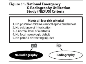
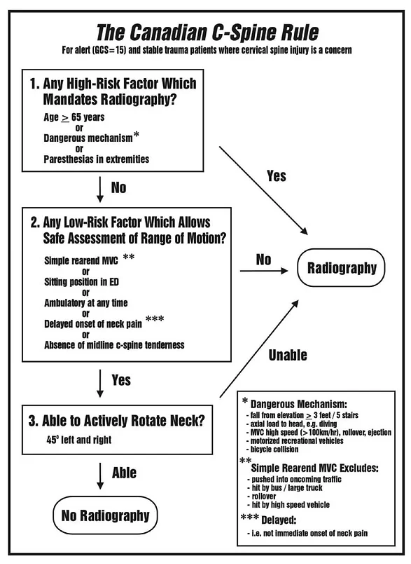
Cervical Spine Clearance
- Can be done clinically for some patients
- Intubated | Sedated | Distracting injury | Booze
- CT neck
- Normal → Neuro exam gross motor
- Ok = Clear
- Can’t do = ASPEN collar and ortho/MRI neck
- Abnormal = ASPEN collar and ortho/MRI neck
- Normal → Neuro exam gross motor
- CT neck
- Awake | No EtOH | No distracting injury
- Clinical assessment
- Midline tenderness or focal neurology
- Yes - X-rays | CT | ortho
- No - clear
- Midline tenderness or focal neurology
- Clinical assessment