- General
- Injuries are often missed, underestimated or have high rate of mortality
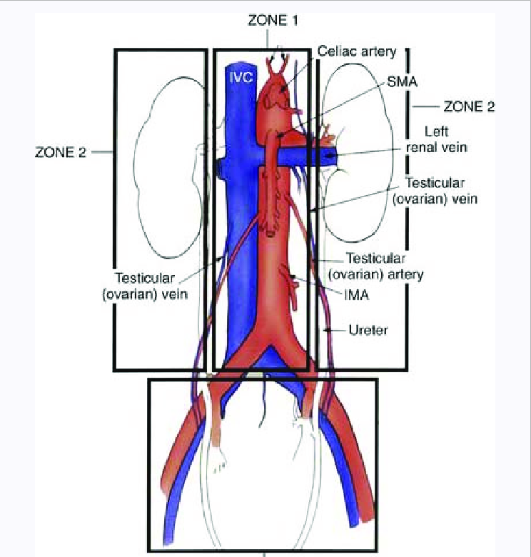
- Zones
- I - Central
- Between both psoas muscles and contains the abdominal aorta, inferior vena cava, pancreas, and midline duodenal structures
- II - Lateral x 2
- Lateral to the psoas muscles on bilateral sides and contains the kidneys, ureters, and portions of the colon
- III - Pelvic
- Management
- Zone 1 - Upper midline central retroperitoneal haematoma
- All should be explored
- Potential sites of injury
- Aorta/IVC
- Kidneys, Ureters, Renal vessels
- Duodenum/Pancreas
- Colon
- Zone 2 - Lateral retroperitoneal haematomas
- Only explore if expanding, thought to have injury to Colon or Ureter
- Usually related to renal bleed which should tamponade
- Zone 3 - Pelvic
- Non - expanding - don’t explore
- Ongoing bleeding
- External fixation of pelvis
- Angioembolization
- If angio is unavailable and enlarging pelvic haematoma/ongoing bleeding
- Extra-peritoneal pelvic packing
- General
- Unstable and peritonitis - Laparotomy
- Observe if stable, not too sore and imaging is reassuring
- Serial abdominal examinations
Retroperitoneal Haematoma
- Zones
- I - central (further subdivision based on apex of haematoma)
- Supracolic
- Juxtarenal or suprarenal aorta, SMA, coeliac
- Infracolic
- Aortic bifurcation/IMA
- Transperitoneal or left medial visceral rotation approach
- Left or right
- If apex is right of midline and primarily venous bleeding
- If apex is left of midline and primarily arterial bleeding
- II - Lateral x 2
- Leave alone if not expanding
- Renal injuries - non operative and selective embolization approach
- If penetrating injury or expanding - explore
- Need to rule out occult post colonic and ureteric injury
- To prevent abscess/urinoma formation
- III - Pelvic
- If stable and CT proven blush - Angioembolise
- If found at surgery and non-expanding - Leave alone
- If found at surgery and expanding - Manage
- Extraperitoneal pelvic packing
- Pelvic binder is on
- Grasp peritoneal edge of inferior 25% of laparotomy wound
- Develop Retzius space (pre-peritoneal plane) all the way round to posterior rectum on both sides
- Evacuate clot
- Packs x 2-3 on each side
- If fails then culprit Internal Iliac Artery ligation
- If works then to IR for assessment/embolization
- Pelvic fixation/binder
Major Abdominal Vessels
Aorta
- Supracoeliac Aortic cross clamping
- Retraction
- Left Liver lobe → Right shoulder
- Body of Stomach → Left hip
- Dissection
- Lesser omental window (Pars flaccida)
- Crus peritoneum incised to expose muscle fibres which are split
- Retract oesophagus to patients left
- Adventitia of Aorta exposed, develop bilateral planes and clamp
- Satinsky clamp placed supracoeliac
- Suprarenal aortic exposure
- First do a supracoeliac clamp
- In context of central supracolic haematoma
- Then left medial visceral rotation
- Exposes coeliac/SMA/L renal vessels/iliacs
- SMA and coeliac nerve plexuses require dissection to access them
- Distal Aorta
- Retraction
- SB right | Transverse Colon superior | Descending Colon left
- Injury Treatment of Aorta
- Aortic injuries need repair
Coeliac Axis
- Proximal control - supracoeliac aortic clamping or anterolateral thoracotomy
- Left medial visceral rotation and may need to mobilise left triangular lig and segments 2+3
- Difficulties
- Dissection coeliac plexus of nerves - dense and fibrous
- Short origins of left gastric, splenic and common hepatic
- All can be tied off if SMA and IMA are intact - collateralization
- How
- 3.0 prolene suture repair
SMA
- Left medial visceral rotation or root of SB mesentery
- Comes off aorta distal to coeliac and proximal to left renal A
- If damage control → Shunt it
- If can’t be repaired then should be replaced with graft to infrarenal aorta
- Graft can kink if proximal end is placed too high
- Keep it away from pancreas
- SMV can be shunted or ligated
IMA
- Can be tied off
- Colon viability needs assessment
Renal Artery
- Repair via infrarenal or medial rotation access
- Down time poorly tolerated - 45mins = Complete loss of function
- Therefore may need nephrectomy at that time
Iliacs
- Proximal and distal control
- Mobilise sigmoid colon and caecum
- May need groin access for distal control
- Beware of the ureters crossing the iliac bifurcation
- Veins closely adhered to back of arteries - Don’t dissect, just enough to clamp
IVC
- Suprahepatic ? chest access
- Not controlled by Pringle’s maneuver
- Controlled with direct downward pressure of liver onto IVC
- Exposure and clamping of infrarenal IVC
- Divide coronary ligaments to displace superomedially and see supra IVC
- Infrahepatic
- Right medial visceral rotation
- Kocher’s will show infrahepatic IVC well and right renal vascular pedicle
- Direct pressure with swabs above and below
- With penetrating injury it may be through and through
- But all posterior injuries do not require repair
- If it does, repair them transcaval as can’t get posterior on outside easily
- Caval injuries below Renal Veins can be ligated if extensive but prefer lateral repair
- Above Renal Veins should be repaired - ligation is unsurvivable
Portal Vein
- In free edge of lesser omentum - Portal Triad
- Pringle’s maneuver
- If more proximal: Kocher’s
- If damage control: Shunt
- Portocaval shunting is a high mortality option to consider
Shunting
- Indications
- Can’t be repaired
- Damage control situation
- Rationale
- Rules
- Diameter of tube - ⅔ circumference of vessel
- Length of tube – 3 x length of defect
- What to use
- NGT/ETT/chest drain/anything
- Other issues
- Ends should be beveled
- Tie silk around tube - ⅓ | ⅔ division
- Technique
- Proximal and distal control
- Proximal control applied
- Place ⅔ end in
- Use suture to pull back to distal end
- Secure with ties
- Last for 2-3 days