Transverse abdominal incision with a midline extension
Diseased liver is mobilised
Because of portal hypertension, the recipient hepatectomy is often the most difficult part, esp. if previous surgery
CBD divided as close to liver as possible
Mobilisation
Dissect the intrahepatic vena and control it with a nylon tape.
Divide the peritoneal attachments of the liver, starting with the left triangular ligament and proceeding to the falciform ligament, thus exposing the anterior surface of the supra-hepatic vena cava.
Completely mobilising the retrohepatic vena cava from the posterior abdominal wall, ligating and dividing the right adrenal vein.
Ligate the portal vein and hepatic artery - maximising the length of the vessels for subsequent re-implantation of the new graft.
Occlusion of the vena cava and portal vein results in a reduction in cardiac output and may necessitate the use of veno-venous bypass
Liver implantation
After placing the donor liver in position
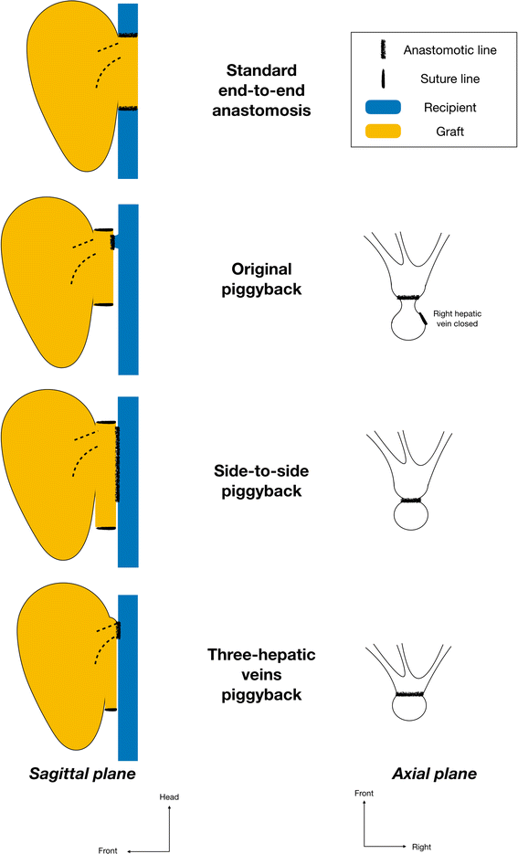
Supra- and infrahepatic caval anastomoses are performed
The portal vein and the hepatic arterial anastomoses are then completed and the graft is reperfused
Finally, biliary drainage is re-established
Duct-to-duct anastomosis, end-to-end (without a T-tube)
Roux-en-Y may be needed for biliary atresia or sclerosing cholangitis
Many pts undergoing liver transplantation are ill and the surgery involved can be very demanding
Optimal peri-operative management crucial for success and presents a major challenge
Blood loss during and after the transplant procedure can be considerable
Management of coagulopathy is particularly important
Measured repeatedly and corrected appropriately with FFP, cryoprecipitate and platelets