Definition
- Also known as “gluten sensitive enteropathy”
- Common immune mediated malabsorptive condition
- Abnormal response to Gliadin (component of Gluten)
- Associated with flattening of the small intestinal villi
Incidence
- Mainly European – 1/1000
- (NB: Ireland 1/300)
- Most common cause of malabsorption
Aetiology
- Genetics
- Immune disorder triggered by environmental agent (gluten) in predisposed individuals
- Association with HLA-DQ2 & HLA-DQ8
- Human Leukocyte Antigens
- Convey susceptibility
- Over 99% affected individuals have one of these
- Risk Factors
- Family history
- 2-5% of 1st degree relatives develop symptomatic Coeliacs
- Strong association with other auto-immune disorders
- Type 1 DM
- Autoimmune Thyroiditis
- Also – Down Syndrome, Turner Syndrome, Pulmonary Haemosiderosis
Clinical Presentation
- Childhood Presentation
- Present at 1-2 yrs
- Previously well then failure to thrive
- May have
- Diarrhoea
- Irritability
- Poor appetite
- May have
- Iron deficiency
- Short stature
- FHx
- Abdo distension
- Clubbing
- Adult Presentation
- Iron-deficiency
- or combined Iron & Folate deficiency
- Weight loss
- Diarrhoea
- Abdo discomfort (pain rare)
- Borborygmi
- Bloating
- Associations:
- Hyposplenism
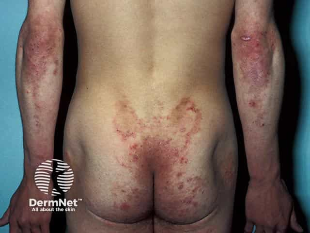
- Dermatitis Herpetiformis
- “Coeliac Disease of the skin”
- Vesicular, crusted, intensely pruritic lesions develop
- Located on the back, buttocks, and elbows
- Secondary infection common
- Iron-deficiency
Pathology
- Involves an abnormal cellular (T-cell mediated) & humoral response to gliadin
- Gliadin is a component of gluten (in wheat, barley), which acts as an epitope that the body reacts to
- Causing mucosal damage
- Get
- Loss of brush border enzymes
- Loss of stimulus for pancreatic & bile secretion
- Low pancreozymin & secretin
- Exudation of protein across denuded mucosa
- Protein losing enteropathy
Histology
- Macroscopically:
- Mucosal Atrophy
- Flattening of mucosal folds
- Fissure, scalloping, prominent submucosal vascularity
- Histological changes
- Increased intra-epithelial lymphocytes
- Villous atrophy / “clubbing”
- Hypertrophy of crypts
- Attempt to increase cell production
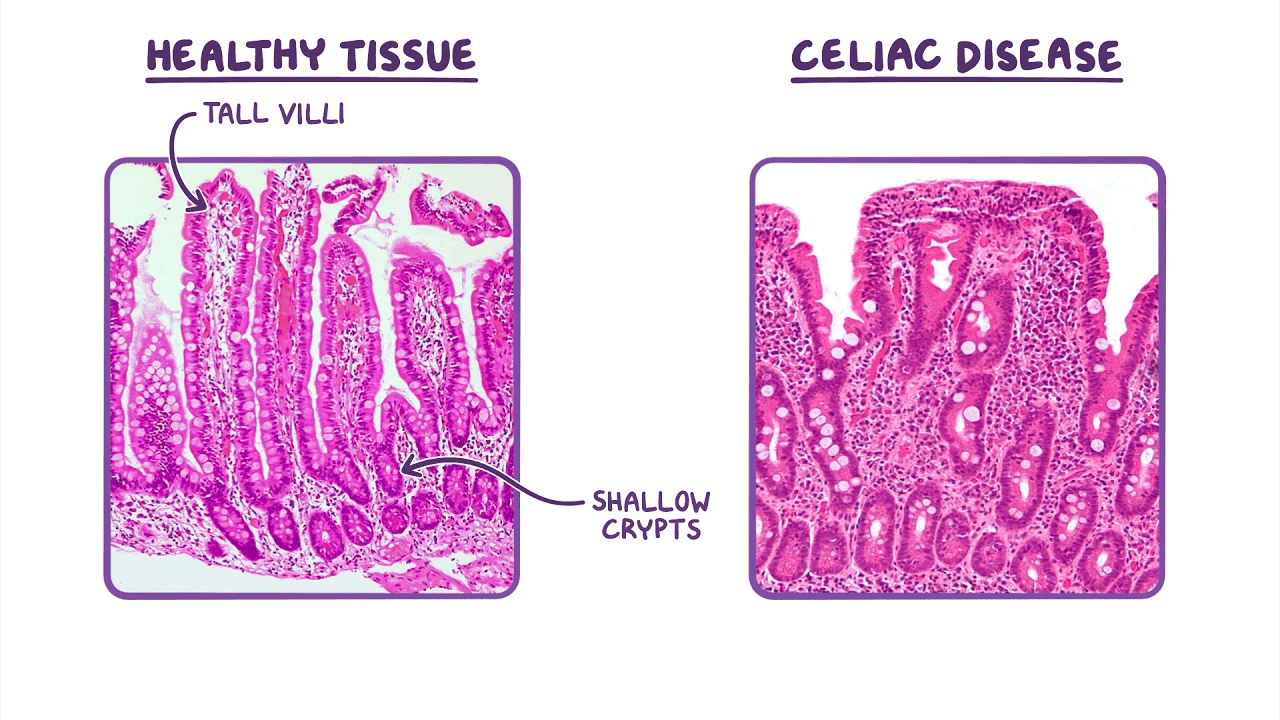
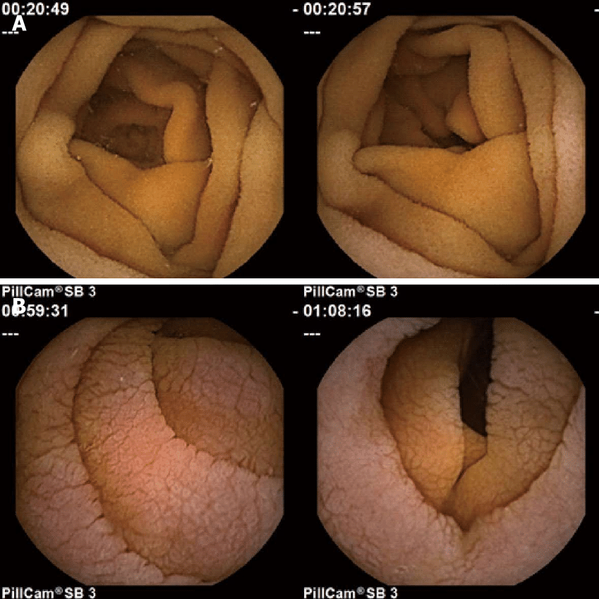 Normal A. Coeliac B
Complications
- Increased risk for lymphoma and GI cancers
- Uncertain whether compliance to gluten-free diet influences rate of cancer
- Lymphoma the most common
- GI cancers include
- Oesophageal SCC
- Small Intestine Adenocarcinoma
- Colorectal
- HCC
- NB: Significantly reduced risk of Breast Cancer
Investigations
- All testing for coeliac disease should be done when a patient is on a gluten-containing diet
- If low probability, screen with serology first, if negative, stop
- If high probability, do both serology and OGD
- Serological blood tests
- IgA anti-Tissue Transglutaminase antibody (anti-tTG)
- Specific test, good NPV
- IgA anti-Endomysial antibodies (EMA)
- Highest diagnostic accuracy
- Costly and not as widely available as anti-tTG
- IgA & IgG anti-Gliadin antibodies (AGA)
- IgA more specific (97%), but poor sensitivity (71%)
- IgG less specific (87%) but more sensitive (87%)
- IgA anti-Tissue Transglutaminase antibody (anti-tTG)
- Endoscopy – if positive serologic testing or high suspicion
- To confirm the diagnosis
- Need at least 4 x biopsies of post-bulbar duodenum
Management
- Strict gluten-free diet
- Avoid wheat, rye, and barley
- NB: Oats are ok
Follow-up
- Repeat biopsy at 3-4 months after dietary treatment
- Check mucosal improvement