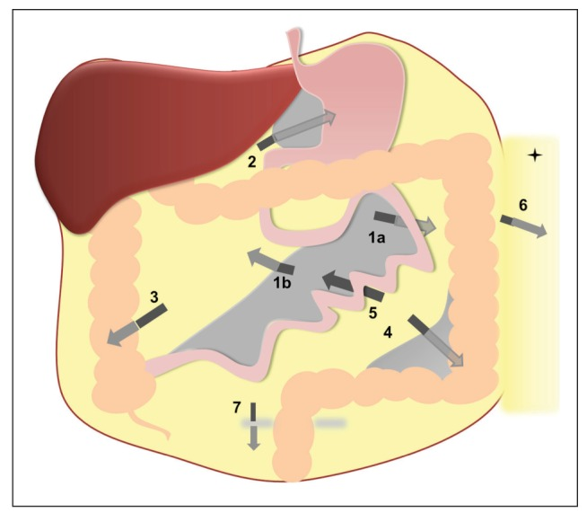
Paraduodenal hernias are internal hernias due to failure of the descending or ascending colonic mesentery to fuse with the posterior parietal peritoneum. Left paraduodenal hernia is more common and can cause closed-loop bowel obstruction and infarction.
Clinical presentation
The patient may have a history of recurrent positional post-prandial pain and can present acutely with pain and vomiting due to small bowel obstruction.
Pathology
Left paraduodenal hernia
- Definition: Protrusion of small bowel through paraduodenal (lateral to 4th part) mesenteric fossa of Landzert (located near ligament of Treitz)
- The more common of the two paraduodenal hernias (75%)
- Small bowel herniates through a single layer of distal colonic mesentery into a retroperitoneal fossa of Landzert (2% incidence at autopsy) that accompanies this abnormality
- There is congenital failure of fusion of the descending colon mesentery to the parietal peritoneum in the left upper quadrant
Right paraduodenal hernia
- Definition: Protrusion of small bowel through jejunal mesentericoparietal fossa of Waldeyer (located inferior to 3rd portion of duodenum)
- The less common of the two paraduodenal hernias (25%)
- Small bowel herniates through a layer of the ascending colon mesentery into the associated retroperitoneal fossa of Waldeyer
- There is congenital failure of fusion of the ascending colon mesentery to the parietal peritoneum in the right lower quadrant
- Associated with small bowel malrotation
Radiographic features
- These hernias usually appear as an encapsulated mass-like cluster of small bowel loops. A closed-loop obstruction may develop and strangulation may occur with engorged vessels, mesenteric oedema and free fluid. This can progress to infarction.
- Vascular landmarks around the neck of the internal hernia help to make a confident diagnosis:
- left paraduodenal hernia
- Encapsulated “cluster” or sac-like mass of small bowel loops located between pancreatic body/tail and stomach to left of ligament of Treitz
- Hernia sac may exert mass effect on posterior wall of stomach, duodenojejunal junction inferiorly and medially, and transverse colon inferiorly/anteriorly
- Crowded, engorged mesenteric vessels supplying bowel loops within hernia sac
- the inferior mesenteric vein (IMV) and ascending branch of the left colic artery are within the anterior neck of the hernial orifice
- Hepatic flexure of colon usually located anterior to hernia sac
- Inferior mesenteric vein (IMV) and left colic artery lie in anterior and medial border of hernia sac, with IMV often displaced to left
- right paraduodenal hernia
- Clustered, encapsulated small bowel loops in right upper abdomen lateral and inferior to descending duodenum
- Ascending colon located lateral to hernia sac
- Mass effect from hernia sac displaces right ureter laterally
- Unusual “looping” course of superior mesenteric artery (SMA) and vein (SMV) to supply bowel in hernia sac
- SMV rotated anteriorly and to left
- Twisted vascular jejunal branches behind SMA extend into hernia sac
- Main trunk of SMA and right colic vein located along anterior medial border of hernia sac
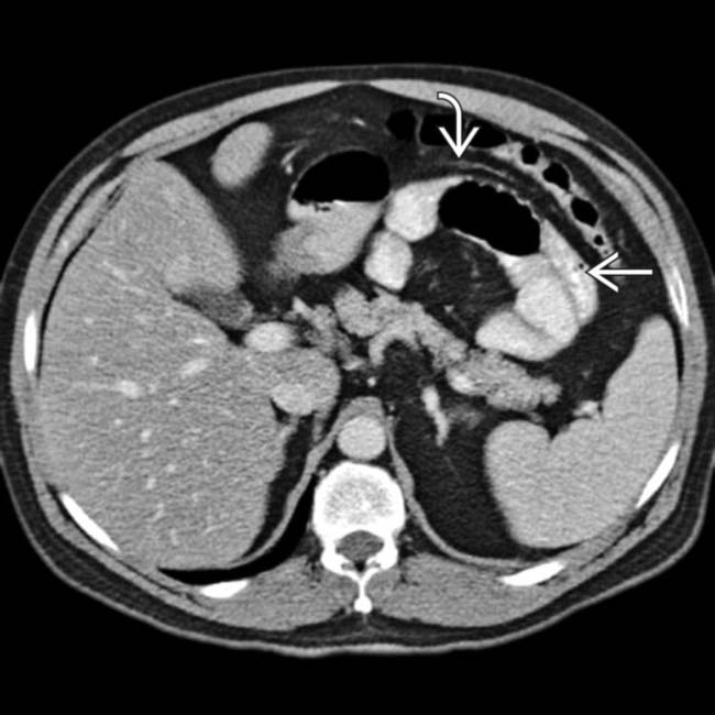 Axial CECT shows a cluster of dilated bowel image interposed between the pancreas and stomach. Note the displaced inferior mesenteric vein image that runs along the anterior edge of the hernia sac. This constellation of findings is characteristic of a left paraduodenal hernia.
- Clustered, encapsulated small bowel loops in right upper abdomen lateral and inferior to descending duodenum