Definition

  • Rotational anomalies and failure of fixation caused by:
    • Abnormal rotation of the embryonic gut

Incidence

  • Midgut volvulus usually present in first month of life
  • But can present into adulthood
  • Other forms of malrotations may present later:
    • Caecal volvulus
    • Internal hernia
    • Incidental finding of non-rotation

Pathophysiology

  • Process of normal rotation and fixation of occurs between 6-10th weeks gestation
    • Gut herniates - Gastrointestinal embryology
      • Revolves 270° anticlockwise around axis of SMA and vitellointestinal duct
      • Returns to abdominal cavity and fixates
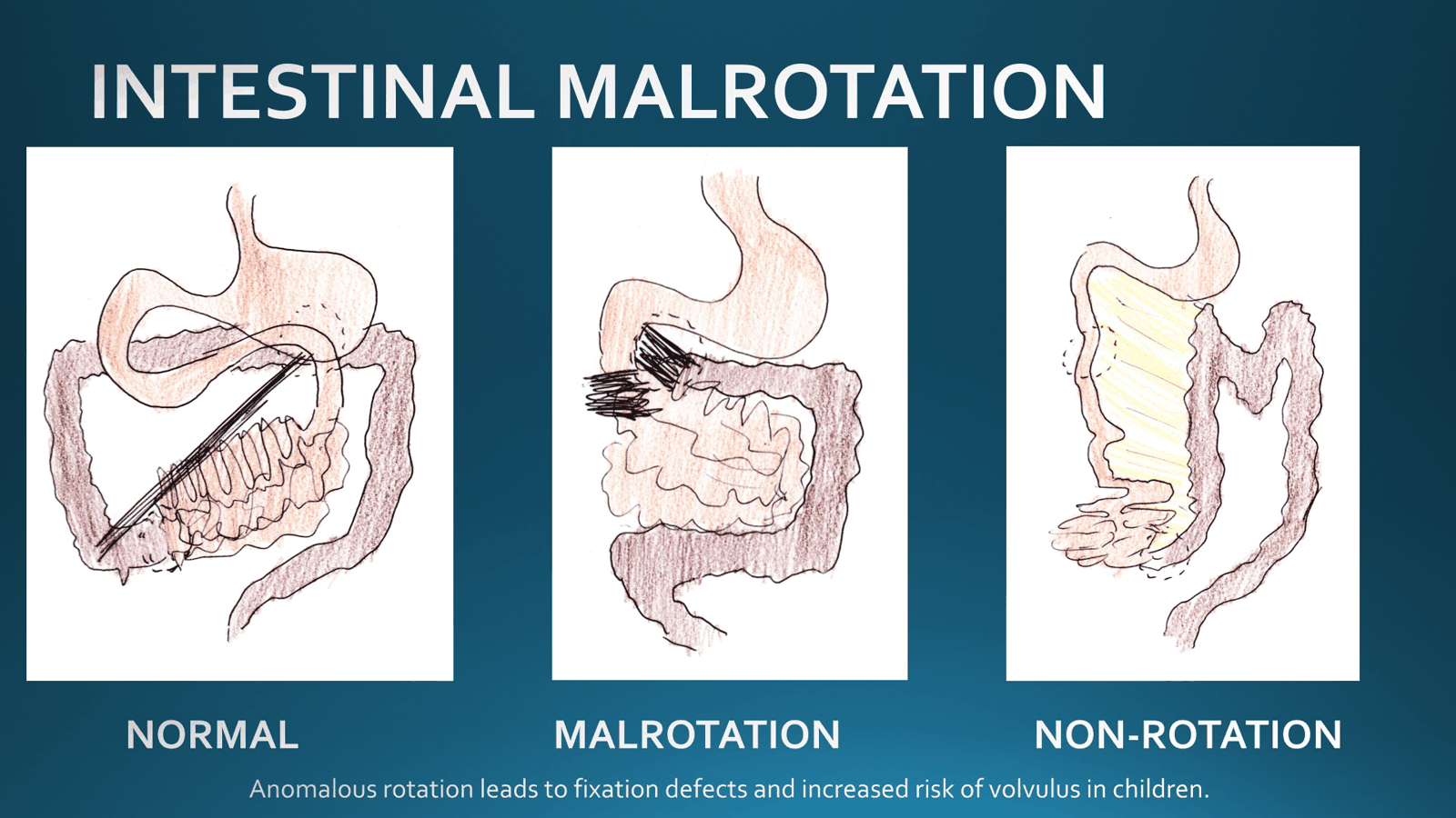
  • Non-rotation
    • No rotation occurs, mesentery wide based, no correction required
  • Malrotation
    • Rotation process interrupted - typically 90 degrees has occurred
    • Caecum and duodenojejunal junction close together
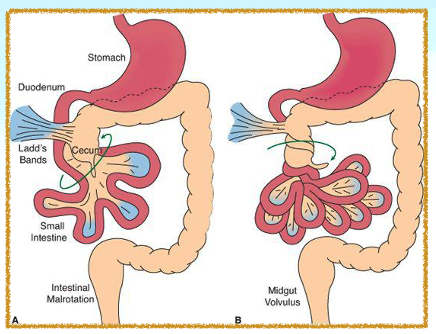
      • This leads to the caecum fusing to the peritoneum of the right lateral abdominal wall via Ladd bands.
    • Narrow based mesentery
    • Midgut volvulus may occur around SMA
      • Leading to intestinal ischaemia
    • Complications
      • Volvulus, e.g. Caecal
      • Paraduodenal hernia
      • Internal hernia (under Ladd’s band)
      • Ectopic appendicitis

Diagnosis

  • AXR
    • Absence of right-sided jejunal markings
    • Absence of stool-filled colon in the RLQ
  • Barium swallow
    • Abnormal DJ junction
      • Fails to cross midline to the left of the left-sided vertebral body pedicle
      • DJ lies inferior to the duodenal bulb
    • Z-shaped course of distal duodenum and proximal jejunum
    • Proximal jejunum on the RIGHT of the abdomen
      • Does not cross midline
  • USS
    • May show Whirlpool Sign
    • Clockwise wrapping of SMV and mesentery around the SMA
    • Switched position of SMA & SMV (i.e. rotation shows SMA on the right and SMV on the left)
  • CT-AP
    • Absence of retro-mesenteric D3
    • Abnormal SMA-SMV relationship
    • LB mostly on the left and SB mostly on the right

Management

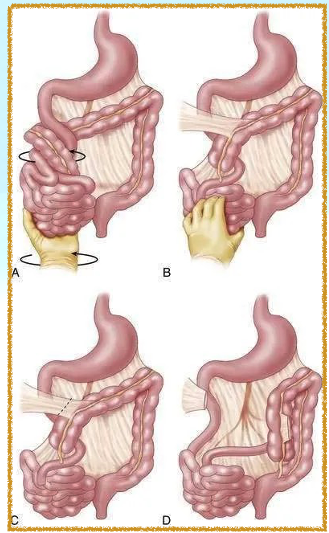
  • Laparotomy + Reduction of volvulus It has 6 key steps:   1)  Division of LADD’S bands   2)   Detorsion of the small bowel   3)  Widening of the mesentery via peritoneal incisions   4)  Assess luminal patency   5) Place bowel back in non-rotated position
    • Small bowel to the right, colon to the left   6) Appendicectomy